Abstract
Griscelli syndrome (GS) type 2 is an autosomal recessive disorder represented by pigment dilution and impaired cytotoxic T lymphocyte (CTL) activity. NK activity has been scarcely investigated in GS patients. Here, we describe a new patient, possessing a hemophagocytic syndrome with a homozygous Q118X nonsense RAB27A mutation. Single specific primer–polymerase chain reaction (SSP-PCR) was developed based on this mutation and is currently used in prenatal genetic analysis. As expected, CTLs in the patient are not functional and NK cytotoxicity against K562 or 721.221 cells is diminished. Surprisingly, however, we demonstrate that CD16-mediated killing is intact in this patient and is therefore RAB27A independent, whereas NKp30-mediated killing is impaired and is therefore RAB27A dependent. We further analyzed the signaling pathways of these 2 receptors and demonstrated phosphorylation of Vav1 after CD16 activation but not after NKp30 engagement. Thus, we identify a novel homozygous mutation in the RAB27A gene of a new GS patient, observe for the first time that some activating NK receptors function in GS patients, and demonstrate a functional dichotomy in the killing mediated by these human NK-activating receptors.
Introduction
Natural killer (NK) cells are the lymphocytes of innate immunity, able to identify and eliminate virus-infected and tumor cells without prior antigen stimulation.1,2 In addition, secretion of cytokines by NK cells recruits and activates additional immune cells and modulates the adaptive immune response.3,4 Unlike other lymphocytes, NK cells do not express any single rearranged receptor such as the T- or B-cell receptors, but rather use a limited number of activating receptors.5,6 The major human NK-activating receptors are NKp30, NKp44, NKp46, NKG2D, and the FcγRIIIA receptor CD16.2,7 Engagement of NK lysis receptors leads to cytoskeleton reorganization, translocation of lytic granules, and directed exocytosis to facilitate the final step of killing. The precise understanding of signal transduction pathways of these receptors is therefore crucial for our basic understanding of how NK cells function.
Griscelli syndrome (GS) was first described by Claude Griscelli et al,8 who described 2 patients with a distinctive silver-gray hair color and impaired immune activity. Since the first description, heterogeneity in clinical manifestations of the syndrome has been recognized in about 100 patients reported so far. Genetic studies found mutations in MYO5A, RAB27A, or MLPH, and patients were thereafter categorized as GS type 1, 2, or 3, respectively.9–11 The abnormal pigmentation is a common feature of all of these patients and immunologic impairments are associated with RAB27A mutations, whereas MYO5A and MLPH have redundant role in lymphoid cells.12 Despite the apparent immunodeficiency, the major clinical difficulty is not an overwhelming infection, but rather a fatal hemophagocytic syndrome caused by inappropriate and excessive lymphoid-cell activation and cytokine release often triggered by seemingly trivial infection.13,14 Diminished cytotoxic T lymphocyte (CTL) activity has been reported in GS patients. However, NK functionality was not studied in detail, and the function of specific NK-activating receptors was not investigated.15–18 In contrast, many studies on T cells reported an inability of CTLs to kill target cells, due to a specific defect in the secretion of lytic granules toward target cells.8,10,19–21 It is therefore logical to assume that molecular mechanisms controlling NK killing would be similar to those found in CTLs. However, profound differences exist between these 2 types of killer cells including the use of redundant signal-transduction pathways in NK cells compared with the essential role of ZAP-70 in T cells.22,23 Therefore, the study of NK cells in GS is important, and may reveal new insights for the mechanisms of killing by NK cells.
NK cells are activated to kill trough-specific receptors.24 Few ligands are known for NK-activating receptors, including influenza hemagglutinin for NKp46 and NKp44,25,26 the cytomegalovirus (CMV)–encoded pp65 for NKp30,27 MICA/B and ULBP1-4 for NKG2D, and the Fc portion of IgG antibodies for CD16.2 The signal of activating NK receptors is transduced through the membrane proximal CD3ζ and FcRγ adaptor molecules for NKp30, NKp46, and CD16 receptors, whereas NKp44 and NKG2D use the DAP12/KARAP and DAP10 adaptor proteins, respectively.7 The downstream signal transduction is not completely characterized and includes an intracellular phosphorylation cascade, resulting in the direct secretion of lytic granule. In humans, CD16, NKp46, and NKp30 are thought to use the same pathways for killing.
In this study, we characterized the NK activity in a new GS patient. A novel homozygous mutation in the RAB27A gene was found and single specific primer–polymerase chain reaction (SSP-PCR) analysis was developed based on this mutation to enable prenatal genetic examination. NK and CTL activity was impaired in this patient. Surprisingly, however, the patient displayed efficient antibody-dependent cellular cytotoxicity (ADCC) mediated via the CD16 receptor, whereas killing mediated via NKp30 was diminished. Therefore, we noted the potential difference in signaling pathways as NKp30-mediated killing is Rab27A dependent, whereas the CD16 is not. Indeed, these differences in the signaling cascade of CD16 and NKp30 were also observed in healthy individuals by comparing the phosphorylation of Vav1 and by demonstrating different sensitivity to specific PI3K and MEK inhibitors. In summary, our results demonstrate a functional dichotomy among human activating NK receptors and reveal a surprisingly potent ADCC ability in the lack of Rab-27A.
Patients, materials, and methods
Cells and antibodies
Peripheral blood mononuclear cells (PBMCs) were obtained by separating blood on ficoll-paque density gradient (GE Healthcare, Uppsala, Sweden). Declaration approval was obtained from the Hadassah Medical School institutional review board for these studies. Informed consent was obtained in accordance with the Declaration of Helsinki. NK cells were isolated from PBMCs using the NK isolation kit II and an AutoMacs instrument (Miltenyi Biotec, Auburn, CA) according to the manufacturer's instructions. The 721.221, K562, and P815 cells were grown in complete RPMI medium supplemented with 10% FCS. Human foreskin fibroblasts (HFFs) were used for propagation of and the infection with human CMV strain AD169 (American Type Culture Collection, Manassas, VA), as previously described.28 The monoclonal antibodies used were anti-CD3 clone T3D, anti-CD16 clone B73.1.1, and anti-NKp30 clone 210845 (R&D Systems, Minneapolis, MN). The anti–phosphotyrosine 4G10 and anti-Vav1 mAb (Upstate Biotechnology, Lake Placid, NY) were used for immune blotting. Anti-Vav1 polyclonal Abs for immunoprecipitation were raised in rabbits against a specific peptide of Vav1, residues 528 to 541.29 Images were visualized using an Olympus BX51 microscope equipped with a Plan 10×/0.25 objective lens at numerical aperture WH10×/22 (Olympus, Center Valley, PA). An Olymphus DP11 camera and Video Studio 8 software were used to acquire and process images.
PCR
The RAB27A ORF was amplified by reverse transcription (RT)–PCR from PBMCs using specific primers on exon 2 caagcggttctctaccctgt and exon 6 catgggccacctgaactact. PCR products were resolved on agarose gel and visualized with EtBr. DNA fragments were purified and sequenced. For genomic analysis, 2 sequence-specific primers in exon 5 were used: wild type, cttatattaataggccagctac and mutation, cttatattaataggccagctat. Reverse primer in the following intron ggctgaggttttgctttaattg was used with either of the former primers in separate reactions. PCR program was as follows: 95°C for 3 minutes; 35 cycles of 95°C for 20 seconds, 60°C for 30 seconds, 72°C for 30 seconds. Reactions were performed in a volume of 50 μL in a MJ PTC-100 thermocycler (MJ Research, Waltham, MA). Specificity was carefully calibrated.
Killing assay
Target cells were labeled with 35S-Met, mixed with PBMC effectors at the indicated effector–target-cell ratios, and incubated for 5 hours at 37°C, as previously described.28 When inhibitors were used, wortmannin (Calbiochem no. 681675; San Diego, CA), PD98059 (Calbiochem no. 513000), damnacanthal (Calbiochem no. 251650), or SP600125 (Calbiochem no. 420119) was incubated with the effector cells for 1 hour before adding the target cells. Inhibitors were at final concentrations of 2.5 μM except wortmannin that was diluted to 50 nM in the assay.
Results
A case study of a new Griscelli syndrome patient
Griscelli syndrome is a result of mutations in either the MYO5A, RAB27A, or the MLPH gene, causing pronounced lack of melanin secretion and neurologic or immunologic disorders.12 The patient described here is a first child to consanguineous parents. She was admitted to the hospital at 4 months of age because of fever and cytopenia associated with hepatosplenomegaly. There was a history of a similar episode 3 weeks prior to her referral that was resolved with antibiotic treatment. No evidence for infection was identified and the bone marrow examination was normal. She had all the clinical and laboratory stigmata consistent with hemophagocytic syndrome, including elevated blood triglycerides, ferritin, and hemophagocytic cells in bone marrow aspirate that otherwise showed normal trilineage hematopoiesis. Immunoglobulin levels were low. Finding of normal bone marrow histology during purportedly active disease is not unusual since hemophagocytosis is difficult to document, and in about half of the patients the hemophagocytic syndrome is evident only at subsequent examinations.30 The unique silver gray hair of the patient (Figure 1A) suggested GS. Indeed, light microscopy examination of the patient's hair showed large unevenly distributed melanosomes along the hair shaft (Figure 1B, normal hair is shown for comparison). Abnormal melanosome transport is indicative but not pathognomonic for GS type 2.14,31 During the course of illness, treatment was commenced with methylprednisolone and cyclosporine. Immunoglobulin was administered intravenously (IVIG). The patient responded to treatment well, organomegaly regressed, and cytopenia resolved. No infective organism was isolated, and there were no neurologic symptoms. Empiric antibiotic administration and steroid dose were discontinued, while cyclosporin was continued to alleviate the excessive lymphocyte activation and resultant excess of cytokines. Allogeneic bone marrow transplantation is the curative treatment for this otherwise fatal disease8,12,14,16,19,20,32 ; no matched donor was found among immediate or extended family members and the search for matching unrelated donor is ongoing.
Griscelli patient with a novel RAB27A mutation. (A) The unique silver hair color characteristic of Griscelli syndrome. (B) Dense melanin spots are visible in the colorless hair shaft compared with normal hair. (C) Stop mutation detected in exon 5 of RAB27A. The full ORF was amplified from cDNA. The lower band is a splice variant completely lacking exon 5, resulting in a frameshift downstream. (D) Sequencing revealed a cytidine to thymidine point mutation, changing glutamine 118 into a stop codon. (E) Genomic analysis for the RAB27A mutation. SSP-PCR revealed homozygosity of the mutation of the patient and heterozygosity of the parents. PCR was performed on genomic DNA with primers differing in the single base mutation as described in “Patients, materials, and methods.”
Griscelli patient with a novel RAB27A mutation. (A) The unique silver hair color characteristic of Griscelli syndrome. (B) Dense melanin spots are visible in the colorless hair shaft compared with normal hair. (C) Stop mutation detected in exon 5 of RAB27A. The full ORF was amplified from cDNA. The lower band is a splice variant completely lacking exon 5, resulting in a frameshift downstream. (D) Sequencing revealed a cytidine to thymidine point mutation, changing glutamine 118 into a stop codon. (E) Genomic analysis for the RAB27A mutation. SSP-PCR revealed homozygosity of the mutation of the patient and heterozygosity of the parents. PCR was performed on genomic DNA with primers differing in the single base mutation as described in “Patients, materials, and methods.”
RAB27A mutation
Mutations in 3 genes (MYO5A, RAB27A, and MLPH) are known to cause GS. We considered the possibility that this patient suffers from GS type 2 (caused by RAB27A mutation) because of the overt hematologic manifestations and absence of neurologic involvement.10,31 Most of the RAB27A mutations in GS patients were found in the coding region.10 Therefore, RT-PCR for RAB27A transcript was performed on PBMCs derived from the patient and from a healthy donor. Analysis of PCR products revealed a 750-bp full-length ORF in the patient, similar in size to the control (Figure 1C). Of interest, a shorter band of about 630 bp was observed only in the patient, but not in the healthy control (Figure 1C). Sequencing of the full ORF revealed a single base C-T transition causing a nonsense Q118X premature stop (Figure 1D). This mutation is upstream of the essential GTP-binding domains and the geranylgeranyl motif, and therefore leads to a loss of Rab-27A function.33,34 The shorter band (Figure 1C) is a splice variant that lacks exon 5, and is frameshifted downstream (data not shown). No other mutations were detected at the splice acceptor/donor sites (data not shown). Since both the short- and full-length transcripts do not code for a functional Rab-27A protein, we concluded that this single point mutation underlies the disease.
Since we observed only a single mutation, a genomic analysis was needed to analyze the other allele. The C597T transition did not result in the generation or in the ablation of any restriction-enzymes sites, thus such analysis could not be used. Therefore, SSP-PCR was designed on the basis of this single base difference between the patient and the healthy controls. This delicate PCR reaction is extremely sensitive to the annealing temperature and was therefore very carefully calibrated. As shown in Figure 1E, a single wild-type band is indeed obtained in the healthy nonrelated control; both parents show the existence of both wild-type and mutated alleles, while only the mutated band is seen in the patient, indicating the presence of a homozygous mutation. (Figure 1E). Of importance, this SSP-PCR method is now used for prenatal genetic analysis in a current pregnancy.
Impaired natural cytotoxicity of the patient
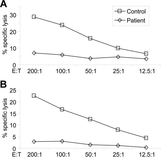
Killing by CTLs and NK cells is mediated by directional secretion of lytic granules toward target cells. Since impaired CTL activity was reported in GS patients,15–18 we wondered whether NK-cell cytotoxicity is also impaired. We therefore first performed direct lysis assay using the classical NK targets 721.221 and K562. In contrast to the pronounced specific lysis obtained by PBMCs derived from a healthy control, the patient demonstrated diminished natural cytotoxicity against 721.221 and K562 cells (Figure 2A and B, respectively). It can be argued that the lack of CTL and NK activities in GS would render the patient susceptible to viral infections, which are often observed in GS type 2 patients.31
Natural cytotoxicity of the patient is diminished. NK target cells 721.221 (A) or K562 (B) were mixed with PBMCs obtained from the patient or from a healthy donor, at the indicated effector-to-target (E/T) ratios. The target cells were radioactively labeled and specific lysis was calculated. Representative data from 1 of 3 experiments are shown.
Natural cytotoxicity of the patient is diminished. NK target cells 721.221 (A) or K562 (B) were mixed with PBMCs obtained from the patient or from a healthy donor, at the indicated effector-to-target (E/T) ratios. The target cells were radioactively labeled and specific lysis was calculated. Representative data from 1 of 3 experiments are shown.
NK cells in the GS patient function in ADCC
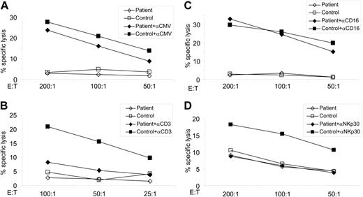
The main NK-activating receptors in humans are NKp30, NKp44, NKp46 (collectively named NCRs), NKG2D, and CD16.7 CD16 can either recognize target cells directly,35 or bind the Fc portion of antibodies to mediate ADCC.36,37 To examine ADCC activity in the GS patient, HFFs were infected with CMV, incubated with or without anti-CMV sera, and incubated with peripheral blood leukocytes (PBLs). In agreement with previous publications,28 in the absence of anti-CMV polyclonal sera, infected HFFs were not killed by PBMCs derived either from the patient or from unrelated healthy control (Figure 3A empty symbols). Efficient ADCC requires the generation of immune complexes that are best achieved when polyclonal antibodies are used.36–38 To generate such immune complexes, we used sera obtained from patients highly infected with CMV. An efficient ADCC activity was observed in NK cells derived from the patient, and this activity was very similar to the ADCC observed in the healthy control (Figure 3A full symbols). Thus, while natural cytotoxicity is impaired (Figure 2), ADCC activity is functional in the GS patient.
GS patient displays normal ADCC and CD16 activation, but lacks CTL activity or NKp30-dependent lysis. (A) HFFs were infected with CMV and incubated with or without anti-CMV sera. The effector-to-target ratios are indicated. Specific lysis is shown from 1 of 2 experiments performed. (B) P815 target cells were incubated with anti-CD3 mAb to redirect CTL cytotoxicity. Lysis assays were performed as above and the effector-to-target ratios are indicated. One of 2 experiments is shown. (C-D) P815 cells were incubated either with anti-CD16 mAb (C) or with anti-NKp30 (D) to redirect NK cytotoxicity. Effector cells were then added in the indicated E/T ratios. Lysis assays were performed as before. One of 3 experiments is shown.
GS patient displays normal ADCC and CD16 activation, but lacks CTL activity or NKp30-dependent lysis. (A) HFFs were infected with CMV and incubated with or without anti-CMV sera. The effector-to-target ratios are indicated. Specific lysis is shown from 1 of 2 experiments performed. (B) P815 target cells were incubated with anti-CD3 mAb to redirect CTL cytotoxicity. Lysis assays were performed as above and the effector-to-target ratios are indicated. One of 2 experiments is shown. (C-D) P815 cells were incubated either with anti-CD16 mAb (C) or with anti-NKp30 (D) to redirect NK cytotoxicity. Effector cells were then added in the indicated E/T ratios. Lysis assays were performed as before. One of 3 experiments is shown.
Previous studies of GS patients reported the profound lack of killing mediated by CD8+ T cells.10,20,21 However, the mutation observed here is novel and NK cells of this patient display efficient ADCC, so we tested the killing by CTLs. For this purpose, we used a redirected lysis assay in which P815 target cells, which express high levels of Fc receptors, are preincubated with monoclonal antibodies specific for an activating receptor of the effector cells. Using this method, we overcame the need for a specific recognition of a peptide-MHC complex by the TCR and in addition we examined the entire CTL population. The anti-CD3–coated P815 cells were killed by CTLs derived from a healthy control, in marked contrast to the minute killing by CTLs derived from the patient (Figure 3B). Thus, the homozygous Q118X mutation in RAB27A impairs cytotoxicity of CTLs but not ADCC by NK.
Direct engagement of CD16 but not NKp30 activates killing in the GS patient
In all functional assays performed so far, we deliberately used PBMCs freshly obtained from the patient and from healthy controls without the use of any external manipulation. This is essential for the genuine assessment of the activity of immune cells in the patient. However, this careful analysis was problematic because we could not obtain the necessary blood samples from the young patient. Therefore, to further investigate NK killing mechanisms in the patient in detail, we had to restrict our research only to certain NK receptors. We chose the CD16 and NKp30 receptors for several reasons. First, we observed that the natural cytotoxic activity against K562 and 721.221 is impaired in the GS patient (Figure 2) and NKp30 but not CD16 is known to recognize these tumor cell lines.27,39 Second, both the CD16 and the NKp30 receptors are thought to use the same adaptor proteins in the killing process.22,23 Third, despite the fact that NKp46 also uses the same adaptor proteins, we did not include this receptor in our analysis because, as reported also by others,40 we did not observe an efficient redirected killing when fresh healthy PBMCs were activated by mAb to the NKp46 receptor (data not shown). Finally, the NKp44 and NKG2D receptors signal through the DAP12/KARAP and the DAP10 adaptor molecules, respectively, unlike CD16 or NKp30,22,23 and the NKp44 is not expressed on fresh NK cells.7
To directly test if the engagement of either CD16 or NKp30 results in an activation of killing, we used P815 cells coated either with anti-NKp30 or anti-CD16 mAb (2 μg/106 P815 cells), and then incubated these target cells with PBMCs derived either from the patient or from a healthy control. In agreement with the potent ADCC activity observed in the patient (Figure 3A), the redirected killing mediated through CD16 was efficient and similar to that observed in the healthy control (Figure 3C), indicating that CD16 engagement could overcome the Rab-27A deficiency. In contrast, no redirected killing was observed when anti-NKp30 mAb was used to stimulate PBLs of the patient, whereas efficient NKp30-mediated killing was observed by healthy donors (Figure 3D). Hence, CD16 can act independently of Rab-27A, while the NKp30 seems to be totally Rab-27A dependent.
High phosphorylation of Vav1 upon CD16 but not NKp30 engagement
Studying the signaling cascade of NK cells derived from the patient was not possible due to the limitation of blood samples. However, it was clear that the mutation in the RAB27A gene could not be responsible for this major difference in the signaling cascade between the NKp30 and the CD16 receptors. The Rab-27A deficiency simply exposed a general phenomenon that also exists in healthy individuals. We therefore went on to examine normal NK cells from healthy individuals. The proximal signal transduction mediated via the CD16 and the NKp30 receptors is considered to be identical,23 and little is known about the downstream signaling cascade of these receptors, especially with regard to NKp30. However, it is known that the general signal transduction pathways of activating NK receptors involve a phosphorylation cascade in which Vav proteins play a central role.23 The Vav1 protein is also involved in cytoskeleton reorganization.21,34 In addition, the Vav1 protein is the only known Rho guanine nucleotide exchange factor (GEF) that has an SH2 domain and conserved tyrosine residues, suggesting that it has a unique GEF activity that is regulated by tyrosine phosphorylation.41
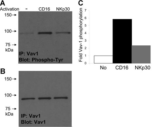
We therefore analyzed the status of Vav1 tyrosine phosphorylation after specific engagement of either the CD16 or the NKp30 receptor. Fresh NK cells were isolated from PBMCs of healthy individuals and stimulated with either anti-CD16 or anti-NKp30 mAb (1 μg/106 fresh NK cells). A strong tyrosine phosphorylation of Vav1 was observed upon the engagement of the CD16 receptor, compared with the basal-level phosphorylation observed in NK cells treated by secondary antibody only (Figure 4A). In contrast, engagement of NKp30 did not result in such high phosphorylation of Vav1 (Figure 4A). To control the experiment, the blots were stripped and reprobed with anti-Vav1 mAb demonstrating comparable levels of total Vav proteins (Figure 4B). Finally, densitometric analysis of phospho-Vav1 showed a pronounced difference between CD16 and NKp30 engagement (Figure 4C). The differences in the Vav1 phosphorylation observed between NKp30 and CD16 engagement could have been due to stronger signals induced by the anti-CD16 mAb versus the anti-NKp30 mAb. We cannot completely rule out this possibility. However, we used saturating concentrations of antibodies and even when we repeated the experiment using higher concentrations of anti-NKp30 (up to 5 μg/106 NK cells), Vav1 phosphorylation was not enhanced (data not shown).
NK cells from healthy donors display high phosphorylation of Vav1 upon CD16 but not NKp30 engagement. (A) Phosphorylation of Vav1 upon CD16 or NKp30 engagement in fresh NK cells. Vav1 was immunoprecipitated with polyclonal rabbit serum; phosphotyrosine was detected by mAb 4G10. Representative data of 1 of 3 experiments is shown. (B) Detection of total Vav1 protein. Blots were striped and reprobed with mAb to Vav1 protein. (C) Quantification of Vav1 phosphorylation. Densitometry of the bands was calculated to the relative specific phosphorylation without activation.
NK cells from healthy donors display high phosphorylation of Vav1 upon CD16 but not NKp30 engagement. (A) Phosphorylation of Vav1 upon CD16 or NKp30 engagement in fresh NK cells. Vav1 was immunoprecipitated with polyclonal rabbit serum; phosphotyrosine was detected by mAb 4G10. Representative data of 1 of 3 experiments is shown. (B) Detection of total Vav1 protein. Blots were striped and reprobed with mAb to Vav1 protein. (C) Quantification of Vav1 phosphorylation. Densitometry of the bands was calculated to the relative specific phosphorylation without activation.
The CD16 receptor uses divergent signaling pathways
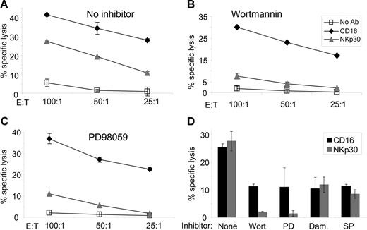
To further investigate whether the CD16-mediated killing pathway is different from that of NKp30, we repeated the redirected lysis assays but added specific inhibitors of known signaling pathways. CD16-mediated killing was only partially inhibited by the PI3K inhibitor wortmannin, while the MEK inhibitor PD98059 had only a minute effect on the killing (Figure 5A-C). In contrast, NKp30-mediated killing was diminished by wortmannin and by PD98059 (Figure 5A-C). Thus, it seems as if NKp30 signaling is PI3K and MEK dependent, whereas CD16 signaling is more robust involving alternative signaling cascades. Notably, as seen in Figure 5A, engagement of CD16 leads to a more efficient killing compared with NKp30. Thus, it might be possible that the inhibitors did not affect CD16-mediated cytotoxicity because of the strength of the CD16-activation signal. We therefore titrated the antibodies to obtain comparable cytotoxicity levels of both CD16 and NKp30 (using 0.5 μg anti-CD16 and 5 μg anti-NKp30 per 106 P815 target cells), and also examined other inhibitors (Figure 5D). All inhibitors used reduced both the CD16- and NKp30-mediated killing. Of importance, CD16 was only partially inhibited by wortmannin or PD98059, while NKp30 was completely abolished (Figure 5D). Inhibition of p56lck (damnacanthal) or JNK (SP600125) had a similar effect on CD16 and NKp30, reflecting similar p56lck and JNK dependency.
CD16 uses additional pathways for killing. PBMCs were incubated with inhibitors for 1 hour prior to the addition of target P815 cells with the indicated mAb. (A) No inhibitor, (B) PI3K inhibitor wortmannin, and (C) MEK inhibitor PD98059. (D) The assay was titrated to obtain similar cytotoxicity by CD16 and NKp30, and examined again with no inhibitor, wortmannin (Wort), PD98059 (PD), p56lck inhibitor damnacanthal (Dam), or JNK inhibitor SP600125 (SP). Lysis assays were performed as before. Representative data of 1 of at least 3 experiments is shown. Error bars indicate standard deviation.
CD16 uses additional pathways for killing. PBMCs were incubated with inhibitors for 1 hour prior to the addition of target P815 cells with the indicated mAb. (A) No inhibitor, (B) PI3K inhibitor wortmannin, and (C) MEK inhibitor PD98059. (D) The assay was titrated to obtain similar cytotoxicity by CD16 and NKp30, and examined again with no inhibitor, wortmannin (Wort), PD98059 (PD), p56lck inhibitor damnacanthal (Dam), or JNK inhibitor SP600125 (SP). Lysis assays were performed as before. Representative data of 1 of at least 3 experiments is shown. Error bars indicate standard deviation.
Thus, by identifying a new GS patient, through the discovery of a novel RAB27A mutation and by studying the NK activity in the GS patient in detail, we demonstrate functional dichotomy among human activating NK receptors.
Discussion
We began this research by identifying a novel nonsense Q118X mutation in the RAB27A gene of GS patient. Genomic PCR analysis showed that the patient is homozygous and both parents are heterozygous for this mutation. A similar Q118 stop mutation was recently described in a single patient that was heterozygous for this transition and had another mutation at the splicing site of the fifth exon.15
Based on this single base transition, we established a sensitive SSP-PCR assay that facilitates the detection of this particular mutation. This SSP-PCR is used for prenatal genetic diagnosis in a current pregnancy of this couple. We propose that similar methods should be used for other point mutations in families of GS patients to help in the planning of their future pregnancies.
Cytotoxic activity of CTLs and NK cells is essential for the removal of virally infected and tumor cells. T and NK cells share many similarities and have a common progenitor during differentiation from hematopoietic stem cells.42 However, their ability to identify and destroy hazardous cells while keeping tolerance toward normal self cells is almost opposite. The population of CTLs is made up of millions of clones, each expressing a single rearranged TCR identifying a specific peptide restricted to a particular MHC class I protein.43 Recognition of a specific class-I MHC molecule loaded with a particular peptide leads to the destruction of target cell by the effector CTL. NK cells, on the other hand, express a small number of germ line–encoded activating receptors that recognize viral-, tumor-, and stress-induced proteins.24 Normal cells are protected from NK killing mainly by the numerous NK inhibitory receptors that recognize normal self class-I MHC.2 Recent studies have demonstrated fundamental differences in effector killing mechanisms by CTLs and NK. For example, the ZAP-70 and Syk kinases are essential for activating signals in T and in B cells, respectively, while double knock-out mice for these proteins still demonstrate a potent NK cytotoxicity.44 Thus, NK cells possess alternative and redundant pathways compared with the other lymphocytes.
While many studies have characterized T-cell cytotoxicity in GS patients, the function of NK cells was not investigated in detail. Klein et al reported reduced lysis of K562 by cells derived from 3 GS patients.16 Decreased lytic activity was also observed in 3 other studies.15,17,18 However, none of these studies assayed specific receptors involved in the killing by NK cells. In agreement with these reports, we observed diminished lysis of K562 and 721.221 tumor cell lines by the cells derived from our patient. Surprisingly, an efficient ADCC activity was observed in our patient. These results point to differences in the pathways used by CD16 and NKp30, in contrast with the current model that describes a similar signaling for these receptors.22,23 In agreement with the results presented here, differences in the signaling cascade of CD16 and other NK receptors were demonstrated in mice.45–47 In humans, ADCC and natural cytotoxicity were reported to differ in patients with mutated CD16,48 TAP,49 and NEMO.50 Recently, it was reported that specific engagement of the NKp46, NKG2D, 2B4 (CD244), DNAM-1 (CD226), or CD2 receptors in fresh NK cells did not result in effective killing, while the CD16 engagement did,40 and NKp30 was not investigated in that study. In agreement with these results, we could not observe efficient killing mediated by NKp46 in freshly isolated normal human NK cells (data not shown). In addition, CD16 activation was demonstrated to cause degranulation of fresh human NK cells even without polarization toward the target cell.51 These data together with the results presented here indicate that CD16 uses a unique signaling pathway to activate killing of fresh NK cells. In the case of GS, such ADCC activity might enable immune protection when antibodies that recognize infected cells are present. It is interesting also to consider the potential activation of CD16 by cellular ligands other than the Fc portions of antibodies that might also help GS patients to confront infections.35
There are many examples, especially regarding viruses, from which we learn about basic immunologic mechanisms from a unique condition. For example, viruses of the herpes family contain several genes evolved to interfere with MHC class I protein processing and presentation.42,52 The molecular studies of these herpes genes led to a better understanding of the classical MHC class I presentation pathways. We therefore assume that NK activity in the GS patient is just a sharper image of the activity of NK cells in healthy individuals. Thus, we thought to examine the functional dichotomy of NK activity regarding CD16 and NKp30 in healthy individuals. Indeed, we observed high phosphorylation of Vav1 only after CD16 but not after NKp30 engagement in cells derived from healthy individuals. Of interest, and in support of our results, it was demonstrated that KO mice of Vav1, Vav2, and Vav3 have differential defects in natural cytotoxicity or in ADCC activities.47 Vav1 was reported to play a major role in an ITAM-independent killing pathway of both mice and human NK cells.53,54 Recently the adaptor protein 3BP2 was shown to recruit Vav1.55 The interaction between Rab-27A and Vav1 is not characterized yet. It would be interesting to examine potential abilities of Vav1 to induce Rab-27A targets, which would explain the ability to bypass Rab-27A in exocytosis. Intriguingly, a recent study showed that in the immunologic synapse of CTLs the centrosome itself translocates to the plasma membrane and the actin cytoskeleton is not present at the final steps of secretion.56 The Rab-27A–dependent translocation of lytic granules to the actin microfilaments could be bypassed by the formation of such a synapse.
To further establish the differential signaling pathways of CD16 and NKp30, we used inhibitors in the functional lysis assays. Diminished NKp30- but not CD16-mediated killing was observed when using PI3K or MEK inhibitors, while p56lck and JNK inhibitors possessed similar effects. Taken together, we demonstrate that NKp30 engagement does not efficiently activate Vav1 and is Rab-27A, PI3K, and MEK dependent, whereas CD16 induces high phosphorylation of Vav1, and is only partially dependent in these signaling pathways. Further studies are needed to completely understand the different pathways by which activating human NK receptors induce cytotoxicity.
Authorship
Contribution: R.G. performed the experiments, analyzed the data, and wrote the paper. M.A. identified the patient, provided the medical information, and planned the study; M.E. performed some of the experiments; H.A. and G.K. handled human blood samples and collected data; D.G.W. provided vital reagents (HFFs, CMV, and anti-CMV sera); S.K. designed some of the experiments and provided vital reagents (anti-Vav1 antibodies); O.M. supervised the work, analyzed the data, and wrote the paper.
Conflict-of-interest disclosure: The authors declare no competing financial interests.
Correspondence: Ofer Mandelboim, The Lautenberg Center for General and Tumor Immunology, The Hebrew University Hadassah Medical School, Ein-Kerem, Jerusalem 91120, Israel; e-mail: oferman@md2.huji.ac.il.
The publication costs of this article were defrayed in part by page charge payment. Therefore, and solely to indicate this fact, this article is hereby marked “advertisement” in accordance with 18 USC section 1734.
Acknowledgment
This work was supported by research grants from the ICRF, the BSF, the Israel Science Foundation (ISF), the AICR, the European Consortium (MRTN-CT-2005 and LSCH-CT-2005-518178), and the Israel Ministry of Health (KKL) (M.O.). R.G. is supported by the Clore foundation.


This feature is available to Subscribers Only
Sign In or Create an Account Close Modal