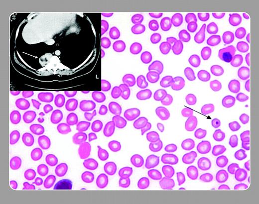
A chest mass was found on a 61-year-old African American male with longstanding anemia. An image from the computed tomography (CT) of the chest is shown as an inset. Prior to a percutaneous fine needle aspiration biopsy (FNA), he was referred for an assessment of his anemia. Hemoglobin level was 92 g/L (9.2 g/dL), mean corpuscular volume, 70 fL; reticulocyte count, 0.049 (4.9%); bilirubin level, 78.7 ümol/L (4.6 mg/dL); haptoglobin level, below 0.1 g/L (below 10 mg/dL); ferritin level, 967 üg/L (967 ng/mL); and soluble transferrin receptor activity (STRA), 65.4 nmol/L. The peripheral smear (main image) showed microcytes, teardrops, a nucleated red cell, and a Howell-Jolly body (arrow).
Interestingly, the hematologist had seen him 20 years earlier for anemia and an enlarged spleen. At that time, a splenectomy was performed. A decade later, his daughter also had her spleen removed. The current hemoglobin electrophoresis on the gentleman reestablished the original diagnosis of β-δ thalassemia.
The hematologist suggested that the retrothoracic, extrapulmonary, paravertebral chest mass was most likely extramedullary hematopoiesis and required no treatment. Nonetheless, the referring physician wanted tissue confirmation, which he received when the FNA was performed.
Many Blood Work images are provided by the ASH IMAGE BANK, a reference and teaching tool that is continually updated with new atlas images and images of case studies. For more information or to contribute to the Image Bank, visit www.ashimagebank.org.


This feature is available to Subscribers Only
Sign In or Create an Account Close Modal