Abstract
Pattern recognition receptors (PRRs) play an essential role in a macrophage's response to mycobacterial infections. However, how these receptors work in concert to promote this macrophage response remains unclear. In this study, we used bone marrow–derived macrophages isolated from mannose receptor (MR), complement receptor 3 (CR3), MyD88, Toll-like receptor 4 (TLR4), and TLR2 knockout mice to examine the significance of these receptors in mediating a macrophage's response to a mycobacterial infection. We determined that mitogen-activated protein kinase (MAPK) activation and tumor necrosis factor-α (TNF-α) production in macrophage infected with Mycobacterium avium or M smegmatis is dependent on myeloid differentiation factor 88 (MyD88) and TLR2 but not TLR4, MR, or CR3. Interestingly, the TLR2-mediated production of TNF-α by macrophages infected with M smegmatis required the β-glucan receptor dectin-1. A similar requirement for dectin-1 in TNF-α production was observed for macrophages infected with M bovis Bacillus Calmette-Guerin (BCG), M phlei, M avium 2151-rough, and M tuberculosis H37Ra. The limited production of TNF-α by virulent M avium 724 and M tuberculosis H37Rv was not dependent on dectin-1. Furthermore, dectin-1 facilitated interleukin-6 (IL-6), RANTES (regulated on activation, normal T expressed and secreted), and granulocyte colony-stimulating factor (G-CSF) production by mycobacteria-infected macrophages. These are the first results to establish a significant role for dectin-1, in cooperation with TLR2, to activate a macrophage's proinflammatory response to a mycobacterial infection.
Introduction
The immune system has the complex task of separating friend from foe. To accomplish this mission, the immune system has evolved receptors that recognize molecules present on pathogenic organisms but which show limited interaction with host components. These receptors, referred to as pattern recognition receptors (PRRs), function to promote an innate immune response and include such members as the mannose receptor (MR), scavenger receptors, and Toll-like receptors (TLRs), among others. PRRs bind to conserved microbial structures called pathogen-associated molecular patterns (PAMPs). The expression of these receptors allows the immune system to recognize a wide variety of pathogens that express one or more of these PAMPs, and their engagement initiates the subsequent immune response. Not surprisingly, the PRRs are expressed on cells of the innate immune system, including macrophages, neutrophils, dendritic cells, and natural killer (NK) cells.1
Binding of microbial products by PRRs elicits a signaling response within the leukocyte, resulting in the production of specific immune modulators. Which PRRs are engaged and in what combination, along with the specific ligands involved, will dictate the overall response by the immune cell. This complexity allows the immune system to tailor its response to a specific pathogen, yet remain flexible enough to recognize a large number of potential pathogens.
One of the most intensively studied PRRs is the MR, which has been implicated in pathogen recognition through binding terminal mannose, fucose, and N-acetylglucosamine residues.2,3 The major ligand for MR on mycobacterial surface is the mannose-capped lipoarabinomannan (ManLAM) from Mycobacterium tuberculosis and M bovis Bacillus Calmette-Guerin (BCG).4-6 Another important group of PRRs are the TLRs, which are also involved in the innate recognition of mycobacteria by the host.7 After ligand binding, TLRs activate signal transduction pathways by recruiting adaptor molecules such as myeloid differentiation factor 88 (MyD88).8,9 Stimulation of TLRs by microbial products activates nuclear factor-κB (NF-κB), mitogen-activated protein kinase (MAPK), and phosphoinositide 3-kinase signaling pathways leading to activation of inflammatory target genes.10 In vitro studies have shown a number of potent agonists on mycobacteria for TLR211,12 and TLR4,13,14 and their role in controlling a mycobacterial infection.15,16 TLRs participate with additional receptors in the innate recognition of microbes, and there is a strong interaction between the signaling pathways induced by these receptors and signal transduction stimulated by TLRs (for review, see Underhill17 and Mukhopadhyay et al18 ). Dectin-1 is one of the receptors recently shown to complement TLR signaling in order to generate a proinflammatory response.19 Dectin-1 is a C-type lectin receptor expressed on monocytes, macrophages, neutrophils, dendritic cells, and Langerhans cells that recognizes fungal wall–derived β-glucans.20,21 Dectin-1 promotes the phagocytosis of live yeast and fungal-derived zymosan particles, as well as promoting zymosan or fungal pathogen-induced proinflammatory response by macrophages and at least in some cases cooperating with TLR2 to mediate this response.19,22 Recent studies have also shown a role for dectin-1 in the phagocytosis of Haemophilus influenzae by eosinophils.23 However, the importance of dectin-1 as a PRR and its role in promoting a proinflammatory response to a mycobacterial infection or to bacterial infections in general remains undefined.
In the present study we assessed the role of various macrophage PRRs in MAPK activation and cytokine production upon mycobacterial infection. We found TLR2 and dectin-1 to work in concert to promote macrophage activation upon mycobacterial infection. These are the first studies to indicate a role for dectin-1 in promoting a macrophage proinflammatory response to a mycobacteria infection.
Materials and methods
Reagents
Unless stated otherwise, all chemical reagents were purchased from Sigma (St Louis, MO).
Mice
Balb/c and C57BL/6 mice were purchased from Harlan (Mutant Mouse Regional Resource Center, Indianapolis, IN). TLR2–/– and TLR4–/– mice were purchased from Jackson Laboratory (Bar Harbor, ME). MyD88–/– (in C57BL/6 background) and CR3–/– (in Balb/c background) were generously provided by Soon-Cheol Hong (Indiana University Medical School, Indianapolis) and Tanya Mayadas-Norton (Harvard Medical College, Cambridge, MA), respectively. MR–/– and C57BL/6 MR+/+ were kindly provided by Michel Nussenzweig (Rockefeller University, New York, NY).
Bacteria
Mycobacteria stocks were generated by using a single colony to inoculate Middlebrooks 7H9 media (Difco, Sparks, MD) supplemented with glucose, oleic acid, albumin, Tween-20, and NaCl (GOATS). For M avium 724 stocks, the mycobacteria were passaged through a mouse to ensure virulence. Bacteria were grown for 3 to 10 days at 37°C with vigorous shaking, centrifuged, resuspended in Middlebrooks/GOATS plus 15% glycerol, aliquoted, and stored at –80°C. Frozen stocks were quantitated by serial dilution onto Middlebrooks 7H10 agar/GOATS. M smegmatis strain MC2155, M bovis BCG, M phlei, M tuberculosis H37Ra (American Type Culture Collection [ATCC], Manassas, VA), M tuberculosis H37Rv, and other M avium strains were cultured and frozen stocks prepared as described. All reagents used to grow mycobacteria were found negative for endotoxin contamination using the E-Toxate assay and the QCL-1000 Endotoxin test (Cambrex Bio Science, Walkersville, MD).
Complement opsonization
Appropriate concentrations of mycobacteria were suspended in macrophage culture media containing 10% horse serum as a source of complement components and incubated for 2 hours at 37°C.24 The same concentration of horse serum was added to uninfected controls for all experiments.
Bone marrow macrophage isolation and mycobacteria infection
Bone marrow–derived macrophages (BMMϕs), used in all experiments, were isolated from 6- to 8-week-old mice as previously described.25 Infection assays evaluated by fluorescence microscopy were performed on each stock of mycobacteria to determine the infection ratio needed to obtain approximately 80% of the macrophages infected. The multiplicity of infection (MOI) required to obtain 80% infection varied from approximately 20:1 to 50:1 mycobacteria to macrophage ratio, and depended on the mycobacterial species and frozen stock of mycobacteria. Briefly, BMMϕs from wild-type (WT) or knock-out (KO) mice were plated on glass coverslips and infected with different doses of mycobacteria in triplicate. Infections were halted at either 1 hour or 4 hours and fixed in 1:1 methanol-acetone, washed with phosphate-buffered saline (PBS), and stained with TB Auramine M Stain Kit (Becton Dickinson, Sparks, MD) in the case of M avium, M tuberculosis, and M bovis BCG, and with acridine orange (Sigma) in case of M smegmatis and M phlei. Slides were visualized using fluorescent microscopy and the level of infection was quantitated by counting the number of cells infected and the approximate number of mycobacteria per cell in at least 4 fields per replicate. No fewer than 100 cells per replicate were counted.
For all experiments, mycobacteria were added to macrophages on ice and incubated for 10 minutes and then incubated at 37°C in 5% CO2 for the specified times. Culture media without antibiotics or L-cell supernatant was used in place of complete media during the infections. For the 24-hour time points, the BMMϕs were incubated for 4 hours with the mycobacteria and washed with PBS 3 times; then, fresh media was added and incubated for an additional 20 hours. All tissue-culture reagents were found negative for endotoxin contamination using the E-Toxate assay.
Antibody treatment
The anti–dectin-1 antibody clone 2A11 and rat immunoglobulin G2b (IgG2b) isotype control were purchased from Serotec (Oxford, United Kingdom). BMMϕs were preincubated with 50 μg/mL 2A11 or isotype control 1 hour prior to infection in Dulbecco modified Eagle medium (DMEM) and then infected with mycobacteria. After 4 hours of incubation, media was removed, cells were washed with PBS 3 times, and fresh media was added and incubated for an additional 20 hours.
Western blot analysis
Blots were performed as previously described.25 Briefly, at designated times, culture media was collected and saved for subsequent enzyme-linked immunosorbent assays (ELISAs), and the cells were washed 3 times with ice-cold PBS containing 1 mM per-vanadate. The cells were then treated for 5 to 10 minutes with ice-cold lysis buffer, and the cell lysates were removed from the plates and stored at –20°C. Equal amounts of protein, as defined using the Micro BCA Protein Assay (Pierce, Rockford, IL), were loaded onto 10% sodium dodecyl sulfate–polyacrylamide gel electrophoresis (SDS-PAGE) gels, electrophoresed and transferred onto PVDF membrane (Millipore, Bedford, MA). The membranes were incubated with the primary antibodies phospho-p38, total p38, phospho–extracellular signal–regulated kinase 1/2 (ERK1/2), or total ERK1/2 from Cell Signaling (Beverly, MA). The blots were washed and incubated with a secondary antibody, either horseradish peroxidase–conjugated anti–rabbit or anti–mouse IgG (Pierce) in Tris-buffered saline plus tween (TBST) plus 5% powdered milk. The bound antibodies were detected using SuperSignal West Femto enhanced chemiluminescence reagents (Pierce).
Cytokine measurements
The levels of cytokines secreted by infected macrophages were measured using the commercially available ELISA reagent kits for TNF-α (PharMingen, San Diego, CA), interleukin-6 (IL-6), RANTES (regulated on activation, normal T expressed and secreted; eBioscience, San Diego, CA) and granulocyte colony-stimulating factor (G-CSF; R&D Systems, Minneapolis, MN). Culture media collected from the macrophages was analyzed for cytokines according to manufacturers' instructions, and the cytokine concentrations were determined against standard curves. A cytokine profile analysis was performed by using the RayBio Mouse Cytokine Array (RayBiotech, Atlanta, GA) with culture supernates from noninfected or infected macrophages.
Statistical analysis
Statistical significance was determined by the paired 2-tailed Student t test with a P level less than .05 denoting significance using InStat/Prism software (Graph-Pad Prism Software, San Diego, CA).
Results
MR is not required for phagocytosis of M avium or M smegmatis by murine macrophages
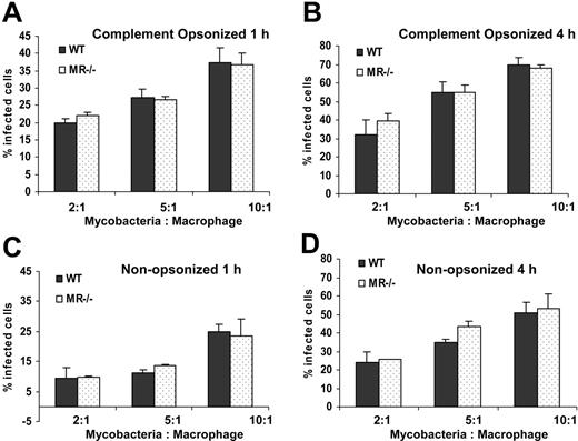
Prior studies have indicated a role for the MR on human alveolar macrophages in the phagocytosis of nonopsonized virulent M tuberculosis strains H37Rv and Erdman but a minimal role in the phagocytosis of the avirulent strain H37Ra.26 With the availability of mice deficient in MR, we compared mouse BMMϕs from WT and MR–/– mice in their ingestion of complement-opsonized and nonopsonized M avium. We found a direct correlation between the MOI and the percentage of cells infected; however, there was no difference in the number of infected cells between WT and MR–/– macrophages at either 1 or 4 hours of infection (Figure 1). Moreover, we did not observe any detectable differences in the number of mycobacteria per infected macrophage (data not shown). As expected, we observed a higher number of infected BMMϕs when M avium was complement-opsonized compared with nonopsonized at both time points and at different MOIs. Similar results were seen with nonpathogenic M smegmatis, as we observed no difference in its phagocytosis by WT and MR–/– BMMϕs at 1 or 4 hours (data not shown). BMMϕs from TLR4–/–, TLR2–/–, and MyD88–/– displayed no difference compared with WT BMMϕs in the uptake of complement opsonized or nonopsonized mycobacteria (data not shown).
MR, CR3, and TLR4 are not required for MAPK activation or TNF-α production by BMMϕs in response to M avium or M smegmatis infection
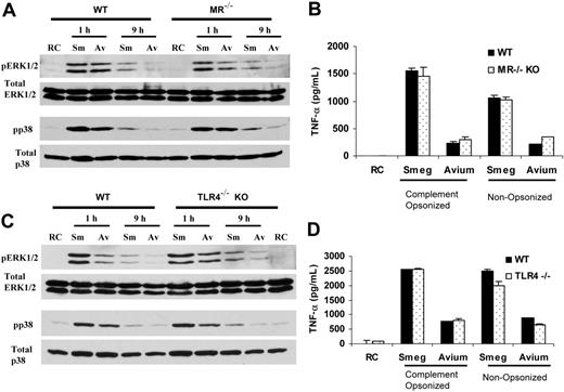
In addition to serving as phagocytic receptors, PRRs function to promote macrophage activation characterized by production of cytokines, chemokines, and oxygen- and nitrogen-reactive species. This macrophage activation requires stimulation of signaling pathways resulting in a transcriptional response. Activation of the MAPK pathways in macrophages has been associated with PRR stimulation resulting in the production of numerous proinflammatory molecules.27-29 To start addressing the role of various PRRs in promoting macrophage activation, we first evaluated the signaling response by WT and MR–/– macrophages to infection with pathogenic M avium and nonpathogenic M smegmatis. Specifically, BMMϕs from WT and MR–/– mice were infected with mycobacteria for 1 hour or 9 hours, and MAPK activation was measured by Western blotting for phospho-p38 and phospho-ERK1/2. We chose to look at 1 and 9 hours, since our previous studies have shown an initial activation of macrophage MAPKs by all mycobacteria, but a differential activation of MAPKs 9 hours after infection in macrophages infected with nonpathogenic mycobacteria compared with M avium.24,25,30 As shown previously, MAPKs are activated upon mycobacterial infection and macrophages infected with M smegmatis show higher levels of MAPK activation at 9 hours compared with noninfected BMMϕs or M avium–infected cells (Figure 2A). However, there was no difference in MAPK activation between WT and MR–/– macrophages following infection with either M smegmatis or M avium 724. Previously we demonstrated the importance of the MAPKs as well as other signaling molecules involved in macrophage's production of TNF-α following a mycobacterial infection.24,25 Therefore, we looked at the TNF-α production in WT and MR–/– BMMϕs infected with M smegmatis or M avium 724. Similar to what we observed with MAPK activation, there was no difference in TNF-α secretion between WT and the KO macrophages (Figure 2B). Moreover, infection using nonopsonized mycobacteria also showed similar levels of TNF-α in WT and MR–/– macrophages (Figure 2B). As previously observed, M smegmatis–infected macrophages produce significantly higher levels of TNF-α relative to M avium–infected cells.25
MR is not essential for the phagocytosis of M avium 724. BMMϕs from WT or MR–/– mice were infected with different ratios of complement opsonized (A-B) or nonopsonized (C-D) mycobacteria for 1 hour (A,C) or 4 hours (B,D). After infection, cells were washed, fixed, and stained with Auramine M. The percentage of phagocytic cells having ingested at least one mycobacterium was measured by fluorescence microscopy as described in “Materials and methods.” Values are expressed as means + SD. Data are representative of 3 separate experiments.
MR is not essential for the phagocytosis of M avium 724. BMMϕs from WT or MR–/– mice were infected with different ratios of complement opsonized (A-B) or nonopsonized (C-D) mycobacteria for 1 hour (A,C) or 4 hours (B,D). After infection, cells were washed, fixed, and stained with Auramine M. The percentage of phagocytic cells having ingested at least one mycobacterium was measured by fluorescence microscopy as described in “Materials and methods.” Values are expressed as means + SD. Data are representative of 3 separate experiments.
BMMϕs from WT and TLR4–/– mice also showed no difference in MAPK activation upon infection with either Mycobacterium (Figure 2C). Moreover, TNF-α production induced upon infection with either M smegmatis or M avium 724 did not differ between TLR4–/– and WT macrophages (Figure 2D). To test the role of CR3 in the macrophage response to a mycobacterial infection, we infected BMMϕs from WT and CR3–/– mice. As expected, BMMϕs from CR3–/– mice showed decreased phagocytosis of complement-opsonized mycobacteria, and required a higher mycobacteria to macrophage ratio in order to obtain a similar infection level as WT macrophages (data not shown). However, there was no difference in MAPK activation or TNF-α production upon infection with complement opsonized or nonopsonized mycobacteria in CR3–/– macrophages compared with WT (data not shown).
Optimal MAPK activation and TNF-α production by mycobacteria-infected macrophages requires TLR2 and MyD88
The data shown in Figure 2 did not support a role for the MR, CR3, or TLR4 in the activation of macrophages by M avium or nonpathogenic M smegmatis. To determine whether the observed BMMϕ's response to M avium and M smegmatis was dependent on TLR2, we infected TLR2–/– and WT BMMϕs with the different mycobacteria. BMMϕs from TLR2–/– mice showed a lack of MAPK activation above noninfected controls following an M avium 724 infection at both 1 and 9 hours after infection (Figure 3A). The low amount of TNF-α produced by M avium 724–infected BMMϕs was also completely lost in TLR2–/– BMMϕs (Figure 3B). In macrophages infected with M smegmatis, no difference in ERK1/2 activation was observed between TLR2–/– and WT macrophages at 1 hour after infection, but by 9 hours a significant decrease in MAPK activation was discerned in the TLR2–/– BMMϕs (Figure 3B). Furthermore, we observed a significant drop but not a complete loss of TNF-α production in M smegmatis–infected TLR2–/– macrophages (Figure 3B). The adaptor protein MyD88 has been shown previously to be critical in TLR signaling.31 To determine whether MAPK activation and TNF-α production was dependent on MyD88, we infected BMMϕs from WT and MyD88–/– mice. Upon infection there was a complete loss of MAPK activation in MyD88–/– macrophages. As shown in Figure 3C, this effect was evident at both 1 and 9 hours after infection. Similarly, there was no TNF-α production detected in MyD88–/– macrophages upon infection with either M smegmatis or M avium 724 (Figure 3D). These findings differ from our results with the TLR2–/– BMMϕs, where there was no loss in ERK1/2 phosphorylation at 1 hour after infection and some TNF-α production in M smegmatis–infected BMMϕs.
MR and TLR4 are not essential for MAPK activation and TNF-α production in BMMϕs upon M smegmatis or M avium 724 infection. BMMϕs from WT and either MR–/– (A-B) or TLR4–/– (C-D) mice were infected with M smegmatis or M avium 724 and screened for MAPK activation at 1 hour and 9 hours (A,C) and TNF-α production at 24 hours (B,D). (A,C) MAPK activation was detected by probing cell lysates of infected or noninfected (RC) BMMϕs by Western blot for activated ERK1/2 and p38 using phospho-specific Abs as described in “Materials and methods.” Total ERK1/2 and total p38 blots were run to show equal protein loading. (B,D) BMMϕs from WT and KO mice were infected with complement opsonized or nonopsonized mycobacteria; 24 hours later, culture supernates were removed and analyzed by ELISA for TNF-α. Values are expressed as means + SD. Data are representative of 3 separate experiments. Sm and Smeg indicate M smegmatis; Av and Avium, M avium; and RC, noninfected BMMϕs.
MR and TLR4 are not essential for MAPK activation and TNF-α production in BMMϕs upon M smegmatis or M avium 724 infection. BMMϕs from WT and either MR–/– (A-B) or TLR4–/– (C-D) mice were infected with M smegmatis or M avium 724 and screened for MAPK activation at 1 hour and 9 hours (A,C) and TNF-α production at 24 hours (B,D). (A,C) MAPK activation was detected by probing cell lysates of infected or noninfected (RC) BMMϕs by Western blot for activated ERK1/2 and p38 using phospho-specific Abs as described in “Materials and methods.” Total ERK1/2 and total p38 blots were run to show equal protein loading. (B,D) BMMϕs from WT and KO mice were infected with complement opsonized or nonopsonized mycobacteria; 24 hours later, culture supernates were removed and analyzed by ELISA for TNF-α. Values are expressed as means + SD. Data are representative of 3 separate experiments. Sm and Smeg indicate M smegmatis; Av and Avium, M avium; and RC, noninfected BMMϕs.
Induction of TNF-α production upon mycobacterial infection is partially dependent on dectin-1
We anticipated that TLR2 and MyD88 were required, at least in part, for the mycobacteria-initiated macrophage activation, and our data support this prediction. However, TLR2 often functions in concert with other receptors, including additional TLRs and non-TLRs, to promote cellular activation.17,18 Which combination of receptors is engaged depends on the ligands involved.32 Dectin-1 is a recently described receptor expressed on BMMϕs,33 which can function together with TLR2 to induce macrophage activation.19,22
MyD88–/– and TLR2–/–BMMϕs show impaired MAPK activation and TNF-α production upon mycobacterial infection. BMMϕs from WT and TLR2–/– mice (A-B) and MyD88–/– mice (C-D) were infected with M smegmatis or M avium 724 and screened for MAPK activation at 1 hour and 9 hours and TNF-α production at 24 hours. (A,C) MAPK activation was detected by preparing cell lysates after 1-hour and 9-hour infections and probed for activated ERK1/2 and p38 using phospho-specific Abs as described in “Materials and methods.” Total ERK1/2 and p38 blots were run to show equal protein loading. (B,D) BMMϕs from WT and KO mice were infected with complement opsonized or nonopsonized mycobacteria; 24 hours later, culture supernates were removed and analyzed by ELISA for TNF-α. Values are expressed as means + SD. Data are representative of 3 separate experiments. Sm and Smeg indicate M smegmatis; Av and Avium, M avium; and RC, noninfected BMMϕs.
MyD88–/– and TLR2–/–BMMϕs show impaired MAPK activation and TNF-α production upon mycobacterial infection. BMMϕs from WT and TLR2–/– mice (A-B) and MyD88–/– mice (C-D) were infected with M smegmatis or M avium 724 and screened for MAPK activation at 1 hour and 9 hours and TNF-α production at 24 hours. (A,C) MAPK activation was detected by preparing cell lysates after 1-hour and 9-hour infections and probed for activated ERK1/2 and p38 using phospho-specific Abs as described in “Materials and methods.” Total ERK1/2 and p38 blots were run to show equal protein loading. (B,D) BMMϕs from WT and KO mice were infected with complement opsonized or nonopsonized mycobacteria; 24 hours later, culture supernates were removed and analyzed by ELISA for TNF-α. Values are expressed as means + SD. Data are representative of 3 separate experiments. Sm and Smeg indicate M smegmatis; Av and Avium, M avium; and RC, noninfected BMMϕs.
Dectin-1, a C-type lectin receptor, has been implicated in the innate recognition of yeasts through its binding to surface β-glucan.34 Recently it has been found to cooperate with TLR2 in eliciting inflammatory responses to zymosan.19 Therefore, we were interested in defining whether dectin-1 plays a role in TNF-α production upon infection with mycobacteria. For this purpose, we used a previously characterized neutralizing antibody against dectin-133 to block the mycobacterial interaction with the macrophage dectin-1 receptor. Pretreating the macrophages with the anti–dectin-1 monoclonal antibody (mAb) 2A11 had no effect on the phagocytosis of M avium or M smegmatis (data not shown). However, pretreating the macrophages with the 2A11 mAb resulted in a significant reduction in TNF-α production upon infection with M smegmatis (Figure 4A). M avium 724 induces little TNF-α production, and thus the effect of 2A11 was difficult to determine. Therefore, we used the avirulent M avium 2151-rough strain, which is known to induce significant TNF-α production.35 Like our findings with M smegmatis, the production of TNF-α by M avium 2151–infected BMMϕs was significantly blocked in the presence of anti–dectin-1 mAb 2A11 (Figure 4A). Similar reduction in TNF-α secretion upon M smegmatis infection was seen in macrophages pretreated for 1 hour with 700 μg/mL laminarin, a soluble β-glucan used for blocking the β-glucan receptor (Figure 4B).36 Pretreatment with the polysaccharide galactan at the same concentration did not affect TNF-α production in M smegmatis–infected macrophages. Blocking dectin-1 in BMMϕs infected with non-opsonized M smegmatis also resulted in inhibition of TNF-α production (Figure 4C). However, there was no difference in TNF-α production in BMMϕs treated with LPS in the presence of 2A11 (Figure 4C).
Dectin-1 functions to promote TNF-α production in M smegmatis– and M avium 2151–infected macrophages. BMMϕs were infected with (A) M smegmatis or M avium 724 or M avium 2151 in the presence of PBS or anti–dectin-1 mAb 2A11 or isotype control Ab, (B) M smegmatis in the presence of laminarin or galactan, and (C) complement opsonized or nonopsonized M smegmatis in the presence of PBS, 2A11, or isotype control Ab. Also shown are LPS-treated BMMϕs with or without antibodies. Culture supernates after 24 hours of infection were analyzed for TNF-α by ELISA. *Significant to M smegmatis plus PBS (P < .01). **Significant to M smegmatis alone and M smegmatis plus galactan (P < .01). Values are expressed as means + SD. Data are representative of 3 separate experiments. Iso-Con indicates isotype control Ab; Smeg, M smegmatis; and RC, noninfected BMMϕs.
Dectin-1 functions to promote TNF-α production in M smegmatis– and M avium 2151–infected macrophages. BMMϕs were infected with (A) M smegmatis or M avium 724 or M avium 2151 in the presence of PBS or anti–dectin-1 mAb 2A11 or isotype control Ab, (B) M smegmatis in the presence of laminarin or galactan, and (C) complement opsonized or nonopsonized M smegmatis in the presence of PBS, 2A11, or isotype control Ab. Also shown are LPS-treated BMMϕs with or without antibodies. Culture supernates after 24 hours of infection were analyzed for TNF-α by ELISA. *Significant to M smegmatis plus PBS (P < .01). **Significant to M smegmatis alone and M smegmatis plus galactan (P < .01). Values are expressed as means + SD. Data are representative of 3 separate experiments. Iso-Con indicates isotype control Ab; Smeg, M smegmatis; and RC, noninfected BMMϕs.
Dectin-1–mediated induction of TNF-α is dependent on TLR2 expression
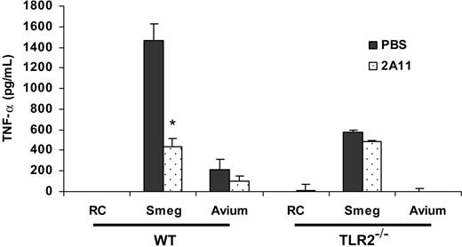
Recent papers have suggested a possible cooperation between TLR2 and dectin-1 in zymosan recognition by HEK 293 cells. Gantner et al showed that NF-κB activation in macrophages by zymosan requires both TLR2 and dectin-1.19 We hypothesized that there is a similar cooperation between dectin-1 and TLR2 in macrophages infected with mycobacteria. As shown earlier, there was a marked diminished TNF-α production in TLR2–/– macrophages compared with WT upon infection with M smegmatis (Figure 5). However in contrast to WT, pretreatment of TLR2–/– BMMϕs with 2A11 did not further reduce TNF-α production upon infection with M smegmatis, suggesting that dectin-1–mediated induction of TNF-α production was dependent on TLR2 (Figure 5). No TNF-α was detected in TLR2–/– BMMϕs upon M avium 724 infection (Figure 5).
Dectin-1–mediated induction of TNF-α by BMMϕs infected with M smegmatis requires TLR2. BMMϕs from WT or TLR2–/– mice were infected with M smegmatis or M avium 724 in the presence of anti–dectin-1 mAb 2A11. After a 4-hour infection, BMMϕs were washed, fresh medium was added to the cells, and infection was continued for a total of 24 hours. Culture supernates were analyzed by ELISA for TNF-α. *Significant to M smegmatis plus PBS (P < .01). Values are expressed as means + SD. Data are representative of 3 separate experiments. Smeg indicates M smegmatis; Avium, M avium; and RC, noninfected BMMϕs.
Dectin-1–mediated induction of TNF-α by BMMϕs infected with M smegmatis requires TLR2. BMMϕs from WT or TLR2–/– mice were infected with M smegmatis or M avium 724 in the presence of anti–dectin-1 mAb 2A11. After a 4-hour infection, BMMϕs were washed, fresh medium was added to the cells, and infection was continued for a total of 24 hours. Culture supernates were analyzed by ELISA for TNF-α. *Significant to M smegmatis plus PBS (P < .01). Values are expressed as means + SD. Data are representative of 3 separate experiments. Smeg indicates M smegmatis; Avium, M avium; and RC, noninfected BMMϕs.
Dectin-1 facilitates TNF-α production in macrophages infected with M phlei, M bovis BCG, and M tuberculosis H37Ra but not H37Rv
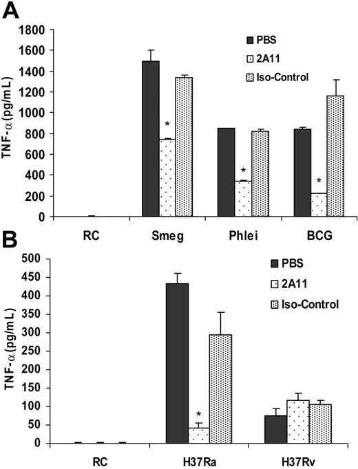
To determine whether the importance of dectin-1 in mediating macrophage activation is limited to the avirulent M avium 2151-rough strain and M smegmatis, we infected BMMϕs with nonpathogenic M phlei, attenuated M bovis BCG, and avirulent M tuberculosis H37Ra and virulent H37Rv. We measured infected BMMϕs for TNF-α production in the presence of 2A11 or isotype control antibody. As observed in previous research,25 macrophages infected with M phlei or M bovis BCG induce high levels of TNF-α production. However, this TNF-α production was reduced significantly in the presence of the 2A11 antibody, while no block was observed with the isotype control antibody (Figure 6A). Moreover, we observed significantly higher levels of TNF-α secreted by M tuberculosis H37Ra–infected BMMϕs compared with H37Rv-infected BMMϕs, as observed previously.37 The TNF-α produced by H37Ra- but not H37Rv-infected BMMϕs was also significantly decreased in cells treated with the 2A11 blocking antibody compared with untreated or isotype control (Figure 6B).
Dectin-1 promotes production of inflammatory mediators in addition to TNF-α in mycobacteria-infected macrophages
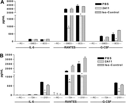
To determine whether the importance of dectin-1 is limited to TNF-α production, we measured the levels of other proinflammatory mediators in the presence of the dectin-1–blocking antibody. We found that in addition to TNF-α, 2A11 significantly blocked the BMMϕ production of IL-6, G-CSF, and RANTES following an M smegmatis or M bovis BCG infection (Figure 7A). A similar 2A11 antibody-mediated block in IL-6, RANTES, and G-CSF production was observed for BMMϕs infected with M avium 2151-rough or M tuberculosis H37Ra (Figure 7B; data not shown). Infection of BMMϕs with M avium 724 or H37Rv did not lead to significant levels of IL-6 or G-CSF in culture supernatants (Figure 7B; data not shown). However, RANTES production was increased in M avium 724–infected BMMϕs, and this was significantly reduced in 2A11-treated cells (Figure 7B). Production of the different mediators was not affected by the isotype control antibody. Together, our data indicate that dectin-1 promotes the macrophage's ability to produce a broad spectrum of proinflammatory mediators upon mycobacterial infection.
Dectin-1 promotes TNF-α production induced in BMMϕs upon infection with nonpathogenic or attenuated mycobacteria but not H37Rv. BMMϕs were infected with M smegmatis, M phlei, or M bovis BCG (A) or M tuberculosis strains H37Rv and H37Ra (B) in the presence of anti–dectin-1 mAb 2A11 or isotype control Ab. After a 4-hour infection, BMMϕs were washed, fresh medium was added to the cells, and the infection was continued for a total of 24 hours. Culture supernates were analyzed by ELISA for TNF-α. *Significant to mycobacteria plus PBS (P < .01). Values are expressed as means + SD. Data are representative of 3 separate experiments. Smeg indicates M smegmatis; RC, noninfected BMMϕs; and Iso-Control, isotype control Ab.
Dectin-1 promotes TNF-α production induced in BMMϕs upon infection with nonpathogenic or attenuated mycobacteria but not H37Rv. BMMϕs were infected with M smegmatis, M phlei, or M bovis BCG (A) or M tuberculosis strains H37Rv and H37Ra (B) in the presence of anti–dectin-1 mAb 2A11 or isotype control Ab. After a 4-hour infection, BMMϕs were washed, fresh medium was added to the cells, and the infection was continued for a total of 24 hours. Culture supernates were analyzed by ELISA for TNF-α. *Significant to mycobacteria plus PBS (P < .01). Values are expressed as means + SD. Data are representative of 3 separate experiments. Smeg indicates M smegmatis; RC, noninfected BMMϕs; and Iso-Control, isotype control Ab.
Discussion
The tailored reaction by macrophages to pathogenic organisms as well as their response to host cell debris and apoptotic cells is dependent on the combination of macrophage receptors engaged which together initiate a specific signaling response. Understanding macrophage biology requires a definition of the macrophage receptors, their ligands, the signaling reactions induced upon receptor engagement, and the consequence of their activation (eg, production of proinflammatory mediators). In this report we focused on a number of macrophage receptors, including CR3, MR, TLR2, TLR4, and dectin-1, that have been characterized as phagocytic and/or signaling receptors in the context of various infection models. The purpose of the present study was to better understand how these receptors are involved in eliciting a macrophage response following an infection with pathogenic and nonpathogenic mycobacteria.
Dectin-1 promotes IL-6, RANTES, and G-CSF production in mycobacterial-infected BMMϕs. Cells were infected with M smegmatis and M bovis BCG (A) or M avium 724 and M avium 2151 (B) in the presence of PBS or anti–dectin-1 mAb 2A11 or isotype control Ab as described in Figure 6. Culture supernatants were analyzed for G-CSF, RANTES, and IL-6 by ELISA. Values are expressed as means + SD. Data are representative of 3 separate experiments. Smeg indicates M smegmatis; 724, M avium 724; 2151, M avium 2151; RC, noninfected BMMϕs; and Iso-Control, isotype control Ab. *Significant to mycobacteria plus PBS.
Dectin-1 promotes IL-6, RANTES, and G-CSF production in mycobacterial-infected BMMϕs. Cells were infected with M smegmatis and M bovis BCG (A) or M avium 724 and M avium 2151 (B) in the presence of PBS or anti–dectin-1 mAb 2A11 or isotype control Ab as described in Figure 6. Culture supernatants were analyzed for G-CSF, RANTES, and IL-6 by ELISA. Values are expressed as means + SD. Data are representative of 3 separate experiments. Smeg indicates M smegmatis; 724, M avium 724; 2151, M avium 2151; RC, noninfected BMMϕs; and Iso-Control, isotype control Ab. *Significant to mycobacteria plus PBS.
The mannose receptor has been well studied and reported to bind a wide variety of pathogens, including Candida albicans,38 Leishmania donovani,39 Pneuomcystis carinii,40 and M tuberculosis.26 It has been shown to bind mannose-capped lipoarabinomannan (Man-LAM) from pathogenic but not the phosphoinositol-capped LAM from nonpathogenic mycobacteria,6 and was revealed to mediate phagocytosis of pathogenic M tuberculosis. However, we observed that the MR was not necessary for the phagocytosis of complement opsonized or nonopsonized M avium 724 or M smegmatis. The observed differences in the MR's role in the present study (using murine BMMϕs) and prior studies (using primary human alveolar macrophages) may in part reflect differences in the expression and function of the MR, or the redundancy of other phagocytic receptors in murine macrophages relative to human macrophages. In addition, the MR on human macrophages was shown to bind terminal mannnosyl residues of LAM,5,6 and studies by Khoo et al indicate a terminal monomannosylation of M avium Man-LAMs compared with a dimannosylation of Man-LAM in M tuberculosis.41 MR on human macrophages was also shown to be involved in the phagocytosis of M smegmatis.42 However, we observed no difference in the uptake of M smegmatis between murine WT and MR–/– BMMϕ (data not shown). Similarly, there was no difference in TNF-α production between WT and MR–/– BMMϕs upon infection with mycobacteria. Therefore, our data do not support a role for the MR in MAPK activation or TNF-α production upon mycobacterial infection of mouse BMMϕs. However, we cannot rule out the contribution of the MR in the activation of other macrophage signaling pathways upon mycobacterial infection or in MAPK activation upon stimulation with other MR ligands.
The TLRs have also been extensively characterized in the context of mycobacterial infections through both in vitro and in vivo studies.7,43 Studies by Feng et al16 indicated that MyD88- and, to a lesser extent, TLR2-deficient mice were highly susceptible to an M avium infection. However, TLR4–/– mice were comparable with WT in controlling an M avium infection.16 This is in agreement with in vitro studies that indicate that M avium can stimulate a response through TLR2 but not TLR4.44 The M avium ligand for TLR2 has recently been defined as glycopeptidolipids.45 This finding contrasts with M tuberculosis, which can engage both TLR2 through the 19-kDa lipoprotein and phosphatidyl-inositol mannoside (PIM),46,47 and TLR4 through heat shock protein 65.48 Nonpathogenic mycobacteria like M smegmatis also express ligands for both TLR2 and TLR4, but whether whole bacteria can activate both receptors has not been defined. At least in the context of MAPK activation and TNF-α production, we failed to observe a role for TLR4 in a macrophage response to an M smegmatis infection.
In contrast, we have observed that both M smegmatis– and M avium–infected macrophages require TLR2 and MyD88 for macrophage activation. The adaptor molecule MyD88 is critical for TLR2 signaling and for signaling by other TLRs, although TLR4 can signal through a MyD88-independent manner as well.9,31 It is interesting that the early MAPK activation and some TNF-α production by M smegmatis–infected BMMϕs was independent of TLR2, but required MyD88, suggesting that M smegmatis can engage other TLRs to initiate the signaling response. This was not the case for M avium, whose MAPK activation and TNF-α production were completely dependent on TLR2. Our results are consistent with previously published in vitro studies indicating that of the TLRs, only TLR2 is stimulated by M avium.16
Nevertheless, our present data demonstrate that TLR2 is vital for an optimal macrophage response to infection by both pathogenic and nonpathogenic mycobacteria. A recent study showed that TLR2 associates with Ras to activate ERK1/2 and TRAF6/TAK1 to activate p38 upon infection with M avium.49 Further experimentation is needed to define how the signaling complexes associated with TLR2 differ in macrophages infected with pathogenic versus nonpathogenic mycobacteria. However, TLR2 often dimerizes with other TLRs such as TLR1 and TLR6 following ligand engagement.50,51 The signaling responses initiated will likely depend on which combinations of TLRs are engaged.
Recent studies indicate that TLR2 can also function in concert with receptors other then TLRs. Of the various receptors that can function in conjunction with TLRs to stimulate a cellular response, we focused our attention on the pattern recognition receptor dectin-1. Dectin-1 is a type II C-type lectin receptor that is expressed on monocytes, macrophages, neutrophils, dendritic cells (DCs), and Langerhans cells. Dectin-1 binds to β-glucans and has been implicated in eliciting an inflammatory response to zymosan and other yeast.21,52 Gantner et al showed that dectin-1 cooperates with TLR2 to induce NF-κB activation and TNF-α production upon zymosan treatment.19 Another recent study showed that dectin-1 could interact with TLR2 during a fungal infection to stimulate TNF-α and IL-6 secretion by macrophages.22 However, it is unclear whether dectin-1 functions in stimulating a macrophage response to organisms other then yeast. Using the neutralizing monoclonal antibody 2A11 to block dectin-1, we observed this PRR to promote macrophage production of TNF-α, RANTES, IL-6, and G-CSF in cells infected with a number of different mycobacteria, including nonpathogenic M smegmatis and M phlei, attenuated M bovis BCG, and avirulent M avium 2151-rough and M tuberculosis H37Ra. However, questions remain as to the importance of dectin-1 in mediating a macrophage-signaling response to pathogenic mycobacteria. The limited release of TNF-α, G-CSF, and IL-6 by M avium 724– and M tuberculosis H37Rv–infected BMMϕs makes it difficult to define a role for dectin-1 in the production of these mediators. Nevertheless, the release of RANTES, which is markedly increased in M avium 724–infected BMMϕs, is significantly diminished in cells treated with the blocking anti–dectin-1 antibody. This suggests that dectin-1 may play a more restricted role in a BMMϕ's signaling response to pathogenic mycobacteria. It is intriguing to speculate that limited engagement/stimulation of dectin-1 by pathogenic mycobacteria is responsible, at least in part, for the minimal macrophage proinflammatory response induced by these mycobacteria. Our data also indicate that TLR2 is required for the dectin-1–mediated proinflammatory response since there was no change in TNF-α production in TLR2–/– BMMϕs infected with M smegmatis in the presence or absence of blocking antibody 2A11.
There have been recent reports that ligation of dectin-1 results in tyrosine phosphorylation of the receptor's immunoreceptor tyrosine-based activation motif (ITAM) signaling motif leading to recruitment of the tyrosine kinase Syk and initiation of the downstream signaling response. For example, dectin-1 engagement by yeast can stimulate IL-2 and IL-10 production in a Syk kinase–dependent but TLR2-independent mechanism.53 Moreover, in a subset of macrophages, dectin-1–mediated production of reactive oxygen species was dependent on Syk kinases.54 The exact mechanism by which dectin-1 promotes a TLR2 proinflammatory response to mycobacteria is unclear but it might function through an effect on NF-κB. Future studies will address this important issue and whether Syk kinases are involved.
What ligand(s) on mycobacteria interact with dectin-1? At present this question remains unanswered. BCG and M tuberculosis express α-glucan within the outer capsule.55,56 In contrast, the presence of β-glucan has not been described in mycobacteria. Nevertheless, M tuberculosis binding to the lectin binding site on CR3 was inhibited by the β-glucan Laminarin.57 However, this may simply indicate that the CR3 lectin site can bind multiple carbohydrates including α and β glucans. Dectin-1 has also been described as binding an endogenous but undefined ligand on T cells,58 indicating that dectin-1 has the potential to bind molecules other then β-glucans. Additional studies will be needed to define the dectin-1 ligand on mycobacteria.
To our knowledge, a role for the pattern recognition receptor dectin-1 in stimulating a macrophage response has not been demonstrated for any bacterial pathogen. Our data suggest that dectin-1 serves to amplify the TLR2-dependent activation of a macrophage's proinflammatory response to nonpathogenic and, to a lesser extent, pathogenic mycobacteria. Therefore, dectin-1 may play a significant role in promoting an immune response against a mycobacterial infection by facilitating macrophage activation. A test of this hypothesis awaits in vivo studies in dectin-1–deficient mice.
Prepublished online as Blood First Edition Paper, July 6, 2006; DOI 10.1182/blood-2006-05-024406.
Supported by grants AI056979 and AI052439 from the National Institute of Allergy and Infectious Diseases (NIAID). We gratefully received the M tuberculosis H37Rv from Colorado State University as part of National Institutes of Health (NIH) NIAID contract no. HHSN266200400091C, titled “Tuberculosis Vaccine Testing and Research Materials.”
The publication costs of this article were defrayed in part by page charge payment. Therefore, and solely to indicate this fact, this article is hereby marked “advertisement” in accordance with 18 U.S.C. section 1734.
We are deeply grateful to Dr Julie Inamine and Dr John Belisle (Colorado State University, Fort Collins, CO) for the M avium 2151 used in this study. The authors have no conflicting financial interests.



This feature is available to Subscribers Only
Sign In or Create an Account Close Modal