Abstract
Thymic plasmacytoid dendritic cells (pDCs) are located predominantly in the medulla and at the corticomedullary junction, the entry site of bone marrow–derived multipotential precursor cells into the thymus, allowing for interactions between thymic pDCs and precursor cells. We demonstrate that in vitro–generated pDCs stimulated with CpG or virus impaired the development of human autologous CD34+CD1a– thymic progenitor cells into the T-cell lineage. Rescue by addition of neutralizing type I interferon (IFN) antibodies strongly implies that endogenously produced IFN-α/β is responsible for this inhibitory effect. Consistent with this notion, we show that exogenously added IFN-α had a similar impact on IL-7– and Notch ligand–induced development of thymic CD34+CD1a– progenitor cells into T cells, because induction of CD1a, CD4, CD8, and TCR/CD3 surface expression and rearrangements of TCRβ V-DJ gene segments were severely impaired. In addition, IL-7–induced proliferation but not survival of the developing thymic progenitor cells was strongly inhibited by IFN-α. It is evident from our data that IFN-α inhibits the IL-7R signal transduction pathway, although this could not be attributed to interference with either IL-7R proximal (STAT5, Akt/PKB, Erk1/2) or distal (p27kip1, pRb) events.
Introduction
In the thymus, T lymphocytes develop from bone marrow–derived multipotential precursor cells. These early thymic precursors, which enter the thymus at the corticomedullary junction,1 are also able to develop into natural killer (NK) cells, conventional dendritic cells (cDCs), and plasmacytoid DC (pDCs)2 (reviewed by Spits3 ). Human thymic precursors express CD34 and lack surface expression of CD1a, which is initiated upon commitment to the T-cell lineage.4 Next to a small portion of TCRγδ+ T cells, mostly TCRαβ+ T cells develop, which sequentially rearrange T-cell receptor (TCR) β genes followed by TCRα genes, up-regulate expression of CD4 and CD8, and undergo positive and negative selection before leaving the thymus as CD3+/hi/TCR-expressing, CD4 or CD8 single-positive T cells (reviewed by Spits3 and Spits et al5 ).
The thymic microenvironment consists of a network of various cell types, including epithelial cells and dendritic cells, which play essential roles in T-cell development. Thymic epithelial cells produce IL-7, the key cytokine for survival, proliferation, and development of T cells in the thymus,6-8 and are involved in positive selection of T lymphocytes. Thymic CD11c+ cDCs, predominantly located in the medulla,9,10 are involved in negative selection (reviewed by Wu and Shortman11 ). CD11c– pDCs are present in the thymic medulla and at the corticomedullary junction,9,10,12 but their contribution to T-cell development remains to be defined.
While the function of thymic pDCs remains elusive, much insight has been obtained on the role of peripheral pDCs. Human pDCs, which are characterized by high surface expression of CD123 (IL-3Rα chain)13 and BDCA2 and BDCA4,14 are present in cord blood and peripheral blood as well as the T-cell areas of lymph nodes. The pDCs express toll-like receptors 7 (TLR7) and 9 (TLR9),15 which can be engaged by enveloped RNA or DNA viruses and distinct CpG oligonucleotides, which mimic bacterial DNA.16,17 Consequently, pDCs produce large amounts of type I interferons (IFN-α/β)18,19 that exert a broad array of biologic functions in innate and adaptive immunity.18,20-22 Type I interferons modulate various aspects of the immune response such as macrophage function, cytotoxic T lymphocyte (CTL) and NK cell activity, Th1 polarization of naive human T cells, and differentiation and maturation of cDCs (reviewed by Colonna M et al,22 Biron,23 and Theofilopoulos et al24 ). Also, IFN-α displays potent antiviral and growth inhibitory functions and is therefore used in the treatment of viral infections, including hepatitis C, and hematologic and solid malignancies (reviewed by Tagliaferri et al25 and Brassard et al26 ).
Thymic pDCs phenotypically resemble peripheral pDCs and are able to produce IFN-α upon stimulation with virus or bacterial DNA in humans10,27 and in mice (reviewed by Wu and Shortman11 ). In the mouse, high concentrations of exogenous IFN-α interfered with T-cell development in vivo, resulting in a reduction of thymic cellularity by more than 80% and a 50% decrease in CD4+CD8+ T cells.28 In murine fetal thymus organ cultures, IFN-α/β inhibited the IL-7–driven expansion of CD4–CD8–CD44+CD25+ pro-T cells.29 Whether IFN-α influences human T-cell development has not been addressed, but because all thymocytes express the IFN-α receptor β chain (CD118), each thymocyte subset is potentially responsive to type I IFNs.30 In addition, it was shown that IFN-α mediates terminal differentiation and subsequent apoptosis of human thymic epithelial cells, which may contribute to thymic atrophy.31
Because activated pDCs are known to produce high amounts of IFN-α, the activation of thymic pDCs could potentially adversely affect T-cell development. Here we examined whether pDCs have an impact on early IL-7–induced human T-cell development. We build on our recent observations that human CD34+CD1a– thymic progenitor cells develop into CD4+CD8+TCRαβ+ and TCRγδ+ T cells as well as BDCA2+CD123hi pDCs upon coculture with the OP9 bone marrow stromal cell line expressing the human Notch ligand Jagged1 (OP9-Jag1) in the presence of IL-7 and Flt3L.32 We observed that after stimulation with either CpG or virus, pDCs impair the development of autologous progenitor cells to T cells. This is reversed by the addition of neutralizing antibodies against type I interferons, implying that T-cell development is impeded by endogenous production of IFN-α/β. Subsequent experiments show that the addition of exogenous IFN-α to cell cultures counteracts IL-7–mediated T-cell development.
Materials and methods
Cell lines
The generation of the OP9 murine bone marrow stromal cell line expressing human Notch ligands Delta-Like1 (OP9-DL1) or Jagged1 (OP9-Jag1) was described before.32 Cells were maintained in culture with MEMα medium (Invitrogen, Carlsbad, CA) with 20% FCS (Hyclone, Logan, UT). PM1 cells were maintained in RPMI 1640 medium (Invitrogen) supplemented with 10% FCS.
Isolation of CD34+ cells from postnatal thymus
The use of postnatal thymus tissue was approved by the Medical Ethics Committee of the Academic Medical Center. From surgical specimens removed from children undergoing open heart surgery, thymocytes were isolated from a Ficoll-Hypaque density gradient (Lymphoprep; Nycomed Pharma, Oslo, Norway). Subsequently, CD34+ cells were isolated using a magnetic-activated cell sorter (MACS) direct CD34 progenitor cell isolation kit (Miltenyi Biotec, Bergisch Gladbach, Germany) resulting in purity of more than 93%. For further purification the CD34+CD1a–CD56–BDCA2– (further referred to as CD34+CD1a–) population was sorted to a purity of more than 99% using a FACSAria (Becton Dickinson, San Jose, CA).
Generation of virus supernatants
PM1 T cells33 were transfected by electroporation with 5 μg of the molecular clone of CXCR4 using HIV-1LAI. The virus-containing supernatant was harvested 5 days after transfection, filtered, and stored at –80°C. The concentration of virus was determined by p24 enzyme-linked immunosorbent assay (ELISA).
Differentiation and stimulation assays
The development of pDCs and T cells was assessed by coculturing 30 000 CD34+CD1a– progenitor cells with 25 000 OP-9-Jag1 (generating pDCs and T cells) or OP9-DL1 (generating T cells only) cells in MEMα medium with 20% FCS, 5 ng/mL IL-7 (R&D Systems, Abingdon, United Kingdom), and 5 ng/mL Flt3L (gift from Dr G. Wagemaker, Erasmus University, Rotterdam, The Netherlands) without or with 10 to 10 000 U/mL IFN-α (Roferon-A; Hoffmann La Roche, Basel, Switzerland) in 24-well plates at 37°C with 5% CO2. The pDCs were stimulated by adding 1 μg/mL CpG oligodeoxynucleotide (CpG) 2216 (ggGGGACGATCGTCgggggG; Sigma-Aldrich, St Louis, MO) or supernatant of HIV-1LAI–infected PM1 T cells (2 to 150 ng p24 per milliliter) to CD34+CD1a– cells cultured on OP9-Jag1 cells for 5 days. CpG 2243 (ggGGGAGCATGCTCgggggG) or supernatant of uninfected PM1 T cells served as negative control. The produced type I IFN was blocked by adding 10 μL/mL of neutralizing sheep antibodies to human type I IFN (gift from Dr I. Julkunen, National Public Health Institute, Helsinki, Finland34,35 ) to the culture 2 to 3 hours before addition of CpG or 1000 units of IFN-α as positive control for MxA induction.
The hybrid murine-human fetal thymic organ culture (FTOC) has been described previously.36 Medium was changed weekly, and IFN-α was added to the culture medium at the following concentrations: day 0, 3000 U/mL; day 3, 2000 U/mL; and days 5, 7, and 10, 1000 U/mL. Cells were analyzed by flow cytometry.
Proliferation and apoptosis assays
Cell proliferation was measured by CFSE (Invitrogen) dilution. CD34+CD1a– cells were labeled with CFSE and cultured as described. Apoptotic cells were detected by annexin V–FITC and 7-aminoactinomycin D (7-AAD) labeling in annexin V binding buffer (BD PharMingen, San Jose, CA). Cells were analyzed by flow cytometry.
IL-7 assays
For detection of phosphorylation of STAT5, Akt, or Erk1/2, MACS-sorted CD34+ postnatal thymoytes were cultured overnight before incubation for 15 to 30 minutes at 37°C in PBS with 5 ng/mL IL-7 and 1000 U/mL IFN-α. For analysis of p27kip1 protein and retinoblastoma (Rb) phosphorylation levels, MACS-sorted CD34+ postnatal thymocytes were cultured 48 to 72 hours with or without 0.5, 1, or 10 ng/mL IL-7 and 5000 U/mL IFN-α.
Flow cytometry
Flow cytometric analyses were performed on an LSRII fluorescence-activated cell sorter (FACS) analyzer (Becton Dickinson) using monoclonal antibodies (mAbs) to CD1a, CD3, CD4, CD8, CD34, CD123, TCRαβ, TCRγδ, streptavidin, and isotype controls conjugated to FITC, PE, PerCP, PE-Cy7, APC, or APC-Cy7 (BD PharMingen) and BDCA2-APC (Miltenyi Biotec).
For detection of intracellular expression of Bcl-2 protein, cells were fixed in cytofix/cytoperm buffer, washed with Perm/Wash buffer (BD PharMingen), and incubated with FITC-conjugated antibody against Bcl-2 (DakoCytomation, Glostrup, Denmark). For detection of phosphorylated proteins, cells were fixed in 1% formaldehyde and permeabilized in ice-cold 90% methanol before incubation with rabbit mAb to phospho-p44/42 MAPK (Thr202/Tyr204, Erk1/2) or phospho-Akt (Ser473) (Cell Signaling Technology, Danvers, MA). As secondary antibody, APC-conjugated donkey anti–rabbit IgG (Jackson ImmunoResearch, Cambridgeshire, United Kingdom) was used. Phosphorylated STAT5 (pSTAT5) was detected by Alexa-conjugated pSTAT5 antibody (BD PharMingen). For intracellular MxA and IFN-α protein, cells were fixed in 1% formaldehyde, permeabilized in 0.2% Tween 20, and resuspended in human AB serum (Cambrex, Verviers, Belgium). Cells were incubated with mouse mAb (M143) directed against human MxA that was obtained from Dr Georg Kochs (Universitätsklinikum Freiburg, Germany)37 or with mouse anti–human IFN-αA (Clone MMHA-2; Biosource, Camarillo, CA). Goat anti–mouse FITC (BD PharMingen) was used as secondary antibody. Irrelevant isotype-matched antibody was used as negative control. Samples were analyzed by flow cytometry.
Immunoblotting
After culturing the cells as described in “Differentiation and stimulation assays,” cell lysates were prepared and equal amounts of protein were analyzed by 15% (for p27kip1) or 7.5% (for Rb) sodium dodecyl sulfate– polyacrylamide gel electrophoresis (SDS-PAGE), transferred onto nitrocellulose membranes, and immunoblotted with the mAbs specific for p27kip1 or Rb (BD PharMingen). Actin levels were measured as loading controls (I19; Santa Cruz Biotechnology, Santa Cruz, CA).
GeneScan analysis of TCRβ and TCRγ gene rearrangement
Genomic DNA was isolated from CD34+CD1a– postnatal thymocytes using a DNA isolation kit (Qiagen, Valencia, CA) directly after sorting or after culturing for 3, 7, 15, and 21 days on OP9-DL1 bone marrow stromal cells with 10 to 10 000 U/mL IFN-α as described. Multiplex polymerase chain reaction (PCR) followed by GeneScan for analysis of TCRβ (Vβ-DJβ and Dβ-Jβ) and TCRγ (Vγ-Jγ) gene rearrangements was performed as described by the BIOMED-2 Concerted Action.38
Results
Notch ligand Jagged1 supports simultaneous development of functional pDCs and T cells from thymic CD34+CD1a– progenitor cells
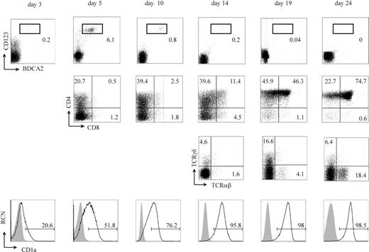
Previously, we demonstrated that both TCRαβ+ and TCRγδ+ T cells and BDCA2+CD123hi pDCs can develop from progenitor cells upon coculture with OP9-Jag1 cells.32 Here we extend our previous findings. CD34+CD1a– human thymic precursor cells were purified and cocultured with the OP9-Jag1 cell line. We found that BDCA2+CD123hi pDCs first appeared after 3 days, reaching a maximum after 5 to 7 days, and disappeared completely after 18 to 20 days (Figure 1). Expression of CD7 (not shown and Dontje et al32 ) and CD1a, indicative of T-cell commitment4 (reviewed by Spits3 ), was up-regulated gradually from the beginning of the coculture. An increasing CD4+ population consisting of immature CD4+ cells and pDCs was detected as early as day 5 of culture. Developing T cells progressed to the CD4+CD8+ T-cell stage by gradual up-regulation of CD8. After 24 days, 75% of the cells had obtained a CD4+CD8+ double-positive phenotype. After 10 days of culture, CD3+ T cells emerged (not shown) and the percentage of CD3+ cells gradually increased. After 3 weeks of coculture, about 70% of the cells expressed either high or intermediate levels of CD3. The arising CD3+ cells first mainly expressed TCRγδ, but TCRαβ+ T cells were detected after 14 days of coculture.
Thymic pDCs have been shown to produce large amounts of IFN-α upon stimulation with viruses or the TLR9 ligand CpG in vitro.27,30 To assess the capacity of pDCs cultured on OP9-Jag1 cells to produce endogenous IFN-α, we stimulated cultures at the peak of pDC development (5 to 6 days) with CpG or virus (HIV-1LAI) and analyzed IFN-α production (Figure 2). Sixteen hours after stimulation with CpG, intracellular IFN-α was detected solely in CD123+ pDCs (Figure 2A), indicating that pDCs generated on OP9-Jag1 cells have the capability to produce IFN-α. Furthermore, we confirmed the secretion of the produced IFN-α by intracellular flow cytometric analysis of MxA, an IFN response protein (reviewed by Grandvaux et al39 ). All human cells in the OP9-Jag1 culture expressed MxA 3 days after the addition of either CpG (2216), recombinant IFN-α (Figure 2B), or HIV-1LAI (Figure 2C). This is consistent with the finding that all cells in the OP9-Jag1 culture expressed the type I interferon (CD118) receptor (data not shown) and are therefore potentially responsive to type I IFN. MxA was not detected when cells were not stimulated or following the addition of either control CpG (2243) or control supernatant from PM1 T cells (Figure 2B). Induction of MxA expression could be blocked completely with neutralizing antibodies to type I IFN. These results indicate that pDCs derived from thymic progenitors cultured on OP9-Jag1 cells are competent to produce IFN-α after CpG or HIV-1LAI stimulation.
The OP9 cell line expressing the human Notch ligand Jagged1 (OP9-Jag1) supports development of CD34+CD1a–human thymic precursor cells into TCRαβ+or TCRγδ+T cells and BDCA2+CD123hipDCs. CD34+CD1a– thymic precursors were cultured for up to 24 days on OP9-Jag1 stromal cells and analyzed by flow cytometry for surface expression of CD123, BDCA2, CD1a, CD4, CD8, TCRαβ, and TCRγδ on the days indicated. Numbers represent the percentages of cells that fall within the electronic gate. In the histograms, shaded curves show isotype controls and open curves show specific stainings. CD1a mean fluorescence intensities (MFIs) are 17.4 (day 3), 44.6 (day 5), 90 (day 10), 244 (day 14), 400 (day 19), 411 (day 24). Results are representative of 3 independent experiments. RCN indicates relative cell number.
The OP9 cell line expressing the human Notch ligand Jagged1 (OP9-Jag1) supports development of CD34+CD1a–human thymic precursor cells into TCRαβ+or TCRγδ+T cells and BDCA2+CD123hipDCs. CD34+CD1a– thymic precursors were cultured for up to 24 days on OP9-Jag1 stromal cells and analyzed by flow cytometry for surface expression of CD123, BDCA2, CD1a, CD4, CD8, TCRαβ, and TCRγδ on the days indicated. Numbers represent the percentages of cells that fall within the electronic gate. In the histograms, shaded curves show isotype controls and open curves show specific stainings. CD1a mean fluorescence intensities (MFIs) are 17.4 (day 3), 44.6 (day 5), 90 (day 10), 244 (day 14), 400 (day 19), 411 (day 24). Results are representative of 3 independent experiments. RCN indicates relative cell number.
Stimulated pDCs hamper T-cell development from thymic progenitors
Previously, we demonstrated that both T cells and pDCs develop from progenitor cells within the thymus microenvironment in vivo.2 We also demonstrated that thymic pDCs produce IFN-α after stimulation with the TLR9 ligand CpG or HIV-1 virus.27 To date, it remains unclear whether stimulated pDCs contribute to T-cell development. Our observations described in “Notch ligand Jagged1 supports simultaneous development of functional pDCs and T cells” allowed us to address this question. Therefore, 5 days after coculture of CD34+CD1a– thymic progenitor cells on OP9-Jag1 cells, cells were stimulated with either CpG or virus for 3 days. Up-regulation of CD1a was used as read-out for T-cell commitment and development on day 8. Addition of either CpG (2216) (Figure 3A) or HIV-1LAI (Figure 3B) to the coculture completely prevented the up-regulation of CD1a expression on the developing thymocytes. In fact, CD1a expression levels did not increase any further than the level observed on day 5. As expected, expression of CD1a on cells treated with either control CpG (2243) or control PM1 supernatant did not differ from untreated control cultures. The absolute cell number or percentage of living cells did not change following 3 days of stimulation with CpG and was only slightly reduced after HIV-1LAI stimulation, indicating that the impaired T-cell development was not due to preferential death of CD1a+ cells (data not shown). Up-regulation of CD1a expression on cells treated with CpG or HIV-1LAI was observed when neutralizing antibodies to type I IFN were added at the time of stimulation, strongly suggesting that the observed block in T-cell development in the presence of activated pDCs was induced by endogenous production of type I IFN. To further confirm these results, recombinant IFN-α was added at day 5 of coculture. Exogenously added IFN-α prevented the up-regulation of CD1a expression to a similar extent as in cultures in which pDCs were stimulated with CpG or virus. This effect of recombinant IFN-α on T-cell development could be counteracted by the addition of the neutralizing IFN-α/β antibodies (Figure 3A).
In vitro–generated pDCs produce IFN-α upon stimulation with either stimulatory CpG or virus. CD34+CD1a– thymic progenitors were cultured on OP9-Jag1 cells. (A) After 6 days of coculture, cells were stimulated with CpG (2216) (10 μg/mL) for 16 hours or left unstimulated. Flow cytometric analysis was performed after intracellular staining of IFN-α and surface staining of CD123. (B-C) After 5 days of coculture, cells were stimulated for 72 hours with (B) CpG (2216) (1 μg/mL) or (C) supernatant of HIV-1LAI–infected PM1 cells (2 ng or 10 ng p24 per milliliter) in the presence or absence of neutralizing antibodies against type I IFNs (10 μL/mL). Control CpG (2243) and supernatant of uninfected PM1 cells served as controls. Flow cytometry was performed after intracellular staining of MxA. (B) Left histogram: MxA protein expression of cultures treated with CpG (2216) (MFI, 114), exogenously added IFN-α (1000 U/mL; MFI, 148) (positive control), control CpG (2243) (MFI, 11), or unstimulated cells (MFI, 12) (negative controls). Right histogram: MxA levels after preincubation with neutralizing antibody to type I IFNs. (C) MxA protein expression in cultures stimulated with 10 μL or 2 μL of control virus supernatant (MFI, 10 and 10, respectively) or HIV-1LAI supernatant in the absence (MFI, 146 and 169, respectively) or presence of neutralizing antibodies (MFI, 85 and 75, respectively). Results are representative of 3 independent experiments.
In vitro–generated pDCs produce IFN-α upon stimulation with either stimulatory CpG or virus. CD34+CD1a– thymic progenitors were cultured on OP9-Jag1 cells. (A) After 6 days of coculture, cells were stimulated with CpG (2216) (10 μg/mL) for 16 hours or left unstimulated. Flow cytometric analysis was performed after intracellular staining of IFN-α and surface staining of CD123. (B-C) After 5 days of coculture, cells were stimulated for 72 hours with (B) CpG (2216) (1 μg/mL) or (C) supernatant of HIV-1LAI–infected PM1 cells (2 ng or 10 ng p24 per milliliter) in the presence or absence of neutralizing antibodies against type I IFNs (10 μL/mL). Control CpG (2243) and supernatant of uninfected PM1 cells served as controls. Flow cytometry was performed after intracellular staining of MxA. (B) Left histogram: MxA protein expression of cultures treated with CpG (2216) (MFI, 114), exogenously added IFN-α (1000 U/mL; MFI, 148) (positive control), control CpG (2243) (MFI, 11), or unstimulated cells (MFI, 12) (negative controls). Right histogram: MxA levels after preincubation with neutralizing antibody to type I IFNs. (C) MxA protein expression in cultures stimulated with 10 μL or 2 μL of control virus supernatant (MFI, 10 and 10, respectively) or HIV-1LAI supernatant in the absence (MFI, 146 and 169, respectively) or presence of neutralizing antibodies (MFI, 85 and 75, respectively). Results are representative of 3 independent experiments.
Thus, we show that in the OP9-Jag1 culture system stimulated pDCs inhibit T-cell development, as determined by the reduced up-regulation of CD1a expression on developing T cells. Our results suggest that the inhibition is solely due to endogenous production of type I IFN by pDCs.
Exogenous IFN-α impairs thymic T-cell development
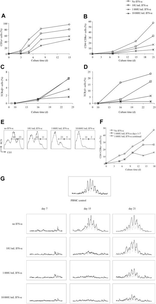
To examine the effect of exogenous IFN-α on human T-cell development in more detail, we employed the coculture system using OP9-DL1 cells with thymic CD34+CD1a– progenitor cells, as previously described.32 This OP9-DL1 culture system supports T-cell development from thymic progenitor cells, while pDC development is strongly inhibited. Increasing concentrations of recombinant IFN-α were added to the coculture, and over time cells were analyzed cells for expression of T-cell–associated surface markers. We observed that addition of IFN-α dose dependently impaired up-regulation of expression of several markers involved in T-cell development, including CD1a, CD4, CD8, TCRαβ, TCRγδ, and CD3 (Figure 4A-E; Table S1, available at the Blood website; see the Supplemental Materials link at the top of the online article). Furthermore, down-regulation of the progenitor-associated marker CD34 was delayed (not shown), strongly suggesting that IFN-α impedes the differentiation of progenitor cells. Upon withdrawal of IFN-α from the cultures, differentiation of progenitor cells progressed normally, demonstrating that the inhibitory effect of IFN-α on T-cell development was reversible (Figure 4F).
These results did not specify whether IFN-α interfered with T-cell development at genomic DNA levels (ie, with TCR rearrangements). To investigate this, a qualitative multiplex PCR method was used.38 Thymic progenitor cells from the OP9-DL1 coculture were analyzed for the status of rearrangement of the TCRγ and TCRβ genes at various times after addition of different concentrations of IFN-α. In line with our previous findings,4 TCRγ rearrangements were already detected in freshly isolated CD34+CD1a– thymocytes (data not shown), and IFN-α did not affect progression of these already-initiated TCRγ rearrangements. In 2 of 3 donors we detected D to J rearrangements of the TCRβ locus in freshly isolated CD34+CD1a– postnatal thymocytes (data not shown), which is consistent with earlier findings.4,40 In the absence of IFN-α, V-DJβ rearrangements were detected first between day 7 and 15 of coculture in this system. Consistent with the phenotypical findings shown in Figure 4A-E, V-DJβ rearrangements were impaired by IFN-α in a dose-dependent manner (Figure 4G). At day 15 of the culture, levels of V-DJβ rearrangements were strongly reduced in the presence of as little as 10 U/mL IFN-α. At 21 days of culture, V-DJβ rearrangements in the presence of low amounts of IFN-α were comparable to those observed in the absence of IFN-α after 15 days, suggesting that IFN-α did not prevent but rather delayed TCRβ rearrangements. Higher concentrations of IFN-α dramatically reduced the level of TCRβ V-DJ rearrangements, even after 3 weeks.
CD1a expression on developing CD34+CD1a–thymic precursors is impaired by type I IFNs endogenously produced by pDCs after stimulation with CpG or HIV-1LAI. Five-day cocultures of CD34+CD1a– thymic progenitors on OP9-Jag1 cells were stimulated with (A) CpG (2216), CpG (2243) (both at 1 μg/mL), recombinant IFN-α (1000 U/mL), or left untreated or with (B) HIV-1LAI (range, 2 to 150 ng p24 per milliliter) or control PM1 supernatant. Stimulations were performed in the presence or absence of neutralizing antibodies against type I IFNs. Three days after stimulation, cells were analyzed by flow cytometry for CD1a surface expression. CD1a expression levels of stimulated cells are shown relative to corresponding controls on day 8, which were set as 1. Average MFI values of at least 3 independent experiments are shown. Error bars represent the range in MFI values of at least 3 independent experiments. Statistical analysis was performed using a paired 2-tailed Student t test; *P < .05, **P < .001.
CD1a expression on developing CD34+CD1a–thymic precursors is impaired by type I IFNs endogenously produced by pDCs after stimulation with CpG or HIV-1LAI. Five-day cocultures of CD34+CD1a– thymic progenitors on OP9-Jag1 cells were stimulated with (A) CpG (2216), CpG (2243) (both at 1 μg/mL), recombinant IFN-α (1000 U/mL), or left untreated or with (B) HIV-1LAI (range, 2 to 150 ng p24 per milliliter) or control PM1 supernatant. Stimulations were performed in the presence or absence of neutralizing antibodies against type I IFNs. Three days after stimulation, cells were analyzed by flow cytometry for CD1a surface expression. CD1a expression levels of stimulated cells are shown relative to corresponding controls on day 8, which were set as 1. Average MFI values of at least 3 independent experiments are shown. Error bars represent the range in MFI values of at least 3 independent experiments. Statistical analysis was performed using a paired 2-tailed Student t test; *P < .05, **P < .001.
Exogenously added IFN-α reversibly interferes with the development of thymic CD34+CD1a–precursors into T cells. Sorted CD34+CD1a– thymic precursors were cultured on OP9-DL1 cells in the presence of IL-7 (5 ng/mL) and increasing concentrations of exogenously added IFN-α. At the time points indicated, percentages of cells expressing (A) CD1a, (B) CD4 and CD8 (ie, double-positive cells), (C) TCRαβ, (D) TCRγδ, and (E) CD3 (numbers indicate the percentages of cells in high and intermediate CD3 expression gates, respectively, on day 18 were determined by flow cytometry. (F) Inhibitory effects of IFN-α on T-cell development are reversible. Seven days after coculture in the presence of IFN-α (1000 U/mL), cells were washed, split, and cultured in the presence or absence of IFN-α. No IFN treatment was used as control. (G) V-DJ rearrangement of the TCRβ chain. Genomic DNA was isolated from CD34+CD1a– thymocytes cultured for the indicated time periods in the presence or absence of IFN-α and analyzed for V to DJ rearrangement by multiplex PCR and GeneScan analysis. Results of “tube A” containing 23 different Vβ primers and 9 different Jβ primers are shown. Black lines represent Vβ-Jβ1 rearrangements; gray lines, Vβ-Jβ2 rearrangements. Genomic DNA of peripheral blood mononucleated cells (PBMCs) served as control for polyclonal V-DJ rearrangement. Results are representative of at least 4 independent experiments.
Exogenously added IFN-α reversibly interferes with the development of thymic CD34+CD1a–precursors into T cells. Sorted CD34+CD1a– thymic precursors were cultured on OP9-DL1 cells in the presence of IL-7 (5 ng/mL) and increasing concentrations of exogenously added IFN-α. At the time points indicated, percentages of cells expressing (A) CD1a, (B) CD4 and CD8 (ie, double-positive cells), (C) TCRαβ, (D) TCRγδ, and (E) CD3 (numbers indicate the percentages of cells in high and intermediate CD3 expression gates, respectively, on day 18 were determined by flow cytometry. (F) Inhibitory effects of IFN-α on T-cell development are reversible. Seven days after coculture in the presence of IFN-α (1000 U/mL), cells were washed, split, and cultured in the presence or absence of IFN-α. No IFN treatment was used as control. (G) V-DJ rearrangement of the TCRβ chain. Genomic DNA was isolated from CD34+CD1a– thymocytes cultured for the indicated time periods in the presence or absence of IFN-α and analyzed for V to DJ rearrangement by multiplex PCR and GeneScan analysis. Results of “tube A” containing 23 different Vβ primers and 9 different Jβ primers are shown. Black lines represent Vβ-Jβ1 rearrangements; gray lines, Vβ-Jβ2 rearrangements. Genomic DNA of peripheral blood mononucleated cells (PBMCs) served as control for polyclonal V-DJ rearrangement. Results are representative of at least 4 independent experiments.
To address whether the effect of IFN-α on T-cell development was due to differential regulation of transcription factors known to be important for proper T-cell development, we analyzed transcript levels of GATA-3, the bHLH family members E12, E47, and HEB, and the Id family members Id2 and Id3 by quantitative reverse transcriptase (RT)–PCR, but no differences between IFN-α–treated and untreated cells were observed (data not shown).
In conclusion, these results demonstrate that IFN-α has a strong negative impact on the transition of developing T cells from CD1a–CD4–CD8– to CD1a+CD4+CD8+ cells. Similarly, surface expression of either the TCRαβ or TCRγδ complex and rearrangements of the TCRβ chain were affected by IFN-α. IFN-α, however, did not abrogate but rather delayed T-cell development, which could be reversed upon withdrawal of IFN-α.
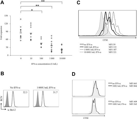
Reduced absolute cell counts after culture of thymic precursors on OP9-DL1 in the presence of IFN-α is due to reduced IL-7–mediated proliferation but not to increased apoptosis. CD34+CD1a– thymic precursors were cultured on OP9-DL1 with IL-7 and different concentrations of exogenously added IFN-α. (A) Absolute cell numbers were determined after 1 week of coculture by counting live cells using trypan blue exclusion. Shown are the fold expansions in cell number compared with the number of cells at the start of the culture of 8 independent experiments. Average values are indicated by a black bar; statistical analysis was performed using a paired 2-tailed Student t test; *P < .05, **P < .001. (B) Three days after culture without or with IFN-α (1000 U/mL), flow cytometric analysis was performed after intracellular staining with anti–Bcl-2 antibody (open curve). Shaded curves represent isotype controls. Numbers indicate MFI of the specific stainings. (C) Directly after sorting, CD34+CD1a– thymic precursors were labeled with CFSE, followed by coculture on OP9-DL1 cells with IL-7 in the presence or absence of different concentrations of IFN-α. On day 6 of coculture, CFSE levels were determined by flow cytometric analysis. (D) Thymic progenitor cells were labeled with CFSE and cultured in cell suspension with recombinant IL-7 (1 ng/mL) in the presence or absence of 100 U/mL IFN-α. After 96 (top panel) and 120 hours (bottom panel), CFSE levels were determined by flow cytometry. Results are representative of 5 independent experiments.
Reduced absolute cell counts after culture of thymic precursors on OP9-DL1 in the presence of IFN-α is due to reduced IL-7–mediated proliferation but not to increased apoptosis. CD34+CD1a– thymic precursors were cultured on OP9-DL1 with IL-7 and different concentrations of exogenously added IFN-α. (A) Absolute cell numbers were determined after 1 week of coculture by counting live cells using trypan blue exclusion. Shown are the fold expansions in cell number compared with the number of cells at the start of the culture of 8 independent experiments. Average values are indicated by a black bar; statistical analysis was performed using a paired 2-tailed Student t test; *P < .05, **P < .001. (B) Three days after culture without or with IFN-α (1000 U/mL), flow cytometric analysis was performed after intracellular staining with anti–Bcl-2 antibody (open curve). Shaded curves represent isotype controls. Numbers indicate MFI of the specific stainings. (C) Directly after sorting, CD34+CD1a– thymic precursors were labeled with CFSE, followed by coculture on OP9-DL1 cells with IL-7 in the presence or absence of different concentrations of IFN-α. On day 6 of coculture, CFSE levels were determined by flow cytometric analysis. (D) Thymic progenitor cells were labeled with CFSE and cultured in cell suspension with recombinant IL-7 (1 ng/mL) in the presence or absence of 100 U/mL IFN-α. After 96 (top panel) and 120 hours (bottom panel), CFSE levels were determined by flow cytometry. Results are representative of 5 independent experiments.
IL-7–mediated proliferation, but not survival, of developing T cells is affected by IFN-α
IFN-α has been used effectively in the treatment of a number of malignancies (reviewed by Tagliaferri et al25 ) because of its ability to inhibit proliferation and/or induce apoptosis in various cell types. Therefore, we examined whether exogenous IFN-α played a role in the proliferation and survival of thymic progenitor cells in the OP9-DL1 coculture system. CD34+CD1a– thymocytes cocultured on OP9-DL1 cells in the presence of IL-7 for 1 week dramatically increased in cell numbers (85-fold ± 24-fold; n = 9, Figure 5A). Addition of 100 U/mL IFN-α or more consistently and significantly (P < .05 for 100 U/mL, P < .001 for 1 000 and 10 000 U/mL) decreased the expansion rate (Figure 5A). Absolute cell numbers remained lower in the presence of IFN-α during at least 28 days. Of note, the addition of 10 U/mL IFN-α did not affect absolute cell numbers, although T-cell differentiation was impaired (Figures 4A-E).
The reduced cell numbers obtained in the presence of IFN-α could be due to either increased apoptosis or reduced cell expansion. To determine whether IFN-α induced apoptosis, flow cytometric analysis was performed after annexin V/7-AAD staining on days 1, 2, 3, 6, and 10 of coculture (data not shown). The percentage of apoptotic cells was not increased at any of the IFN-α concentrations tested (4% ± 2.1% annexin V positive; 4.1% ± 1% 7-AAD positive). Consistent with these findings, no change in expression of the antiapoptotic protein Bcl-2 was detected after 3, 6, or 10 days of coculture in the presence of IFN-α (Figure 5B and data not shown). Furthermore, the expression of 35 other regulators of apoptosis, simultaneously quantified by a multiplex ligation-dependent probe amplification method (RT-MLPA),41 in thymic precursors did not significantly change in cells cultured in the presence or absence of IFN-α for 1, 2, 3, or 6 days on OP9-DL1 cells (data not shown). These findings suggest that in this culture system IFN-α did not impinge on pathways involved in either cell survival or cell death.
To address whether IFN-α affected cell proliferation, sorted CD34+CD1a– thymic progenitor cells were labeled with CFSE and cocultured for 6 days on OP9-DL1 cells. Higher CFSE levels were observed when cells were cultured in the presence of increasing concentrations of IFN-α, indicating that IFN-α inhibited cell proliferation (Figure 5C).
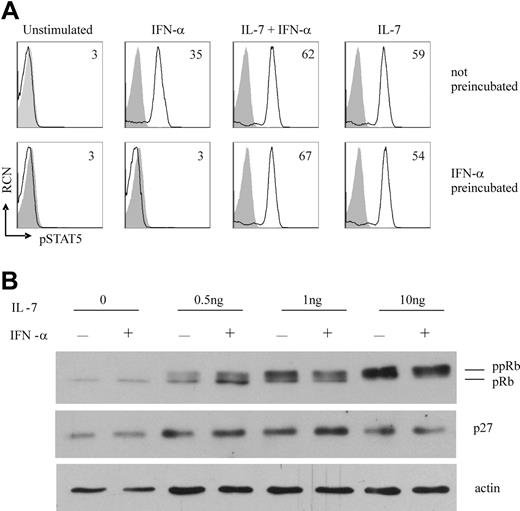
Influence of IFN-α on IL-7R proximal and distal signal transduction events. (A) IFN-α has no effect on IL-7–induced phosphorylation of STAT5. MACS-enriched CD34+ thymocytes were preincubated overnight in culture medium only or in culture medium with IFN-α (1000 U/mL). The following day, cells were stimulated for 15 minutes with IL-7 (1 ng/mL), IFN-α (1000 U/mL), or both, stained for intracellular phospho-STAT5, and analyzed by flow cytometry. Shaded curves represent isotype controls; open curves, specific staining. Numbers indicate MFI of specific stainings. (B) IFN-α does not prevent IL-7–mediated down-regulation of p27kip1 protein or hyperphosphorylation of Rb. MACS-enriched CD34+ thymocytes were cultured for 48 hours with IL-7 in the absence or presence of IFN-α (5000 U/mL). Total cell lysates were analyzed by Western blot analysis using antibodies against either p27kip1 or Rb. Actin blotting was performed to ensure equal protein loading. Results are representative of 2 independent experiments.
Influence of IFN-α on IL-7R proximal and distal signal transduction events. (A) IFN-α has no effect on IL-7–induced phosphorylation of STAT5. MACS-enriched CD34+ thymocytes were preincubated overnight in culture medium only or in culture medium with IFN-α (1000 U/mL). The following day, cells were stimulated for 15 minutes with IL-7 (1 ng/mL), IFN-α (1000 U/mL), or both, stained for intracellular phospho-STAT5, and analyzed by flow cytometry. Shaded curves represent isotype controls; open curves, specific staining. Numbers indicate MFI of specific stainings. (B) IFN-α does not prevent IL-7–mediated down-regulation of p27kip1 protein or hyperphosphorylation of Rb. MACS-enriched CD34+ thymocytes were cultured for 48 hours with IL-7 in the absence or presence of IFN-α (5000 U/mL). Total cell lysates were analyzed by Western blot analysis using antibodies against either p27kip1 or Rb. Actin blotting was performed to ensure equal protein loading. Results are representative of 2 independent experiments.
To gain insight into whether IFN-α specifically reduces the IL-7– or Notch-mediated proliferation rate, thymic progenitor cells were cultured with either IL-7 (1 ng/mL) or OP9-DL1 in the presence or absence of IFN-α (Figure 5D and data not shown). We observed that the IL-7–induced proliferation rates were lower in the presence of IFN-α, both after 96 and 120 hours. Culture of thymic progenitor cells on OP9-DL1 cells in the absence of exogenous IL-7 led to massive cell death (more than 90%), which prevented conclusive assessment (data not shown). Collectively, we conclude that IFN-α (at least 100 U/mL) reduces the IL-7–induced expansion, but not survival, of thymic precursors.
Involvement of IL-7R proximal or distal events in the IFN-α–induced inhibition of T-cell development
To elucidate the molecular mechanism of IFN-α–induced impairment of IL-7–dependent T-cell development, we analyzed several known downstream targets of IL-7R signaling, including signal transducer and activator of transcription 5 (STAT5), PI3K, and MEK-Erk pathways.7,42 Sorted CD34+CD1a– thymocytes were stimulated with IL-7 in the presence or absence of IFN-α, and the phosphorylation status of STAT5, extracellular-regulated kinase 1 and 2 (Erk1/2), and Akt (PKB) proteins was determined by flow cytometry. As expected, IL-7 induced phosphorylation of STAT5, Erk1/2, and Akt (PKB) (Figure 6A and data not shown). However, no reduction in the level of phosphorylation of any of these proteins was observed when both IL-7 and IFN-α were added. Consistent with observations in cell lines,43 IFN-α alone induced the phosphorylation of STAT5 in CD34+CD1a– thymocytes. To exclude indirect effects of IFN-α on IL-7R–mediated phosphorylation, we also preincubated CD34+CD1a– thymocytes with IFN-α before IL-7 stimulation. This did not affect the IL-7–induced phosphorylation of STAT5, Erk1/2, or Akt (PKB), although STAT5 phosphorylation upon repeated stimulation with IFN-α alone was prevented (Figure 6A and data not shown). Together these findings strongly support the notion that the IFN-α–induced block in T-cell development was not imposed by obstruction of IL-7R proximal signaling events.
We further evaluated whether IFN-α inhibits IL-7R distal events. It has been reported that activation of PI3K-PKB inhibits forkhead transcription factor FKHR-L1 activity, consequently down-regulating transcription of the cyclin-dependent kinase inhibitor (CKI) p27kip1.44 In addition, IL-7 induces hyperphosphorylation of the Rb protein, eventually leading to cell cycle progression.45 Using immunoblotting, we addressed whether IFN-α interfered with the IL-7–induced p27kip1 down-regulation or pRb hyperphosphorylation in CD34+ thymocytes (Figure 6B). As expected, IL-7 reduced the levels of p27kip1 protein and increased the level of Rb phosphorylation. No significant impact of IFN-α on these IL-7–mediated events was observed at 48 hours (Figure 6B) or 72 hours (data not shown). Consistent with these findings, no changes in the IL-7–induced increase in cell size were detected by flow cytometric analysis after addition of IFN-α (data not shown).
In conclusion, while IFN-α clearly impeded IL-7–induced T-cell development and proliferation, this could not be attributed to inhibition of either IL-7R proximal events, including the phosphorylation of STAT5, PI3K, or Erk1/2, or IL-7R distal events, such as down-regulation of p27kip1 or hyperphosphorylation of Rb.
Discussion
We show that IFN-α, when produced endogenously by pDCs upon stimulation by either bacterial or viral compounds or added exogenously as recombinant protein, has a deleterious effect on early T-cell development. IFN-α interfered with IL-7–mediated T-cell differentiation of progenitor cells, resulting in a delay in the transition from CD4–CD8– to CD4+CD8+ stage, and inhibited rearrangement of the TCRβ chain and up-regulation of the T-cell–associated surface proteins CD1a, CD3, TCRαβ, and TCRγδ. In addition, IFN-α affected proliferation but not survival of the developing precursors. This inhibitory effect was reversible, as withdrawal of IFN-α restored normal T-cell differentiation.
For the present study we made use of a recently described in vitro assay for T-cell development.46 In the presence of the cytokines IL-7 and Flt3L, the OP9-DL1 cell line supports the development of CD4+CD8+TCRαβ+ and TCRγδ+ T cells from CD34+CD1a– human thymic progenitor cells while inhibiting pDC development.32 Previously, we reported that human thymic precursors cultured in the presence of only IL-7 develop into early CD4+CD8+ thymocytes that lack TCR expression.47 We show here that thymic progenitors cultured on OP9-DL1 undergo TCR rearrangements leading to complete, diverse TCR rearrangements of the β and γ loci. Continuous Notch signaling of at least 1 to 2 weeks was required to induce a polyclonal TCRβ repertoire, which is consistent with findings in the mouse.48
In contrast to OP9-DL1 cells, the OP9-Jag1 culture system supports both T-cell and pDC development from the same progenitor cells32 (Figure 1). This allowed us to directly assess the impact of stimulated pDCs on early T-cell commitment and differentiation. In the thymic environment, pDCs are located at the corticomedullary junction and in the medulla.12 It has been reported that in mice thymic precursors enter the thymus at the corticomedullary junction,1 which allows for interactions of progenitor cells with pDCs. Here we show that stimulation of pDCs by either CpG or virus arrested early T-cell development of thymic progenitor cells cocultured with OP9-Jag1 cells. The effect was mainly due to endogenous production of type I IFN, because T-cell development could be rescued completely by the addition of blocking antibodies against type I IFNs. Moreover, addition of exogenous IFN-α to developing thymic precursors on OP9-DL1 cells or in mouse FTOC also impaired T-cell development (Figures 4 and S1). These findings further supported our notion that stimulated pDCs hamper T-cell development by endogenous production of IFN-α.
Because IL-7 is critical for survival, proliferation, and development of T cells in the thymus,6-8 we anticipated that IFN-α counteracted IL-7–induced signal transduction. In mice, a negative correlation between IL-7 and IFN-α signaling was suggested previously, because IFN-α inhibited IL-7–driven proliferation of CD3–CD4–CD8– thymocytes in vitro.29 In addition, treatment of newborn mice with high doses of IFN-α resulted in a reduction of thymic cellularity by more than 80% and a 50% decrease in CD4+CD8+ cells.28 Importantly, the window of sensitivity of the developing thymocytes to IFN-α corresponded closely to the stages during which IL-7 signaling is essential. While here we describe the effects of IFN-α on early human T-cell development, we also observed that IFN-α interfered with IL-7–induced maturation at later stages of thymocyte development, at the transition of CD3+CD1+ to CD3+/hiCD1– (data not shown). This indicates that IFN-α also in humans affects those stages of T-cell development in the thymus that are dependent on the presence of IL-7.49 Unraveling the molecular mechanisms of IFN-α–mediated effects on early T-cell development is complicated. Phosphorylation of known downstream effector molecules of the IL-7R signal transduction pathway, including Akt/PKB, STAT5, and Erk1/2,7,42 was not affected by addition of IFN-α, suggesting that the negative effects of IFN-α were not due to IL-7R proximal signaling events. Further downstream signaling events induced by IL-7 include Bcl-2 up-regulation, which is critical for survival of immature thymocytes,6 and down-regulation of the CKI p27kip1 followed by phosphorylation of the Rb protein and E2F-driven transcription, which mediate cell cycle progression.45 In the primary human thymocytes analyzed here we observed that Bcl-2 was expressed in IL-7–treated cells. In addition, we observed that IL-7 destabilized p27kip1 protein and phosphorylated Rb. These events, however, were either not or only weakly affected by IFN-α. Therefore, we consider it unlikely that IFN-α interferes with these IL-7R–induced distal events.
Almost all leukocytes can produce IFN-α, but pDCs secrete the highest amounts of IFN-α. In vivo IFN-α is produced locally after stimulation by bacterial or viral infections, and elevated levels are found in peripheral blood during chronic immune activation, as observed in chronic viral infections and autoimmune diseases such as systemic lupus erythematosus. As primary producers of IFN-α, pDCs likely play an important role in the immune response against viruses. In this respect, we have shown recently that if stimulated to produce IFN-α, pDCs can slow HIV-1 replication.27 The IFN-α response generated by HIV-1 alone, however, was insufficient to completely control virus replication and therefore failed to prevent HIV-1 pathogenesis in the thymus. Our data indicate that despite its favorable short-term antiviral effects, continual virus-induced production of IFN-α by pDCs in the thymus can be harmful. HIV-1–induced IFN-α may up-regulate major histocompatibility complex (MHC) class I on thymic epithelial cells30 and result in preferential selection of dysfunctional CD4–CD8+ thymocytes.50 We show here that pDC-derived endogenous IFN-α directly impedes early T-cell lymphopoiesis. The presence of high levels of IFN-α may prevent T-cell regeneration in HIV-1 infection and contribute to the depletion of developing T cells by HIV-1.51
IFN-α is widely used for treatment of hematologic and solid malignancies and viral infections, particularly chronic hepatitis C.25,26 Exogenous IFN-α as a therapy may enhance the impact of locally produced IFN-α by pDCs in the thymus. In fact, leukopenia is a common side effect of IFN-α treatment (reviewed by Sleijfer et al52 ). The effects of exogenous IFN-α on early T-cell development that we describe here may explain in part the side effects of IFN-α treatments and therefore need to be considered when applying IFN-α therapy to patients, especially in children, where peripheral T cells are predominately maintained through the production of naive T cells by the thymus.53
Authorship
The authors declare no competing financial interests.
Prepublished online as Blood First Edition Paper, August 17, 2006; DOI 10.1182/blood-2006-02-004978.
The online version of this article contains a data supplement.
The publication costs of this article were defrayed in part by page charge payment. Therefore, and solely to indicate this fact, this article is hereby marked “advertisement” in accordance with 18 USC section 1734.
This work was supported by the National Institutes of Health/National Institute of Allergy and Infectious Diseases (NIH/NIAID) grant R01-AI52002-02 (principal investigator, C.H.U.) and the Universitywide AIDS Research Project (UARP) ID03-LA-001 (principal investigator, C.H.U.). We thank Berend Hooijbrink for his help with FACS sorting and maintenance of the FACS facility, Eric Eldering for analyzing regulators of apoptosis by RT-MLPA, and Maho Nagasawa for technical support. Dr M. Hazekamp and staff at the Leiden University Medical Center and the Academic Medical Center, University of Amsterdam, are acknowledged for providing postnatal thymus tissue.


This feature is available to Subscribers Only
Sign In or Create an Account Close Modal