

Abstract
INTRODUCTION: Oligocloanality is a phenomenon shown in up to 40% of patients with childhood B-lineage acute lymphoblastic leukemia (ALL), and it might be associated with a poorer prognosis.
OBJECTIVE: To present a case of oligoclonality based on the detection of IGH gene rearrangements in a patient with ALL and the correlation of the results with those of flow cytometry immunophenotyping and chimerism quantification.
PATIENT AND METHODS: 26 years old female, pre-B ALL diagnosed in her childhood, in relapse after autologous and allogeneic HLA-identical unrelated stem cell transplantation (SCT). Immunophenotyping was done by multiparametric flow-cytometry, chimerism quantification was performed using fluorescent in situ hybridization (FISH) for the sex chromosomes, and IGH gene rearrangements were detected by nested-PCR revealed by capillary electrophoresis. Disease associated leukocyte lineages (CD19+, CD19+CD34+ and CD19+CD34−) were purified using immunomagnetic technology.
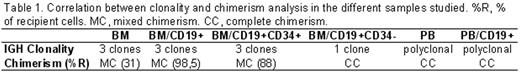
RESULTUS: Flow-cytometry analysis in PB and BM samples at diagnosis showed expression of CD45 (weak), CD34, CD38, CD10, CD19, CD20, CD22, CD66, CD79acyt, TdT and aberrant expression of CD33, with a detectable clonal IGH rearrangement. At relapse, two distinct CD19+ cell populations were revealed from BM samples: one immature (CD34+, 15% of the total leukocytes) with similar phenotype at diagnosis, and another one mature (CD34−, 10% of the total leukocytes). FISH analysis showed mixed chimerism (MC)(31% recipient) in BM samples and complete chimerism (CC) in PB samples. Molecular analysis showed three different clones in BM and a polyclonal pattern in PB. In order to increase sensitivity, CD19+ cells were isolated from BM and PB. Cells selected from BM showed MC (98,5% recipient) and the three clonal IGH gene rearrangements while those from PB showed CC and a polyclonal pattern. Further studies exhibited persistence of the CD19+CD34+ population and significant reduction of the mature CD19+CD34− cells. A persistent three clone pattern was present in BM samples. In order to clarify the origin of the different IGH gene rearrangements, two consecutive immunomagnetic selections, one with CD19+ and other with CD34+ antibodies, were performed from BM samples to purify both CD19+CD34+ and CD19+CD34− populations. CD19+CD34+ cells showed all the three IGH clones and MC (88% recipient). Moreover, CD19+CD34− cells showed a monoclonal pattern with an unique peak, not detected in previous studies, and CC (Table 1).
CONCLUSIONS: The clonal diversity of IGH gene rearrangements detected at relapse after SCT was generated from the original malignant CD34+ clone, possibly due to clonal evolution, and seems to be associated with the poor outcome of this case. After multiple therapy modalities, the CD19+CD34+ cells showed resistance to treatment. Surprisingly, the CD19+CD34− cells showed a new clonal rearrangement although its donor origin confirmed its reactive nature in the context of SCT. This observation has relevance in the further follow up and minimal residual disease detection in this case.
Correlation between clonality and chimerism analysis in the different samples studies. %R, % of recipient cells. MC, mixed chimerism. CC, complete chimerism.
Disclosure: No relevant conflicts of interest to declare.

This feature is available to Subscribers Only
Sign In or Create an Account Close Modal