Abstract
Introduction: MRI is gaining increasing importance for the noninvasive quantification of organ iron burden. To date, MRI validation studies have not systematically examined the effects of different iron chelators. Since transverse relaxation rates depend on iron distribution as well as iron concentration, physiologic and pharmacologic processes that alter iron distribution could change MRI calibration curves. This paper compares the effect of three iron chelators, deferoxamine, deferiprone, and deferasirox on R1 and R2 calibration curves according to two loading and chelation strategies.
Methods: 33 Mongolian gerbils underwent iron dextran 500 mg/kg/wk for 4 weeks followed by 4 weeks of chelation therapy using deferoxamine, deferiprone and defersirox. An additional 32 animals received less aggressive iron loading (200 mg/kg/week) for 10 weeks, followed by 12 weeks of chelation therapy. R1 and R2 measurements were obtained immediately post euthanasia using an NMR relaxometer. Calibration curves from 28 unchelated animals loaded with 200 mg/kg/week from 2 to 48 weeks were used as the reference standard for both chelated groups, using Bland-Altman analysis.
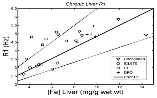
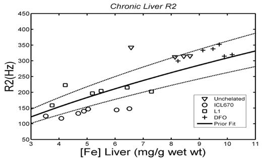
Results: In the liver, R2-iron calibration became more variable over time regardless of whether chelation was performed or not (mean COV 28% versus 12%); no significant changes were observed in the heart R2-iron relationship. Variability in R1 measurements did not change for either heart or liver. Two systematic chelator-specific changes in liver iron calibration curves were noted:
deferiprone treated animals exhibited signficantly higher R1 values (Figure 1) and
deferasirox treated animals demonstrated lower R2 values for given iron concentration (Figure 2).
Both changes were associated with obvious changes in water content or iron distribution.
Discussion: The acuity of the iron loading process affects the variability but not the bias of MRI-iron calibration curves. In contrast, iron chelation can produce systematic shifts in MRI calibration curves compared with the unchelated state, reflecting gross changes in tissue hydration and iron distribution. Since the rate of iron-loading and extraction performed in animals is more extreme than occurs in humans, limiting tissue requilibration, it is possible that the present studies overestimate the potential for chelator-specific calibration bias. Nonetheless, caution should be used in extrapolating calibration curves derived from patients using deferoxamine therapy to others being treated with deferiprone and deferasirox. Careful, longitudinal assessment of MRI calibration curves of patients receiving oral chelation therapies is warranted.
Disclosures: Dr. Nick is an employee of Novartis Pharma.; Dr. Wood is a consultant for Novartis Pharma and Apotex.; Dr. Wood has received research support from Novartis Pharma and Apotex.; Dr. Wood has received speakers honoraria from Novartis Pharma and Apotex.; Dr Wood contributed to the Exjade Speakers Bureau curriculum.
Author notes
Corresponding author


This feature is available to Subscribers Only
Sign In or Create an Account Close Modal