Abstract
Introduction: Targeted disruption of the hemojuvelin gene in mice (HJV−/−) has recently been reported to cause markedly increased liver, pancreas and heart iron deposition, and act as a model for juvenile hemochromatosis (Niederkofler et al. 2005, Huang et al. 2005). Deferasirox, a novel tridentate oral iron chelator, is effective in the treatment of patients with transfusional iron overload and it has not been investigated in primary iron overload. We sought to examine spontaneous iron loading and the effect of deferasirox on the removal of iron in HJV−/− mice with iron overload.
Methods: Inductively-Coupled Plasma Optical Emission Spectrometry (ICP-OES, measurement of elemental iron) determinations in liver, heart, spleen and pancreas were performed at week 8 (study start), 14, 20 and 28 in HJV −/− and wild-type (wt) mice (n=6–8), subsequent to MRI measurements in heart and liver. Starting at week 20 three groups of HJV−/− animals received daily 0 (vehicle), 30 or 100 mg/kg deferasirox, control wt-mice remained untreated. Iron loading was observed up to 28 weeks (including vehicle group) and the effects of deferasirox were assessed by comparing the 28-week groups. MRI R2* measurements were performed using a 4.7T MR imager. For the myocardial and hepatic R2* assessments single-slice, ECG gated gradient-echo images and single-slice transversal gradient-echo images were acquired, respectively.
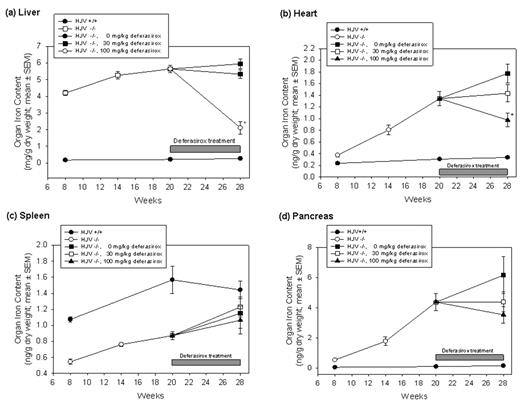
Results: Iron loading of the liver and the heart was 21- and 5-fold higher in HJV−/− than in wt mice. Conspicuously, pancreas showed the most extreme difference in iron load (34-fold). Visual inspection of the time courses of iron loading between week 8 and 28 suggest a delayed loading of the heart and pancreas as compared with liver.
Effects of deferasirox on organ iron load were modest at 30 mg/kg but quite pronounced in liver and heart at 100 mg/kg. This may seem contradictory to data from human studies, which show effects between 20 and 30 mg/kg in iron overload, but may be explained by the approximately 6–9 times lower exposure of deferasirox in mice. At 100 mg/kg deferasirox, liver iron was reduced by 63% and heart iron by 41% as compared with the (HJV−/−)-vehicle treated group. Pancreas showed a clear trend to lower levels at the 100 mg/kg dose. Contrary to liver, heart and pancreas, iron burden of the spleen was lower in HJV−/− as compared to wt mice. This behavior is expected because the same (enhanced) iron efflux mechanism that allows iron to enter the circulation from enterocytes is also in place in reticuloendothelial cells.
MRI R2* values are more variable than organ iron concentrations determined by ICP-OES. However, the profiles of the time courses of R2* of liver and heart in HJV−/− mice were similar to the time courses of liver and heart iron as determined by ICP-OES, underlining the correlation between R2* values and organ iron.
Conclusion: Deferasirox significantly reduced liver and heart iron in the hemojuvelin knock-out mouse and showed a trend to improvement of pancreatic iron load within 8 weeks of treatment.
Disclosures: H Nick, P Allegrini, L Fozard, U Junker, L Rojkjaer, R Salie and T O’Reilly are Novartis employees.
Author notes
Corresponding author


This feature is available to Subscribers Only
Sign In or Create an Account Close Modal