Polymorphonuclear leukocyte (PMN) recruitment to vascular endothelium during acute inflammation involves cooperation between selectins, G-proteins, and β2-integrins. LFA-1 (CD11a/CD18) affinity correlates with specific adhesion functions because a shift from low to intermediate affinity supports rolling on ICAM-1, whereas high affinity is associated with shear-resistant leukocyte arrest. We imaged PMN adhesion on cytokine-inflamed endothelium in a parallel-plate flow chamber to define the dynamics of β2-integrin function during recruitment and transmigration. After arrest on inflamed endothelium, high-affinity LFA-1 aligned along the uropod-pseudopod major axis, which was essential for efficient neutrophil polarization and subsequent transmigration. An allosteric small molecule inhibitor targeted to the I-domain stabilized LFA-1 in an intermediate-affinity conformation, which supported neutrophil rolling but inhibited cell polarization and abrogated transmigration. We conclude that a shift in LFA-1 from intermediate to high affinity during the transition from rolling to arrest provides the contact-mediated signaling and guidance necessary for PMN transmigration on inflamed endothelium.
Introduction
Neutrophils are recruited at vascular sites of acute inflammation by the sequential binding of selectins, CXC chemokines, and β2-integrins that function cooperatively to elicit rolling, arrest, and transmigration. From observations of neutrophil recruitment in the murine microcirculation and on endothelial monolayers grown in tissue culture, a number of rules of engagement have emerged. First, Mac-1 (αMβ2) and LFA-1 (αLβ2) are necessary and sufficient for neutrophil arrest and transmigration, with each subunit providing distinct adhesive contributions throughout the process from rolling to transmigration.1-3 Second, polymorphonuclear leukocyte (PMN) rolling on a monolayer of cells coexpressing E-selectin and intercellular adhesion molecule-1 (ICAM-1) is sufficient to induce selectin ligand clustering (PSGL-1 and L-selectin) and to signal a shift in LFA-1 and Mac-1 from low to high affinity to bind ICAM-1. This process is synergistic with chemokine signaling on rolling PMN to amplify the efficiency of arrest.4,5 LFA-1 appears to function early in this process in that it participates in tethering to ICAM-1 as it shifts from low to intermediate and high affinity.6,7 How changes in conformation of the heterodimer result in changes in affinity to interact with ICAM-1 and mediate rolling, arrest, and outside-in signaling is only partially defined.8
Structural studies of LFA-1 reveal that extension and activation of ICAM-1 binding involves an inserted or I-domain on the α-subunit and an I-like domain on the β-subunit that exerts a pull on the C-terminal α7-helix of the α-subunit, leading to the open shape of the heterodimer and high-affinity ligand binding.9,10-12 This conformational shift to high affinity can be stabilized by binding of Mg2+ or Mn2+ or by inside-out signaling by chemokine receptors.9,13,14 Once activated by a chemokine such as IL-8 or SDF-1, extension and opening of the LFA-1 heterodimer initiate rapid arrest on ICAM-1, as do activated I-domain mutants.6,15-18 Counteracting a shift to high affinity, small molecule allosteric anti-inflammatory inhibitors of LFA-1 function by effectively stabilizing the low-affinity state and antagonize binding to ICAM-1 and leukocyte adhesion.19,20 XVA143 is one such molecule that targets the I-like domain on the β-subunit and stabilizes the intermediate affinity and extended conformation of LFA-1.21,22 Treatment with XVA143 supports lymphocyte rolling but not stable adhesion on ICAM-1 in shear flow.17 Another small molecule, BIRT377, binds with high affinity (Kd = 26 nM) to the I-domain on the α-subunit and stabilizes a low-affinity conformation, thereby preventing firm adhesion of leukocytes to ICAM-1.15-17,22,23 To date, all allosteric antagonists that target the α-subunit I-domain, including BIRT377, LFA703, A-286982, and lovastatin (Novartis, Basel, Switzerland), stabilize a closed, low-affinity conformation.19 In this study we characterized IC487475, a P-arylthiocinnamide or diarylsulfide analog that binds to the I-domain allosteric site (IDAS) of LFA-1.24 We have recently reported that IC487475 hastens ICAM-1 dissociation from activated LFA-1, increasing the rate from 0.003 second-1 to 0.03 second-1. This correlated with a downshift of affinity from high to intermediate while leaving the integrin in an extended state.20 Allosteric stabilization with small molecules provides a tool to determine how LFA-1 conformation and corresponding affinity correlate with leukocyte adhesion and migratory function.
Coinciding with a shift to high affinity, the stimulation of leukocytes elicits redistribution of LFA-1 into submicron clusters necessary for efficient arrest and transmigration at the region of contact with ICAM-1.14,18,25-27 Current data suggest that these high-density clusters of LFA-1 provide outside-in signaling of subsequent functions, including recruitment of high affinity LFA-1, F-actin polymerization, microtubule rearrangements, Rho GTPase activation, and up-regulation of β1-integrin, ultimately leading to cell polarization and migration.9,28-33 Induction of this migratory phenotype correlates with redistribution of LFA-1 to the leading edge of lymphocytes.34 Although membrane redistribution of LFA-1 on chemokine-stimulated leukocytes is well documented, how dynamic shifts of LFA-1 affinity, membrane topography, and outside-in signaling function in coordinating the multistep process of rolling to arrest and subsequent transendothelial migration remains ill defined.
In this study we used phase and fluorescence microscopy to observe in real time the dynamics of neutrophil recruitment in shear flow on inflamed human venular endothelium. These studies define how rolling velocity and its variance change as a function of LFA-1 affinity regulated through the I-domain. We discovered that a shift in LFA-1 from intermediate to high affinity plays a regulatory role in the steps leading from neutrophil arrest to cell polarization and diapedesis.
Materials and methods
Isolation of human neutrophils
Whole blood was obtained from healthy adults by venipuncture into sterile syringes with heparin (10 U/mL blood; Elkins-Sinn, Cherry Hill, NJ) according to protocol number 200311635-6 of the University of California at Davis institutional review board. Informed consent was provided according to the Declaration of Helsinki. Neutrophils were isolated in an unactivated state (Figure S1, available on the Blood website; see the Supplemental Figure link at the top of the online article) using a one-step Ficoll-Hypaque density gradient (Robbins Scientific, Sunnyvale, CA). Neutrophils were maintained at room temperature in a calcium-free HEPES buffer and were assessed for viability by trypan blue exclusion (greater than 98% viable).
Cell culture and reagents
Human umbilical vein endothelial cells (HUVECs; Cascade Biologics, Portland, OR) were maintained in Medium 200 (Cascade Biologics, Portland, OR). Transfected L cells coexpressing human E-selectin and ICAM-1 (L-E/I), as described previously, were maintained in a modified RPMI medium (Invitrogen, Carlsbad, CA).5
Agonists, inhibitors, and antibodies
Recombinant human ICAM-1/IgG (ICOS, Bothell, WA) is a chimer with two full-length ICAM-1 spliced onto the Fab domains and was used at 10 μg/mL.18 Recombinant LFA-1 (rLFA-1) heterodimer is composed of a leucine zipper motif inserted between the αL and β2 subunits and was provided by ICOS, as was anti-leucine zipper mAb 324C. Human IL-8 and IL-1β were purchased from R&D Systems (Minneapolis, MN). Blocking antibodies targeting CD18 were anti-Mac-1 mAb 2LPM19c (10 μg/mL) (DAKO, Carpinteria, CA) and anti-LFA-1 mAb TS1/22 (10 μg/mL) (ICOS). mAb R3.1 binds to the open I-domain of LFA-1 and was provided by ICOS.23,35 For real-time immunofluorescence, nonblocking mAbs TS2/4 (20 μg/mL) (ICOS) to LFA-1, ICRF44-phycoerythrin (PE) (BD Biosciences PharMingen, San Diego, CA) to Mac-1 (20 μg/mL), and 327C (20 μg/mL) (ICOS) to neoepitopes expressed on the high-affinity conformation of CD18 were used.36 mAb KIM127 (10 μg/mL) was provided by Martin Robinson (CellTech, Brighton, United Kingdom).10 327C, ICAM-1/IgG, R3.1, and TS2/4 were conjugated with Alexa-488 or Alexa-546 (Molecular Probes, Eugene, OR), as indicated, according to the manufacturer's protocol. KIM127 was secondarily labeled with FITC-conjugated goat anti-mouse F(ab)2 (KPL, Gaithersburg, MD). IC487475 is a small molecule of the P-arylthiocinnamides series that targets the I-domain allosteric site of LFA-1.37 BIRT is an allosteric small molecule antagonist that is structurally analogous to and functionally identical with BIRT377.23,37,38 Lovastatin sodium was purchased from Calbiochem (La Jolla, CA). Monoclonal antibody 240Q binds CD18 and induces an active conformation, as previously described.18,39 Goat anti-mouse IgG RPE was purchased from Martek Biosciences (Columbia, MD).
Flow cytometric detection of PMN activation and adhesion
To examine specific adhesion by the β2-integrins, neutrophils were sheared with fluorescent beads derivatized with ICAM-1/IgG, as previously described.18,40 Briefly, 106 neutrophils/mL were labeled with LDS-751 8 ng/mL (Molecular Probes) and were preincubated with adhesion blocking mAbs or IC487475 for 10 minutes at 23°C. Fluorescent beads coated with ICAM-1/IgG (1 μm; 2 × 10⁷ beads/mL; Molecular Probes) were then added, along with a magnetic stir bar, as neutrophil suspensions were stimulated with 1 nM IL-8 and analyzed for adhesion by FACScan flow cytometry (Becton Dickinson, San Jose, CA) in a sheared mixing chamber at 37°C. The magnetic stir device created a shear field (shear stress, approximately 1.0 dyne/cm2) within the test tube and initiated collisional interactions of approximately 25 milliseconds. Neutrophil-bead adhesion was quantitated by green bead fluorescence on fluorescence histograms. Quantal increases in fluorescence appeared as peaks in the fluorescence histogram corresponding to populations of neutrophils binding increasing numbers of beads.40 To distinguish relative levels of bead capture within the stimulated neutrophil population, neutrophil-bead interactions were quantitated as the average number of beads per neutrophil according to the following equation:
where N represents the number of nonadherent neutrophils and NBi represents the number of neutrophil-bead aggregates bound to 1 to 6 or more beads. To assess high-affinity binding to ICAM-1, neutrophils were preincubated with 2LPM19C or IC487475 for 10 minutes at 23°C. Alexa-488 ICAM-1/IgG was then added as cell suspensions were stimulated with 5 μg/mL mAb 240Q for 10 minutes at 37°C. Binding of ICAM-1 to neutrophils was detected by FACScan flow cytometry. To assess CD18 extension, neutrophils were preincubated with 1 μM IC487475, 1 μM BIRT or vehicle, and KIM127. Suspensions were then activated with 1 nM IL-8 and incubated with FITC-conjugated secondary goat anti-mouse F(ab′)2 for 5 minutes at 37°C, followed by fixation and analysis by flow cytometry. To determine the effect of allosteric inhibitors on the expression of the open I-domain, rLFA-1-derivatized beads were prepared as previously described.37 Briefly, amino microsphere latex beads (6-μm diameter) (Biosciences, Piscataway, NJ) were mixed with a sulfosuccinimidyl maleimide-N-hydroxysuccinimide ester cross-linker (Pierce, Rockford, IL), according to the manufacturer's protocol. Sulfhydrylated anti-leucine zipper mAb 324C-SH was prepared by incubating mAb 324C with a sulfhydryl linker (Pierce) according to the manufacturer's protocol. 324C-SH was then mixed with the amino cross-linker beads for 2 hours at 8°C. rLFA-1 (20 μg/mL) was mixed with 324C-SH beads (106/mL) at 37°C for 30 minutes. rLFA-1 beads were then mixed for an additional 5 minutes at 37°C with allosteric inhibitors IC487475 (1 μM), BIRT (5 μM), and lovastatin (1 mM) in the presence of 3 mM MgCl2. Beads were then incubated with 10 μg/mL mAb R3.1 for 25 minutes at 37°C, followed by 10 μg/mL goat anti-mouse IgG RPE, before washing and flow cytometry.
Analysis of neutrophil rolling, arrest, and transmigration kinetics
Neutrophil adhesion to IL-1β-activated HUVECs (10 U/mL, 4 hours) was measured in a parallel-plate flow chamber.41 To assess rolling, arrest, and transmigration, isolated neutrophils were preincubated with adhesion blocking mAbs, IC487475 (1 μM), or BIRT (1 μM) for 10 minutes at room temperature, then perfused into the flow chamber over activated HUVECs by syringe pump (Harvard Apparatus, Holliston, MA) at 2 dyne/cm2 for 5 minutes. During this time, five 30-second digital image sequences were captured over unique fields of view every minute. Neutrophil interactions with the monolayer substrates were imaged using a Nikon TE200 inverted microscope equipped with a Plan Fluor 20 × (NA = 0.45) phase-contrast objective, an analog charge-coupled device (CCD) camera (Dage-MTI, Michigan City, MD) and a digital frame grabber (Scion, Frederick, MD). Image sequences were captured at a frame rate of 0.250 per second and analyzed using Image Pro Plus version 4.5 (Media Cybernetics, Silver Spring, MD). From spatial coordinates acquired throughout the video sequence, an instantaneous velocity was calculated for individual cells by determining the migration step distance between (x2, y2) and (x1, y1) every 0.250 second according to the following formula:
Quantitation of neutrophil polarization
To differentiate the roles of E-selectin and IL-8 in eliciting neutrophil shape change, a novel polydimethylsiloxane (PDMS) microflow channel device was constructed from a photo-etched silicon master with dimensions of 200 μm × 100 μm × 2 mm (3 μL volume). Neutrophils were incubated with IC487475 (1 μM), BIRT (1 μM), or anti-LFA-1 for 10 minutes at 23°C, then perfused into the microflow channels over L-E/I using a syringe pump (Harvard Apparatus) at 1 dyne/cm2 for 3 minutes. The perfusion buffer was then adjusted to 1 nM IL-8, as indicated. Polarization of adherent neutrophils on HUVECs was assayed using the parallel-plate flow chamber, as described for rolling, arrest and transmigration kinetics. To determine the rate and extent of neutrophil polarization on HUVECs or L-E/I, individual neutrophil membrane perimeters were defined using an automated polygon region of interest tool at 1-second intervals throughout the video sequence. The aspect ratio of the cell was then quantitated using Image Pro Plus version 4.5 (Media Cybernetics) software by the following equation: aspect ratio = major radius/minor radius.
A neutrophil was considered polarized if the aspect ratio exceeded 1.4. The automated polygon region of interest tool was also used to track the centroid of vehicle- or IC487475-treated neutrophils at 5-second intervals at the start of migration. Centroid movement was tracked for 30 seconds and analyzed for total migration distance, net migration distance, and average velocity. Total migration distance was determined by summing migration step distances every 5 seconds throughout the video sequence. Net migration distance was quantitated as the direct distance between the start of migration and the end of migration.
Real-time immunofluorescence microscopy
The topography of LFA-1, Mac-1, and high-affinity CD18 was imaged in real time during neutrophil rolling and arrest in shear flow. Neutrophils were preincubated with nonblocking fluorescent mAbs and allosteric inhibitors, as indicated, at 37°C for 10 minutes and then perfused into the parallel-plate flow chamber at 2 dyne/cm2. Inhibitors were present in the perfusion buffer throughout the period of acquisition. Immunofluorescence microscopy coupled with phase-contrast microscopy images were acquired at 1 frame/s using a Nikon TE2000-S inverted microscope with a 60 × Plan-Apo oil immersion objective (NA = 1.4), an automated bright-field source shutter (Vincent Associates, Rochester, NY), and a optical excitation filter wheel (Sutter Instrument, Novato, CA) with filters appropriate for Alexa-488, Alexa-546, and PE labels. Images were captured with a digital CCD camera (ORCA, Hamamatsu Photonics KK, Hamamatsu, Japan) and Simple PCI acquisition software (Compix, Imaging Systems, Cranberry, PA). A 2-D blind deconvolution algorithm was applied to all fluorescent images using Simple PCI software (Compix). Fluorescent pixel intensity of CD18 at leading cell projection and the uropod were quantitated using Image Pro Plus version 4.5 (Media Cybernetics) software. Pixel bit maps were averaged to produce a mean fluorescence intensity at either the cell front or uropod and then were normalized by the total fluorescence intensity over the entire cell surface. Fluorescence intensity of 327C at contact on arrest was determined by quantitating the pixel intensity within defined regions around fluorescent clusters, with a maximum value set at 255.
Statistical analysis
Data analysis was performed using GraphPad Prism version 4.0 software (GraphPad Software, San Diego, CA.). All data are reported as mean ± SD or mean ± SE, as indicated. Nonparametric group data were analyzed by analysis of variance (ANOVA) and secondary analysis for significance with Tukey posttests. Gaussian-distributed mean values were analyzed by Student t test. Group comparisons were deemed significant for 2-tailed P values below .05.
Results
IC487475 inhibits LFA-1 adhesion to ICAM-1 by stabilizing an extended conformation
To evaluate the specificity and dose dependence of allosteric inhibition with IC487475, human neutrophils were sheared with fluorescence-labeled dimeric ICAM-1/IgG- or ICAM-1-coated latex beads, and binding was assayed by flow cytometry (Figure 1). Activation of CD18 was initiated allosterically by binding of mAb 240Q, as previously described.18 In the absence of 240Q activation, ICAM-1/IgG was not detected on PMNs, nor did treatment with IC487475 or anti-Mac-1 induce binding (Figure 1A). In contrast, ICAM-1/IgG binding increased uniformly on populations of activated PMNs, which were inhibited approximately 50% in the presence of anti-Mac-1 or IC487475 alone and were blocked to baseline in combination (Figure 1A-B). PMNs stimulated with IL-8 and sheared in suspension captured ICAM-1-derivatized latex beads through LFA-1 (Figure 1C). Treatment with anti-LFA-1 reduced bead capture to unstimulated control, whereas anti-Mac-1 blocked only residual ICAM-1 adhesion.18 Targeting the IDAS with IC487475 blocked ICAM-1 bead capture with an IC50 of approximately 50 nM and reduced PMN adhesion to baseline at 100 nM (Figure 1C). IC487475 was specific for LFA-1 because Mac-1-dependent capture of albumin-coated latex beads (ACLBs) was not affected by IC487475 treatment (data not shown).18
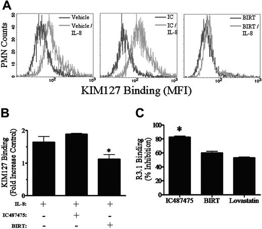
We next examined the effect of the allosteric inhibitors IC487475 and BIRT on shifting the conformation of β2-integrin by detecting the binding of mAb KIM127, which recognizes a neo-epitope expressed on the extended β-subunit of LFA-1 and Mac-1.42 IL-8 stimulation up-regulated KIM127 expression by 1.7-fold over unstimulated control (Figure 2A-B). Pretreatment of PMNs with IC487475 did not significantly diminish the up-regulation of KIM127 in stimulated PMNs. In contrast, treatment with BIRT decreased KIM127 binding uniformly on populations of PMNs by 50% to a level significantly less than treatment with IC487475 or vehicle alone (Figure 2A-B). Residual binding of KIM127 was blocked to unstimulated baseline by the addition of anti-Mac-1 to BIRT-treated PMNs (data not shown). These data are consistent with previous reports that BIRT377 effectively downshifts the β-subunit of LFA-1 to a low-affinity conformation state.17,23 We next tested the binding of mAb R3.1, which has previously been reported to bind an epitope on the I-domain and to become displaced in response to an allosteric shift to the low-affinity conformation.23 Treatment with BIRT and lovastatin, another IDAS allosteric inhibitor, reduced R3.1 binding to LFA-1 by approximately 60% (Figure 2C). Similarly, IC487475 inhibited R3.1 binding by approximately 80% (Figure 2C). These data, in conjunction with a recent report characterizing the binding activity of IC487475 to LFA-1,37 demonstrate that IC487475 targets the IDAS and stabilizes the I-domain in an intermediate-affinity state for ICAM-1. Moreover, this demonstrates in wild-type LFA-1 that a decrease in ICAM-1 affinity can be allosterically induced at the I-domain while CD18 is maintained in an extended state, as defined by the KIM127 epitope.
Allosteric inhibition of LFA-1 binding to ICAM-1. (A) Neutrophils were preincubated with Fc fragments (all samples), anti-Mac-1 (2LPM19c), and IC487475, as indicated. ICAM-1/IgG-Alexa-488 was then added to neutrophil suspensions and was followed immediately by activation with mAb 240Q. Cells were fixed and analyzed by flow cytometry. Fluorescence histograms are representative of the average population response from 4 separate experiments. (B) Data represent the mean ± standard error of the mean (SEM) from 4 separate experiments, as described in panel A. *Significance compared with mAb 240Q + anti-Mac-1 (P < .05). **Significance compared with mAb 240Q (P < .05). (C) Isolated human neutrophils were incubated over a dose range with IC487475 or TS1/22. Suspensions were then activated with IL-8 and immediately analyzed by FACScan flow cytometry for binding of ICAM-1/IgG-coated latex beads (approximately 25 sites/μm2). Data are representative of the average response from 4 separate experiments.
Allosteric inhibition of LFA-1 binding to ICAM-1. (A) Neutrophils were preincubated with Fc fragments (all samples), anti-Mac-1 (2LPM19c), and IC487475, as indicated. ICAM-1/IgG-Alexa-488 was then added to neutrophil suspensions and was followed immediately by activation with mAb 240Q. Cells were fixed and analyzed by flow cytometry. Fluorescence histograms are representative of the average population response from 4 separate experiments. (B) Data represent the mean ± standard error of the mean (SEM) from 4 separate experiments, as described in panel A. *Significance compared with mAb 240Q + anti-Mac-1 (P < .05). **Significance compared with mAb 240Q (P < .05). (C) Isolated human neutrophils were incubated over a dose range with IC487475 or TS1/22. Suspensions were then activated with IL-8 and immediately analyzed by FACScan flow cytometry for binding of ICAM-1/IgG-coated latex beads (approximately 25 sites/μm2). Data are representative of the average response from 4 separate experiments.
LFA-1 conformation regulates the microkinetics of PMN rolling
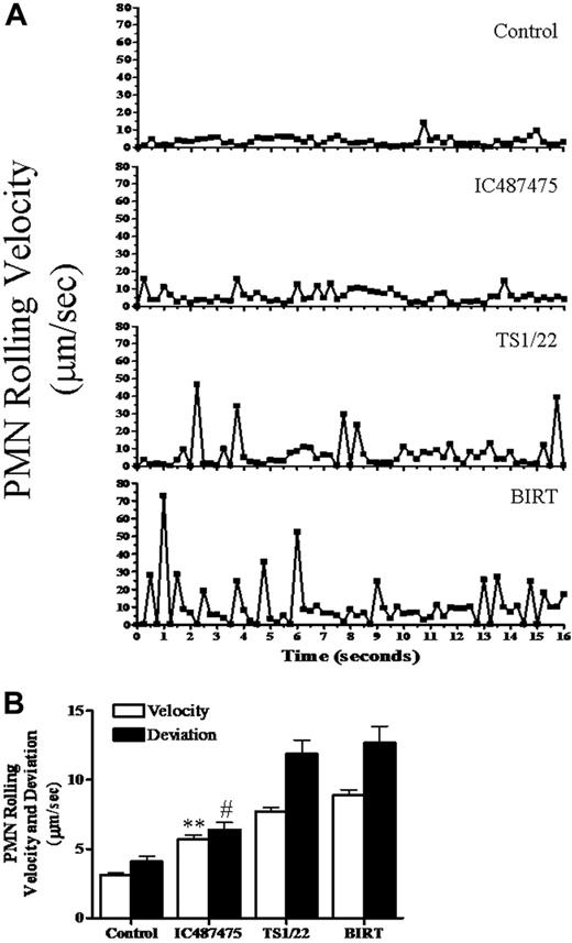
We next examined the influence of steric and allosteric inhibition of LFA-1 on the microkinetics of PMN rolling on IL-1-stimulated HUVECs in a parallel-plate flow chamber. Untreated PMNs rolled in the direction of fluid flow at an average velocity of approximately 3 μm/s and exhibited subsecond deviations in velocity averaging approximately 4 μm/s (Figure 3). Allosteric stabilization of LFA-1 at intermediate affinity in the presence of IC487475 increased rolling velocity by approximately 1-fold and velocity deviation by approximately 50%. Low-affinity LFA-1 in the presence of BIRT increased mean velocity and its deviation approximately 2-fold above control and 50% above IC487475 treatment. Steric inhibition of LFA-1 by blocking of the I-domain with mAb TS1/22 elicited rolling kinetics equivalent to BIRT treatment, with frequent subsecond excursions at velocity greater than approximately 30 μm/s. Despite these alterations in the rolling kinetics, the efficiency of capture as determined by the total number of PMNs interacting with the cell monolayer was comparable between untreated and antibody or allosteric inhibition. In fact, the increase in rolling velocity and velocity deviation in the presence of TS1/22 and BIRT was equivalent to PMN rolling on an L-cell monolayer transfected to express only E-selectin.5 Thus, LFA-1 conformation and availability are key parameters in the regulation of PMN rolling velocity and stability.
PMN arrest and transmigration are regulated by LFA-1 conformation
The efficiency of PMN recruitment, as determined by the conversion from rolling to arrest in postcapillary venules, was recapitulated on cytokine-stimulated HUVECs in that approximately 80% of rolling PMNs transitioned to arrest within seconds of capture in the parallel-plate flow chamber (Figure 4A-B). To determine the role of LFA-1 conformation and affinity in neutrophil recruitment, we preincubated PMNs with mAbs targeting β2-integrin ligand-binding domains or the allosteric inhibitors IC487475 and BIRT. Treatment with anti-Mac-1 slowed the rate of arrest but did not significantly alter the number of PMNs arrested. LFA-1 inhibition primarily decreased the efficiency of PMN arrest and transmission electron microscopy (TEM) but did not diminish the total number of PMN tethering and rolling, which is selectin dependent.43 Blocking its function with mAb TS1/22 or allosterically stabilizing a low or intermediate affinity diminished the rate of arrest by approximately 75% compared with untreated PMNs, indicating LFA-1 function as the primary braking receptor. Residual arrest was mediated by Mac-1 as arrest was abrogated in the presence of antibodies to LFA-1 and Mac-1 or to IC487475 and anti-Mac-1 (data not shown). Transendothelial migration proceeded within minutes of arrest and continuously increased over the time course of interaction on inflamed HUVECs (Figure 4C). Treatment with anti-Mac-1 reduced the initial rate of TEM by approximately 40%; however, by 5 minutes it was not significantly different from that of control. In contrast, anti-LFA-1 diminished the rate of TEM and blocked the extent by approximately 60% relative to control. Allosteric inhibition of LFA-1 abrogated TEM, which was equivalent to treatment with antibody blocking of LFA-1 and Mac-1 (Figure 4C). These data highlight a significant difference in that antibodies to LFA-1 and Mac-1 abrogated arrest altogether, whereas allosteric inhibition at best blocked only 50% of PMNs arrested. Taken together, these data confirm previous reports that activated LFA-1 functions early in the multistep process of PMN recruitment and cooperates with Mac-1 in achieving arrest in shear flow.44 The current study shows for the first time that allosterically stabilizing LFA-1 in a low- or intermediate-affinity conformation is sufficient to impair PMN arrest and abrogate TEM.
I-domain allosteric regulation of LFA-1 extension during neutrophil activation. (A-B) Isolated human neutrophils were preincubated with 1 μM IC487475, 1 μM BIRT or vehicle, and the CD18 extension reporter mAb KIM127. Suspensions were then activated with 1 nM IL-8 and were incubated with FITC-conjugated secondary goat anti-mouse F(ab′)2 before fixation and analysis by flow cytometry. Fluorescence histograms (A) are representative of the average population response from 6 separate experiments. Bar graph data (B) represent the mean ± SEM from 6 separate experiments. *Significance in KIM127 binding between BIRT and both IC487475 (P < .01) and vehicle-treated PMNs (P < .05). (C) rLFA-1 heterodimer was derivatized to latex microspheres and incubated with allosteric inhibitors IC487475, BIRT, and lovastatin. rLFA-1 beads were then incubated with MgCl2 and mAb R3.1, followed by secondary labeling with fluorescent goat anti-mouse F(ab′)2 and flow cytometry. Data are given as mean ± SEM for 4 separate experiments and are presented as percentage inhibition relative to maximal binding by vehicle-treated rLFA-1 beads. *Significant inhibition of R3.1 binding compared with vehicle (P < .001).
I-domain allosteric regulation of LFA-1 extension during neutrophil activation. (A-B) Isolated human neutrophils were preincubated with 1 μM IC487475, 1 μM BIRT or vehicle, and the CD18 extension reporter mAb KIM127. Suspensions were then activated with 1 nM IL-8 and were incubated with FITC-conjugated secondary goat anti-mouse F(ab′)2 before fixation and analysis by flow cytometry. Fluorescence histograms (A) are representative of the average population response from 6 separate experiments. Bar graph data (B) represent the mean ± SEM from 6 separate experiments. *Significance in KIM127 binding between BIRT and both IC487475 (P < .01) and vehicle-treated PMNs (P < .05). (C) rLFA-1 heterodimer was derivatized to latex microspheres and incubated with allosteric inhibitors IC487475, BIRT, and lovastatin. rLFA-1 beads were then incubated with MgCl2 and mAb R3.1, followed by secondary labeling with fluorescent goat anti-mouse F(ab′)2 and flow cytometry. Data are given as mean ± SEM for 4 separate experiments and are presented as percentage inhibition relative to maximal binding by vehicle-treated rLFA-1 beads. *Significant inhibition of R3.1 binding compared with vehicle (P < .001).
Microkinetics of neutrophil rolling is regulated by LFA-1. (A) The instantaneous velocity of rolling neutrophils was quantitated as the distanced traversed by individual neutrophil centroids every 0.250 second for the length of time the neutrophil remained in the field of view. Data are representative of the average response of 30 individual neutrophils from 6 to 8 separate experiments. (B) LFA-1 activation regulates neutrophil rolling dynamics. The average rolling velocity and standard deviation from the mean velocity for neutrophils on IL-1β activated HUVECs were quantitated at 0.250-second intervals for the length of time an individual rolling neutrophil remained in the field of view. Data represent mean ± SEM for 30 neutrophils in 4 to 6 separate experiments. **Significance in velocity between IC487475 and both TS1/22 (P < .01) and BIRT (P < .001). #Significance in deviation between IC487475 and both TS1/22 (P < .01) and BIRT (P < .001).
Microkinetics of neutrophil rolling is regulated by LFA-1. (A) The instantaneous velocity of rolling neutrophils was quantitated as the distanced traversed by individual neutrophil centroids every 0.250 second for the length of time the neutrophil remained in the field of view. Data are representative of the average response of 30 individual neutrophils from 6 to 8 separate experiments. (B) LFA-1 activation regulates neutrophil rolling dynamics. The average rolling velocity and standard deviation from the mean velocity for neutrophils on IL-1β activated HUVECs were quantitated at 0.250-second intervals for the length of time an individual rolling neutrophil remained in the field of view. Data represent mean ± SEM for 30 neutrophils in 4 to 6 separate experiments. **Significance in velocity between IC487475 and both TS1/22 (P < .01) and BIRT (P < .001). #Significance in deviation between IC487475 and both TS1/22 (P < .01) and BIRT (P < .001).
LFA-1 and CXCR cooperate to signal polarization of arrested PMNs in shear flow
Previous reports suggest that leukocyte rolling on inflamed HUVECs facilitates G-protein-coupled chemotactic receptor (GPCR) signaling and activation of CD18 binding to ICAM-1.45 Binding of high-affinity LFA-1 to ICAM-1 appears to provide an important co-signal that activates T-cell migration.29 We examined the role of LFA-1 affinity and binding to ICAM-1 in providing a co-signal leading to cytoskeletal activation and contact guidance during PMN transendothelial migration. Within seconds of arrest on cytokine-stimulated HUVECs, virtually all PMNs destined to transmigrate exhibited significant shape changes (Figure 5A). By 2 minutes, virtually all PMNs adopted a polarized morphology as they projected and retracted multiple pseudopods during migration to a site of TEM. Blocking LFA-1 with TS1/22 did not alter the rate or extent of shape change in PMNs that achieved arrest, despite the efficacy of TS1/22 in diminishing arrest and TEM by approximately 60% (Figure 5A). In contrast, allosteric inhibition of LFA-1 with IC487475 or BIRT significantly retarded the onset of shape change and ultimately inhibited polarization in 50% of arrested PMNs.
We next examined the kinetics of shape change after PMN recruitment on L-E/I (Figure 5B-C). Studies were performed in a custom microfluidic flow channel engineered to introduce PMNs onto the monolayer, followed by injection of IL-8 at a defined position in the flow field. This model system provided a means of observing PMN rolling to arrest on L-E/I after activation through GPCR ligation. As with the interaction on inflamed HUVECs, most rolling PMNs achieved arrest on L-E/I in the microchannels.5 A significant difference was that PMNs remained spherical after arrest, indicating that neither tethering on E-selectin nor adhesion through high-affinity CD18 binding to ICAM-1 was sufficient to activate PMN shape change (Figure 5B-C). Perfusion of IL-8 into the microchannel elicited rapid and uniform shape changes such that within 1 minute virtually the entire PMN population achieved polarization. As observed on HUVECs, allosteric inhibition significantly decreased the rate and extent of IL-8-induced polarization (Figure 5B-C). It is important to note that PMNs activated with chemokine in suspension retained the capacity to form pseudopods in the presence or absence of IDAS inhibitors (data not shown). We conclude that a conformational shift in LFA-1 to high affinity cooperates with CXCR signaling in the activation of PMN polarization after rolling and arrest on E-selectin and ICAM-1.
High-affinity LFA-1 coordinates PMN directional guidance during migration
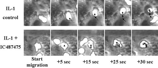
Allosteric inhibition of LFA-1 was found to attenuate PMN polarization and effectively abrogate subsequent TEM, suggesting that LFA-1 may provide a regulatory function in contact guidance to the site of diapedesis. To further examine how LFA-1 affinity regulates this process, PMN migration on inflamed HUVECs was tracked for untreated PMN and PMN with LFA-1 stabilized with IC487475 at intermediate affinity. In a 30-second interval, PMNs traveled a mean total distance of 9.1 μm, with a point-to-point net migration distance of 7.0 μm (Figure 6; Table 1). A representative sequence illustrates that PMNs migrated to the site of TEM with high persistence (Figure 6). In contrast, allosteric inhibition with IC487475, or BIRT (data not shown), diminished the total migration distance to 7.2 μm and the net migration distance to 4.9 μm (Figure 6; Table 1). PMNs treated with anti-LFA-1 mAb TS1/22 migrated to the site of TEM with trajectories similar to those of untreated control, whereas allosteric inhibition resulted in less persistent, more random migration, as indicated by reduced net movement. Migration velocity over the time course was equivalent between untreated PMNs and in the presence of LFA-1 inhibitors (Table 1). These data clearly demonstrate that a high-affinity conformation of LFA-1 is necessary for persistent migration and subsequent diapedesis.
Neutrophil locomotory parameters on inflamed HUVECs
. | Total migration distance, μm . | Net migration distance, μm . | Average migratory velocity, μm/s . |
|---|---|---|---|
| Control | 9.1 ± 1.4 | 7.0 ± 0.9 | 0.29 ± 0.04 |
| IC487475 | 7.2 ± 0.6 | 4.9 ± 0.8* | 0.22 ± 0.02 |
| Anti–LFA-1 | 10.8 ± 3.8 | 8.5 ± 1.5 | 0.34 ± 0.05 |
. | Total migration distance, μm . | Net migration distance, μm . | Average migratory velocity, μm/s . |
|---|---|---|---|
| Control | 9.1 ± 1.4 | 7.0 ± 0.9 | 0.29 ± 0.04 |
| IC487475 | 7.2 ± 0.6 | 4.9 ± 0.8* | 0.22 ± 0.02 |
| Anti–LFA-1 | 10.8 ± 3.8 | 8.5 ± 1.5 | 0.34 ± 0.05 |
Neutrophils were preincubated with IC487475, anti–LFA-1 (TS1/22), or vehicle and then perfused into the parallel-plate flow chamber over IL-1β–activated HUVECs at a shear stress of 2 dyne/cm2. After arrest, the geometric coordinates of neutrophil centroids were determined at 2-second intervals for 30 seconds during migration. The total migration distance is the sum of the migration step distance over 30 seconds. Net migration distance is the direct distance between the start of migration and the end of migration after 30 seconds. Average migratory velocity was determined every 2 seconds during migration and then averaged over the 30 seconds of migration. Data are presented as mean ± SEM of 10 neutrophils quantified in 4 separate experiments.
Significance compared with control (P < .05)
Kinetics of neutrophil rolling, arrest, and transmigration on inflamed endothelium. Isolated human neutrophils were incubated with IC487475, BIRT, TS1/22, 2LPM19c, or vehicle, as indicated. Neutrophil suspensions were then perfused into a parallel-plate flow chamber over IL-1β activated or unstimulated control HUVEC monolayers at a shear stress of 2 dyne/cm2. (A) The number of rolling neutrophils, defined as those moving more than 1 cell diameter in 30 seconds, was quantitated in a field of view every minute throughout the interaction. Data represent the mean ± SEM from 5 to 10 separate experiments. (B) The number of adherent neutrophils, defined as those moving less than 1 cell diameter in 30 seconds, was quantitated in a field of view every minute throughout the interaction. Data represent the mean ± SEM from 5 to 10 separate experiments. (C) The number of transmigrated neutrophils was quantitated in a field of view every minute throughout the interaction. Data represent the mean ± SEM from 5 to 10 separate experiments.
Kinetics of neutrophil rolling, arrest, and transmigration on inflamed endothelium. Isolated human neutrophils were incubated with IC487475, BIRT, TS1/22, 2LPM19c, or vehicle, as indicated. Neutrophil suspensions were then perfused into a parallel-plate flow chamber over IL-1β activated or unstimulated control HUVEC monolayers at a shear stress of 2 dyne/cm2. (A) The number of rolling neutrophils, defined as those moving more than 1 cell diameter in 30 seconds, was quantitated in a field of view every minute throughout the interaction. Data represent the mean ± SEM from 5 to 10 separate experiments. (B) The number of adherent neutrophils, defined as those moving less than 1 cell diameter in 30 seconds, was quantitated in a field of view every minute throughout the interaction. Data represent the mean ± SEM from 5 to 10 separate experiments. (C) The number of transmigrated neutrophils was quantitated in a field of view every minute throughout the interaction. Data represent the mean ± SEM from 5 to 10 separate experiments.
Kinetics of neutrophil shape change on HUVECs and L cells expressing E-selectin and ICAM-1. The membrane aspect ratio was quantitated as a measure of cell shape change for individual neutrophils immediately after arrest on IL-1β-activated HUVECs in the parallel-plate flow chamber (A) or L-E/I in the microfluid flow channel device (B-C) at 2 dyne/cm2 shear stress. Neutrophils were preincubated with IC487475, BIRT, TS1/22, or vehicle, as indicated, then perfused in the respective flow chamber. On L-E/I, IL-8 was perfused into the microflow channel after neutrophils reached maximum adhesion, 3 minutes after injection. (A-B) Data are presented as the fraction of all arrested neutrophils in the field of view exhibiting a membrane aspect ratio greater than 1.4, a value deemed to be a threshold in membrane skewness based on empirical observations. Data represent the mean ± SEM from 4 separate experiments. (C) Images are 20 × phase contrast and are representative of adherent neutrophils on L-E/I under the conditions described in panel B.
Kinetics of neutrophil shape change on HUVECs and L cells expressing E-selectin and ICAM-1. The membrane aspect ratio was quantitated as a measure of cell shape change for individual neutrophils immediately after arrest on IL-1β-activated HUVECs in the parallel-plate flow chamber (A) or L-E/I in the microfluid flow channel device (B-C) at 2 dyne/cm2 shear stress. Neutrophils were preincubated with IC487475, BIRT, TS1/22, or vehicle, as indicated, then perfused in the respective flow chamber. On L-E/I, IL-8 was perfused into the microflow channel after neutrophils reached maximum adhesion, 3 minutes after injection. (A-B) Data are presented as the fraction of all arrested neutrophils in the field of view exhibiting a membrane aspect ratio greater than 1.4, a value deemed to be a threshold in membrane skewness based on empirical observations. Data represent the mean ± SEM from 4 separate experiments. (C) Images are 20 × phase contrast and are representative of adherent neutrophils on L-E/I under the conditions described in panel B.
Allosteric inhibition of LFA-1 diminishes persistence of migration. Isolated human neutrophils were incubated with IC487475 or vehicle, then perfused into a parallel-plate flow chamber over IL-1β-activated HUVECs at a shear stress of 2 dyne/cm2. After arrest, the centroid of migrating neutrophils was tracked at 5-second intervals for 30 seconds. Arrows outline the trajectory of migrating neutrophils, with the arrowhead positioned at cell centroid at the conclusion of each time segment. Micrographs are representative of approximately 100 individual neutrophil observations over 4 to 6 separate experiments.
Allosteric inhibition of LFA-1 diminishes persistence of migration. Isolated human neutrophils were incubated with IC487475 or vehicle, then perfused into a parallel-plate flow chamber over IL-1β-activated HUVECs at a shear stress of 2 dyne/cm2. After arrest, the centroid of migrating neutrophils was tracked at 5-second intervals for 30 seconds. Arrows outline the trajectory of migrating neutrophils, with the arrowhead positioned at cell centroid at the conclusion of each time segment. Micrographs are representative of approximately 100 individual neutrophil observations over 4 to 6 separate experiments.
LFA-1 redistributes to high-affinity sites of CD18 during PMN arrest and polarization
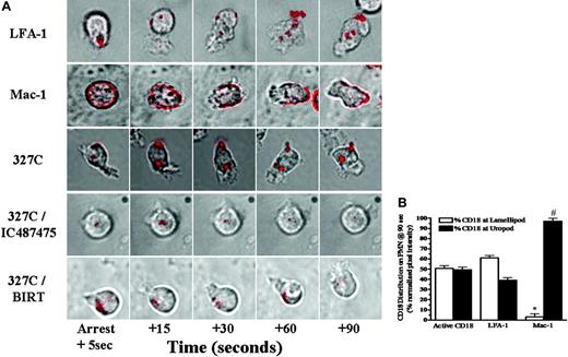
A final set of experiments was initiated to image in real time the dynamics of β2-integrin activation and membrane redistribution during PMN recruitment and activation on IL-1-stimulated HUVECs in shear flow. During the interval from rolling to arrest and shape change, LFA-1, Mac-1, and activated CD18 were imaged by real-time immunofluorescence of nonblocking, fluoro-conjugated mAbs. Within seconds, LFA-1 formed focal clusters in the plane of adhesive contact of arrested PMNs, as depicted in the representative image of Figure 7A. Subsequently, approximately half the total LFA-1 fluorescence redistributed into submicron clusters that tracked to the base of extending and retracting lamellipodia during PMN migration to the site of TEM (Figure 7B). A consistent observation was a second site of clustered LFA-1 that appeared more stable and that localized to the uropod of polarized PMNs. In contrast, anti-Mac-1 fluorescence exhibited a different topography in that it was distributed uniformly around the cell periphery and subsequently diffused to the uropod of polarized PMNs (Figure 7).
Membrane distribution of high-affinity CD18 was imaged with a fluoroconjugate of mAb 327C (Figure 7A). High-affinity CD18 typically formed clusters at sites of contact that aligned along the lamellipod-uropod major axis of polarized PMNs. This topography corresponded to that of LFA-1 at the base of forming lamellipodia, and less mobile focal clusters of LFA-1 and Mac-1 stabilized at the uropod. Active CD18 was equally distributed at the front and rear of the PMN during the process of migration, suggesting its role in orientation and guidance during diapedesis (Figure 7B). Treatment with allosteric inhibitors reduced 327C fluorescence intensity at arrest by approximately 3-fold compared with control but did not abrogate expression of 327C within the adhesive contact of arrested PMNs (Figure 7A). Despite this accumulation of active CD18, bipolar clusters of 327C did not form in the presence of BIRT or IC487475. As depicted in the bottom sequences of Figure 7A, neither active CD18 nor LFA-1 (data not shown) was observed to redistribute during lamellipod extension. These data reveal a pattern of LFA-1 membrane redistribution at the site of adhesive contact that is associated with a shift to high affinity and subsequent signaling and contact guidance of PMNs to the site of transendothelial migration.
Discussion
In the present study, we demonstrate that the affinity of LFA-1 is a key determinant in the efficiency of PMN arrest and transmigration on inflamed endothelium. We show that (1) a shift in LFA-1 from low to intermediate to high affinity cooperates with selectins in reducing the velocity of PMN rolling in shear flow, (2) a shift in LFA-1 to high affinity during slow rolling is necessary for efficient arrest, and (3) high-affinity LFA-1 redistributes along the major axes of polarized PMNs and is necessary in contact guidance and subsequent TEM. A key observation was that antibody blocking of LFA-1 binding did not alter PMN shape change, nor did it significantly diminish migration, which was partially supported by Mac-1. However, allosterically stabilizing the I-domain with IC487475 in an intermediate-affinity, or locking LFA-1 in a low-affinity, state with BIRT377 abrogated TEM by perturbing PMN polarization. These data are the first to demonstrate that dynamic shifts in LFA-1 affinity for ICAM-1 can serve as a gatekeeper by providing a critical outside-in signal to direct contact guidance during the transition from arrest to transmigration in shear flow.
Experimental approaches using site-directed mutagenesis and allosteric small molecule targeting have begun to reveal how shifts in the conformation of LFA-1 mediate profound changes in equilibrium binding of ICAM-1. Soluble monomeric ICAM-1 binding to low-affinity, wild-type LFA-1 or to an I-domain mutant locked in a closed conformation dissociates at rates of 5.5 seconds-1 and 2.8 seconds-1, respectively.46 Stabilizing an intermediate-affinity conformation of LFA-1 by displacement of the β6-α7 loop of the I-domain in cysteine mutants lowered the dissociation rate an order of magnitude to approximately 0.7 second-1.20 On cell activation, LFA-1 transitions from a low- to a high-affinity state, which corresponds to an off rate for soluble monomeric ICAM-1 binding of approximately 0.02 second-1, 3 orders of magnitude slower than the low-affinity I-domain.20,46 One class of low molecular-weight allosteric inhibitors of LFA-1 stabilizes the so-called extended conformation of LFA-1 and interferes with β I-like domain regulation of α I-domain affinity.21,22,47 A second class of small molecule inhibitors binds within the α-subunit IDAS and stabilizes LFA-1 in a bent, low-affinity conformation by preventing downward movement of the C-terminal α-helix.48,49 Members of this class include LFA703, LFA878, LFA451, Lovastatin (all Novartis) and BIRT377, and all prevented LFA-1-dependent rolling or adhesion on ICAM-1.22,23,38,47
β2-Integrin activation and redistribution on polarized and migrating neutrophils. Neutrophils were preincubated with IC487475, BIRT, nonblocking anti-LFA-1-Alexa 546 (TS2/4), nonblocking anti-Mac-1-PE (ICRF44), or mAb 327C-Alexa 488, as indicated. Cell suspensions were then infused into the parallel-plate flow chamber at a shear stress of 2 dyne/cm2 over a monolayer of IL-1β-activated HUVECs. (A) On neutrophil rolling to arrest, real-time detection of β2-integrin distribution was visualized by sequential collection of phase-contrast and immunofluorescent images every second for 90 seconds using a 60 × oil immersion objective. Micrographs are presented as phase-contrast (gray) and deconvolved immunofluorescence (red) overlays representative of approximately 100 individual neutrophil observations in 4 to 6 separate experiments. (B) Fractional distribution of β2-integrin on polarized neutrophils after 90 seconds was determined by quantitating pixel intensity of the fluorescence signal at the leading projection and uropod. Data are presented as mean ± SEM and are representative of the β2-integrin distribution of approximately 10 cells in 4 to 6 separate experiments. *Significance compared with active β2-integrin and LFA-1 at the leading projection (P < .01). #Significance compared with active CD18 and LFA-1 at the uropod (P < .01).
β2-Integrin activation and redistribution on polarized and migrating neutrophils. Neutrophils were preincubated with IC487475, BIRT, nonblocking anti-LFA-1-Alexa 546 (TS2/4), nonblocking anti-Mac-1-PE (ICRF44), or mAb 327C-Alexa 488, as indicated. Cell suspensions were then infused into the parallel-plate flow chamber at a shear stress of 2 dyne/cm2 over a monolayer of IL-1β-activated HUVECs. (A) On neutrophil rolling to arrest, real-time detection of β2-integrin distribution was visualized by sequential collection of phase-contrast and immunofluorescent images every second for 90 seconds using a 60 × oil immersion objective. Micrographs are presented as phase-contrast (gray) and deconvolved immunofluorescence (red) overlays representative of approximately 100 individual neutrophil observations in 4 to 6 separate experiments. (B) Fractional distribution of β2-integrin on polarized neutrophils after 90 seconds was determined by quantitating pixel intensity of the fluorescence signal at the leading projection and uropod. Data are presented as mean ± SEM and are representative of the β2-integrin distribution of approximately 10 cells in 4 to 6 separate experiments. *Significance compared with active β2-integrin and LFA-1 at the leading projection (P < .01). #Significance compared with active CD18 and LFA-1 at the uropod (P < .01).
We describe here the function of IC487475, a class of P-arylthio cinnamides that targets the IDAS and stabilizes the I-domain with intermediate affinity, corresponding to an ICAM-1 dissociation rate of approximately 0.14 second-1.37 This is the first report of an α allosteric antagonist that downshifts I-domain affinity independent of global shifts in the α-β heterodimer conformation. Closing of the I-domain was confirmed by blocking the binding of mAb R3.1, which reports on I-domain occupancy, in the presence of BIRT377 or IC487475. IC487475 did not alter heterodimer extension, as determined by binding of KIM127 to activated CD18. In contrast, BIRT induced KIM127 displacement from activated CD18. The activity of IC487475 is similar to that of XVA143, which also stabilizes an intermediate affinity of LFA-1 by reportedly altering α-β intersubunit signaling.12,47 XVA143 enhanced lymphocyte rolling on purified ICAM-1 and high endothelial venules and virtually abrogated lymphocyte arrest by stabilizing LFA-1 in the extended conformation.22 Other P-arylthio cinnamides or diarylsulfide analogs are also capable of stabilizing the I-domain into low-affinity conformation, as determined by nuclear magnetic resonance (NMR) and X-ray crystallography (O.E.S., unpublished data, June 2005). Our data demonstrate for the first time that the affinity of wild-type LFA-1 can be downshifted allosterically by a small molecule binding to the IDAS independently of global conformational changes in the α-β heterodimer.
Neutrophils treated with IC487475 or BIRT were recruited to HUVECs with equal efficiency, yet rolling velocity supported by each was dramatically different. LFA-1 locked in a low-affinity conformation by BIRT or blocked from binding ICAM-1 by TS1/22 exhibited unsteady rolling with frequent velocity spikes. Published data show that LFA-1, or isolated I-domain, can support cell rolling on ICAM-1 at shear stress in excess of 10 dyne/cm2; however, in all cases, this required an initial interaction period of static or very low shear (less than 0.3 dyne/cm2).15-17 In this regard, mice deficient in E- and P-selectin lack the capacity for leukocyte capture and rolling.50 Thus, selectins are necessary for capture at venular shear rates, yet optimal rolling requires cooperativity with LFA-1 because leukocyte rolling on monolayers coexpressing E-selectin and ICAM-1, or in vivo within the microcirculation of ICAM-1(-/-) mice, is markedly increased relative to ICAM-1 expressed alone.5,51 Our data suggest that intermediate-affinity LFA-1 stabilized by IC487475 can participate in neutrophil capture and rolling; however, further up-regulation to high affinity by other activation mechanisms, including selectins, chemokines, and inflammatory lipids, is critical for the transition to arrest.5,18,52
Ligation of neutrophil chemotactic receptors is thought to be a primary mechanism directing polarization and contact-mediated guidance to sites of transendothelial migration.53,54 Integrins also function as transmembrane signaling receptors that can activate kinases, cytoskeletal reorganization, and cell motility.55 In particular, LFA-1 binding to ICAM-1 on lymphocytes in Mg2+-containing buffer induces conformational extension of extracellular domains, unclasping of integrin cytoplasmic domains, F-actin polymerization, microtubule reorganization, and activation of signaling molecules such as PKC-β and Rho kinase.9,28,29,33 In contrast to lymphocytes, PMNs are not activated by contact with ICAM-1 even in the presence of Mg2+; rather, they require signaling through chemokine or selectin ligation to induce rapid integrin activation.18 On cytokine-activated HUVECs, PMNs roll 10-fold more slowly (approximately 7 μm/s) than lymphocytes, and soluble chemokine immobilized on the substrate or through selectin-mediated signaling can trigger the induction of high-affinity LFA-1.
A recent report from Shamri et al6 supports a model in which chemokine and ICAM-1 must be juxtaposed on the substrate of a flow channel to activate LFA-1 and to arrest lymphocytes in shear. In this model, only chemokine immobilized on a substrate, but not in solution, effectively stabilized activated LFA-1 on T cells, as reported by binding to activation reporters such as 327C and KIM127. Shamri et al6 conclude that chemokine signals only transiently convert low-affinity LFA-1 to an extended intermediate-affinity conformation in which it is poised to bind ICAM-1. Interaction of extended I-domain with ICAM-1 must then occur within a fraction of a second to mediate shear-resistant stable adhesion. This model of T-cell adhesion highlights several potential differences between LFA-1 function on lymphocytes and that on PMNs. Stimulation of PMNs in suspension with chemokine, or rolling on E-selectin, is sufficient to activate high-affinity LFA-1 and stable adhesion to ICAM-1 in shear flow. Our data support a model in which LFA-1 in intermediate, but not low, affinity promotes deceleration during PMN rolling. A conversion to high affinity and clustering of LFA-1 within the site of contact is essential for efficient transition to arrest with endothelial ICAM-1, and this can be elicited by rolling on E-selectin.56 Synergistic with the latter process is chemokine signaling, which rearranges high-avidity LFA-1 to function in coordinating PMN adhesion, cell polarization, and migratory guidance to the site of TEM.
Several proposed mechanisms may explain a role for LFA-1 as a gatekeeper in PMN transmigration through its documented association with other leukocyte integrins and cytoplasmic proteins, including (1) transdominant crosstalk between β1- and β2-integrins, whose activity is required for optimal polarization and transmigration,31,32 (2) assembly of cytoskeletal attachment proteins such as talin, paxillin, vinculin, and α-actinin on the cytoplasmic domain of high-affinity ligated LFA-1,57-59 and (3) modulation of signaling molecules such as calpain, PIP kinase, PKC-β, PLCγ-1, Rho GTPase, PI3 kinase, and Ca2+ as a function of the LFA-1 affinity state.25,33,60-63
We hypothesize that outside-in signaling by LFA-1 is a vital component in the hierarchy of events leading to neutrophil polarization and transmigration. Upon polarization, high-affinity LFA-1 forms dynamic clusters at the base of newly forming lamellipodia and more stable clusters at the uropod. This distribution on migrating neutrophils is different from the uniform distribution observed at the front of the extended lamellipodia on lymphocytes, which reflects the role of LFA-1 in establishing a synapse with antigen-presenting cells.34,64 By comparison, Mac-1 redistributes with slower kinetics to cap at the uropod, an observation supported by previous reports of translocation of albumin-coated latex beads to the uropod of activated neutrophils.65,66 This is the first report of dynamic redistributions of high-affinity, CD18-forming clusters aligned with the major axis of polarized neutrophils. In this context, several studies have shown that cleavage of talin and dissociation of LFA-1 from the cytoskeleton are not critical to the induction of high affinity during tethering but may be required for outside-in signals leading to focal clustering and oligomerization of LFA-1 bonds to ICAM-1. Thus, calpain-mediated cleavage of talin and other LFA-1 cytoskeletal tail-binding proteins, such as RAPL,34 may be involved in the dynamic membrane redistributions of LFA-1 along the major axes of migrating PMNs, providing for dynamic regulation of integrin bond lifetime and adhesion strength.14,37,67
A more complete model for neutrophil recruitment that accounts for cooperativity among selectins, integrins, and CXC receptors during neutrophil recruitment is emerging. A critical checkpoint in the process appears to be the ligation of high-affinity LFA-1 at axial contacts on polarized neutrophils, thus enabling migration to the endothelial junction and diapedesis.68 Studies in our laboratory are focused on correlating the dynamics of LFA-1 and G-protein-coupled activation with recruitment of adapter proteins within the adhesive contact region to elucidate the precise regulatory role of each during the multistep process of neutrophil emigration.
Prepublished online as Blood First Edition Paper, November 8, 2005; DOI 10.1182/blood-2005-06-2303.
Supported by National Institutes of Health grant AI47294. C.E.G. is supported by National Institutes of Health/National Heart, Lung, and Blood Institute-National Research Service Award (NIH/NHLBI-NRSA) Postdoctoral Training Fellowship 2-T32-HL07013-26.
D.E.S. is employed by a company, ICOS, whose potential product was studied in the present work.
The online version of this article contains a data supplement.
The publication costs of this article were defrayed in part by page charge payment. Therefore, and solely to indicate this fact, this article is hereby marked “advertisement” in accordance with 18 U.S.C. section 1734.
S.I.S. is an Established Investigator of the American Heart Association. D.E.S. has declared a financial interest in a company (ICOS) whose potential product was studied in the present work. D.E.S. has declared financial interest in a competitor of a company whose product was studied in the present work. The authors studied an analog of BIRT377, BIRT, which was made by ICOS based on the published structure of BIRT377, originally developed by Boehringer Ingelheim. These data are consistent with what is already published regarding the efficacy of BIRT377, so BIRT serves as a positive control in these studies. This approach is commonly applied in studies of receptor inhibitors and should not generate any commercial or competitor issues. D.E.S. is a Senior Scientist at ICOS and is the only author who has a potential commercial interest. However, none of the compounds in the current article are in clinical trials; the IC487475 compound is a generation behind the preclinical compounds currently investigated by ICOS. None of the authors hold patents related to the work described in this manuscript. Aside from reagent provisions, funding for this research was provided wholly by the NIH. The results submitted in this manuscript are intended only for research purposes to facilitate understanding of the mechanisms by which LFA-1 contributes to neutrophil recruitment.


This feature is available to Subscribers Only
Sign In or Create an Account Close Modal