Abstract
Dendritic cells (DCs) and chemokines are important in linking innate and adaptive immunity. We previously reported that Fas ligation induced interleukin 1β (IL-1β)–dependent maturation and IL-1β–independent survival of DCs, with extracellular signal–regulated kinase (ERK) and nuclear factor–κB (NF-κB) signaling pathways involved, respectively. We describe here that Fas ligation induced DCs to rapidly produce both CXC and CC chemokines, including macrophage inflammatory protein 2 (MIP-2), MIP-1α, MIP-1β, monocyte chemoattractant protein 1 (MCP-1), RANTES (regulated on activation normal T cell expressed and secreted), and TARC (thymus and activation-regulated chemokine), resulting in enhanced chemoattraction of neutrophils and T cells by Fas-ligated DCs in vivo or by its supernatant in vitro. These chemokines work synergistically in chemoattraction of neutrophils and T cells with MIP-2 more important for neutrophils, MIP-1α and TARC more important for T cells. Moreover, Fas-ligated DCs increased endocytosis by neutrophils and activation and proliferation of antigen-specific naive T cells. Fas ligation-induced DC secretion of chemokines involves Ras/Raf/mitogen-activated protein kinase kinase (MEK)/ERK activation and is ERK, but not NF-κB, dependent. Activation of caspases, including caspase 1, but not IL-1 autocrine action, is involved in this process. These data indicate that Fas signaling provides a key link between innate response and adaptive immunity by promoting DC chemokine production.
Introduction
Fas-FasL (Fas ligand) interaction has been long viewed as a means to achieve immune down-regulation by mediating apoptosis of activated T cells and other sensitive cells by providing immune privilege for organs such as eyes and testis, and by inhibiting immune response in the immune escape of tumors.1-4 However, when FasL was transfected into various murine transplants, with the expectation that it would promote immune tolerance, it was found to cause neutrophil infiltration and to exacerbate allograft destruction.5-7 Increasing evidence demonstrates that Fas ligation is involved in the induction of proinflammatory responses, eliciting secretion of proinflammatory cytokines (interleukin 1 [IL-1], tumor necrosis factor-α)8,9 and chemokines (IL-8, monocyte chemoattractant protein 1 [MCP-1], macrophage inflammatory protein 2 [MIP-2], keratinocyte-derived chemokine)9-13 in various cell types, with or without apoptosis induction. It has been suggested that Fas-induced chemokines are directly responsible for neutrophil infiltration; however, there is no direct evidence supporting this hypothesis, and the mechanisms by which Fas signal induced chemokine production remain to be fully understood.
Dendritic cells (DCs), professional antigen-presenting cells with a unique capacity to prime naive T cells, play an essential role in linking innate and adaptive immune responses.14,15 In recent years, increasing data support the idea that DCs are resistant to Fas-mediated apoptosis, despite Fas expression on their surface.16,17 Our previous work demonstrates that Fas ligation on DCs induces maturation and survival rather than apoptosis of DCs, consistent with the report of another group.16 We also show that extracellular signal-regulated kinase (ERK) and nuclear factor–κB (NF-κB) pathways play different roles in Fas-induced DC maturation and survival.18 Given that the proinflammatory function of Fas ligation is exerted in many cell types and chemokines are very important in the activation of innate and adaptive immunity, we wondered whether Fas signal can enhance chemokine production in DCs and whereby increase chemoattraction of immune cells around DCs, thus creating an ideal microenvironment for activation of adaptive immunity.
In this study, we report that Fas ligation induces DCs to rapidly produce higher amounts of CXC and CC chemokines, including MIP-2, MIP-1α, MIP-1β, MCP-1, RANTES (regulated on activation normal T cell expressed and secreted), and TARC (thymus and activation-regulated chemokine). Fas-ligated DCs or their supernatant chemoattract more neutrophils as well as naive and activated CD4+, CD8+ T cells in vivo and in vitro and enhance their functions. Moreover, Fas-induced chemokine production in DCs is transcription independent, but depends on the activation of Ras/Raf/mitogen-activated protein kinase kinase (MEK)/ERK signaling and caspase cascades. Taken together, our data demonstrate that Fas ligation promotes DC chemokine production, which subsequently results in enhanced recruitment and functions of neutrophils and T cells and more efficient activation of innate and adaptive immunity.
Materials and methods
Reagents and mice
Recombinant mouse granulocyte-macrophage colony-stimulating factor (GM-CSF) and IL-4 were purchased from PeproTech (London, United Kingdom). Recombinant mouse IL-1β (rmIL-1β), recombinant mouse FasL (rmFasL), NA/LE (no azide/low endotoxin) anti–mIL-1β antibody (Ab), anti–IL-1 receptor (IL-1R), and mouse anti-6 × histidine Ab were obtained from R&D Systems (Minneapolis, MN). NA/LE anti-mFas Ab Jo-2 (15400D) and isotype Ab (11150D) were from BD Pharmingen (San Diego, CA). PDTC (Pyrrolidinecarbodithoic acid), an inhibitor of NF-κB; PD98059, an inhibitor of MEK1; Ac-YVAD (Tyr-Val-Ala-Asp-chloromethylketone)–CMK (40012), a relatively specific inhibitor of caspase 1; FTI-277 (344555), the inhibitor of Ras and the Raf1 kinase inhibitor I (553008), were obtained from CalBiochem (Darmstadt, Germany). The pan-caspase inhibitor benzyloxycarbonyl-Val-Ala-Asp-fluoromethyl ketone (ZVAD-fmk; G7232) was from Promega (Madison, WI). Anti–phospho-Raf Ab(ser259), anti-Raf Ab, anti–phospho-MEK1/2 Ab, and anti-MEK1/2 Ab were from Cell Signaling (Beverly, MA). The Ras activation assay kit was from Upstate (New York, NY). Lipopolysaccharide (LPS; Escherichia coli, O26:B6) was purchased from Sigma (St Louis, MO). C57BL/6J mice were obtained from Joint Ventures Sipper BK Experimental Animal (Shanghai, China). Enhanced green fluorescent protein (EGFP) transgenic mice, Fas-deficient B6.MRL-Tnfrsf6lpr mice, OT-1 OVA257-264–specific T-cell receptor (TCR) transgenic mice and DO11.10 OVA323-339–specific TCR transgenic mice were obtained from JAX Mice (Bar Harbor, ME) and bred under specific pathogen-free conditions.
DC preparation and stimulation
Bone marrow–derived DCs from different mice were generated as previously described.19 Unless otherwise mentioned, DCs derived from C57BL/6J mice were used. Following 7 days of culture with 10 ng/mL GM-CSF and 1 ng/mL IL-4, DCs were harvested and purified using anti-CD11c (N418)–coated microbeads (Miltenyi-Biotec, Auburn, CA), then a highly enriched population of DCs (90%-95% CD11c+ cells) was obtained. CD11c+ DCs (5 × 10⁵/mL) were stimulated with agonistic anti-Fas Ab (Jo-2), isotype Ab, or LPS for 18 hours at the stated concentrations. In some experiments, DCs were concurrently stimulated with 1 ng/mL rmIL-1β or pretreated with 0.5 μg/mL anti–mIL-1β Ab, or anti–IL-1R Ab, or different inhibitors for 30 minutes prior to Jo-2 treatment.
Detection of cytokines or chemokines
Presence of chemokines in the supernatants or lysates of the resting and stimulated DCs, and IL-2 and interferon γ (IFN-γ) in the supernatant of the DC-T coculture system was assayed using enzyme-linked immunosorbent assay (ELISA) kits (R&D Systems). For reverse transcriptase–polymerase chain reaction (RT-PCR) analysis of chemokine expression in DCs, total RNA was isolated from DCs with TRIzol (Gibco BRL, Carlsbad, CA), and then cDNA was synthesized from 1 μg total RNA by extension with oligo(dT)18 primer and SuperScript II (200 units; Gibco BRL). The primers used for PCR amplification were 5′-GCA TCC TCA CCC TGAAGT AC-3′ and 5′-TTC TCC TTA ATG TCA CGC AC-3′ (β-actin), 5′-ATC ATG AAG GTC TCC ACC AC-3′ and 5′-TCT CAG GCA ATC AGT TCC AG-3′ (mMIP-1α), 5′-CTG CGT GTC TGC CCT CTC TCT CCT C-3′ and 5′-CTG CTC AGT TCA ACT CCA AGT CAC T-3′ (mMIP-1β), 5′-ATG AAA GTC TCT GCC GCC CTT C-3′ and 5′-AGT CTT CGG AGT TTG GGT TTG-3′ (mMCP-1), 5′-TCT GCA GCT GCC CTC ACC ATC ATC-3′ and 5′-TGC CCA TTT TCC CAG GAC CGA GTG-3′ (mRANTES), 5′-CAG GAA GTT GGT GAG CTG GTA TA-3′ and 5′-TTG TGT TCG CCT GTA GTG CAT A-3′ (mTARC), 5′-CAG AAG TCA TAG CCA CTC TCA AGG-3′ and 5′-CAA CTC ACC CTC TCC CCA GAA AC-3′ (mMIP-2). PCR amplification from cDNA was performed in a final volume of 50 μL containing 2.5 mM magnesium dichloride, 1.25 units Ex Taq polymerase (TaKaRa, Dalian, China), and 1 μM specific primers. Cycling conditions were 94°C for 30 seconds, 58°C for 30 seconds, and 72°C for 60 seconds (geneAmp 9600 PCR system; Perkin-Elmer, Wellesley, MA). All PCR products were resolved by 1.5% agarose gel electrophoresis and visualized by staining with ethidium bromide.
In vivo chemoattraction of naive T-cell and phagocyte populations by Fas-ligated DC supernatant
A model for in vivo chemoattraction of inflammatory and immune cells by DCs was established using EGFP+ DCs. CD11c+ DCs derived from EGFP transgenic mice (EGFP+CD11c+ DCs) were treated as indicated overnight, then washed with ice-cold phosphate-buffered saline (PBS). Two hours following intraperitoneal injection with 1 × 10⁶/0.5 mL DCs or 0.5 mL PBS, mice were killed, and peritoneal washout cells (PWCs) were harvested in 10 mL ice-cold PBS containing 2 mM EDTA (ethylenediaminetetraacetic acid). The collected samples were stained with anti-CD4–PE (phycoerythrin), Mac1-PE, or Gr1-PE in the presence of 2.4G2 and then analyzed using a flow cytometer (Becton Dickinson, Mountain View, CA). To compare the numbers of collected cells, the duration of the data acquisition period was kept constant, at a constant flow rate, for each sample.
In vitro chemotaxis assay for neutrophils and T cells
Cell migration was performed in 3.0-μm pore size polyethylene terephthalate (PET) track-etched membrane cell-culture inserts (BD Labware, Franklin Lakes, NJ). Splenocytes stained with CD3-PE and bone marrow cells stained with Gr1-FITC (fluorescein isothiocyanate) from a mouse were mixed. Mixed cells (2 × 10⁵) in 200 μL RPMI 1640 without fetal calf serum (FCS) were then added to the transwell insert. Ten-fold diluted DC conditioned supernatant (600 μL) that had been incubated with or without the neutralizing antibody to MIP-1α, TARC, or MIP-2 for 30 minutes was added to wells of a 24-well plate, and transwell inserts were placed into wells. After 2 hours of incubation at 37°C, cells that had migrated into wells were harvested and counted by FACSCalibur flow cytometer and analyzed by CellQuest software (both from Becton Dickinson, Mountain View, CA).
Preparation of Th1 and Th2 cells
Splenocytes from DO11.10 transgenic mice were purified using anti-CD4 (L3T4)–coated microbeads (Miltenyi-Biotec). CD4+ T cells were then cocultured with DCs as antigen-presenting cells (APCs) in the presence of 200 nM OVA323-339 peptide and either 400 ng/mL mIFNγ plus 10 μg/mL anti–mIL-4 Ab or 10 ng/mL mIL-4 plus 10 μg/mL anti-mIFNγ Ab to obtain T helper 1 (Th1) or Th2 cells, respectively.20 After 4 days, cells were collected for use in the chemotaxis assay.
Preparation of activated CD8+ T cells
Splenocytes from OT-1 transgenic mice were purified using anti-CD8 (Ly-2)–coated microbeads (Miltenyi-Biotec). CD8+ T cells were then cocultured with DCs as APCs in the presence of 200 nM OVA257-264 peptide. After 4 days, cells were collected as activated CD8+ T cells for use in the chemotaxis assay.
Assay for endocytosis capability of neutrophil
Neutrophils were isolated from the bone marrows of C57BL/6J mice as described by Weinmann et al.21 Briefly, the cells from bone marrow were incubated with anti-Gr1–FITC, then anti-FITC microbeads, and isolated by magnetic cell sorting (MACS). Purity was greater than 90% as measured by analysis of Gr1+ cells using flow cytometry. DCs (2 × 10⁴) treated as indicated for 12 hours were incubated with 2 × 10⁵ neutrophils for 20 hours in 96-well plate. Then, 2 μL Fluospheres per well (Molecular Probe, Eugene, OR) was added into the coculture system, and 3 hours later the cells were collected and stained with CD11c-PE to avoid the influence of DCs. Cells were detected by fluorescence-activated cell sorting (FACS), and the FL1 fluorescence of the CD11c- cells (neutrophils) was analyzed. Meanwhile, we seeded Jo-2–treated DCs on the upper chamber, whereas neutrophils were seeded onto the bottom of a 1.0-μm pore size transwell, and then detect the endocytosis by neutrophils, to determine whether DC-neutrophil contact is required for the enhanced endocytosis by neutrophils.
Assays for activation of antigen-specific CD4+ T cells by Fas-ligated DCs
The proliferation of antigen-specific T cells were assayed as described previously.22 Briefly, splenic CD4 T cells from DO11.10 OVA323-339-specific TCR transgenic × C57BL/6 F1 hybrid mice were positively selected by MACS, then cocultured with DCs treated as indicated in the presence of OVA323-339 peptide at a ratio of 1:10 (DC/T) in round-bottom 96-well plates (1 × 10⁵ T cells/200 μL/well) for 5 days. Supernatants were collected at 24 hours for assay of cytokines, and cells were double stained with anti-CD4–FITC and 7-amino-actinomycin D (7-AAD), resuspended in exactly 300 μL PBS, and cellular data were acquired for 56 seconds using a flow cytometer (1 × 10⁵ 3-μm PE-labeled beads were added to each well as an internal control prior to antibody labeling). The number of CD4+7-AAD- live cells and control bead events acquired were analyzed, and the total cells in each well were calculated according to the formula: NumberTotal = (Numberlive CD4/Numberbeads) × 105.
For enzyme-linked immunospot (ELISpot) assay, purified CD4 T cells from DO11.10 OVA323-339-specific TCR transgenic × C57BL/6 F1 hybrid mice were cocultured with DCs treated as indicated in the presence of OVA323-339 peptide at a ratio of 1:10 (DC/T) in the IFN-γ ELISpot plate (3 × 10⁴ T cells/200 μL/well) for 24 hours (R&D Systems). Photos were taken from the developed microplates using a dissection microscope (Olympus SZX12; Olympus, Tokyo, Japan).
Western blot analysis of Ras, Raf-1, and MEK1/2 activation
Ras activation was detected using the Ras activation assay kit according to manufacturer's instructions. For detection of Raf-1 and MEK1/2 activation, blots were probed for 1 hour with 1:1000 diluted anti–Phospho-Raf or anti–Phospho-MEK1/2 and then incubated with 1:5000 diluted horseradish peroxidase (HRP)–conjugated anti–rabbit immunoglobulin G (IgG) for 1 hour at room temperature. Proteins were visualized using SuperSignal West Femto Maximum Sensitivity Substrate as instructed by the manufacturer (Pierce, Rockford, IL).
Statistical analysis
Statistical comparisons between experimental and control groups were performed using Student t test analysis, with P less than .05 regarded as a statistically significant difference. Data are presented as mean ± SD.
Results
Fas ligation induces DCs to rapidly produce CXC and CC chemokines in a transcription-independent manner
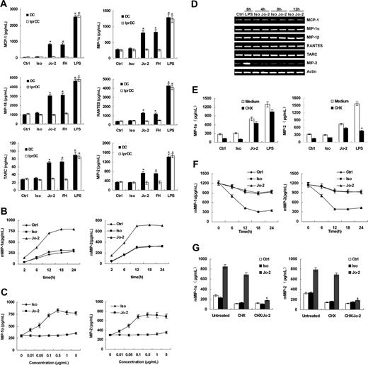
CD11c+ DCs derived from either lpr mice (lprDCs) or C57BL/6J mice (DCs) were incubated with 0.5 μg/mL anti-Fas antibody Jo-2 or 5 μg/mL his-tagged rmFasL in the presence of anti-6 × histidine antibody (FH) for 18 hours. After Fas ligation by either Jo-2 or rmFasL, wild-type DCs secreted more CC chemokines MCP-1, MIP-1α, MIP-1β, RANTES, and TARC, and CXC chemokine MIP-2 than unstimulated DCs but less than LPS-stimulated DCs (Figure 1A). In contrast, neither Jo-2 nor FasL enhanced chemokine secretion from lprDC, demonstrating the Fas-specificity of this effect. Because there was no significant difference in the outcomes of Jo-2 and rmFasL mediated Fas-crosslinking on DCs (P > .05), we chose to use Jo-2 to stimulate DCs in the subsequent experiments, because the antibody is more stable; thus, we could better control the amount used between experiments. We selected MIP-1α and MIP-2 as the representatives of the CC and CXC chemokines for the following experiments. Fas ligation rapidly induced chemokine secretion by DCs. For example, MIP-1α and MIP-2 were detected 2 hours after Jo-2 stimulation (Figure 1B). In addition, Fas ligation induced chemokine production in DCs in an anti-Fas monoclonal antibody dose-dependent manner. Even at very low concentrations (0.01 μg/mL), Jo-2 induced significantly more MIP-1α and MIP-2 than control antibody, indicating that Fas ligation efficiently induces chemokine release from DCs (Figure 1C). This rapid secretion of chemokines in DCs following Jo-2 stimulation indicates that it might not require de novo gene expression. RT-PCR results showed that Jo-2 stimulation had little effect on mRNA expression of chemokines in DCs (Figure 1D), suggesting that Fas ligation–induced chemokine secretion is independent of gene transcription. To test this, DCs were incubated with 0.5 μg/mL CHX for 30 minutes prior to stimulation with Jo-2, isotype, or LPS. As expected, the level of MIP-1α and MIP-2 induced by Jo-2 stimulation remained unchanged, indicating that Fas ligation–induced DC chemokine secretion is independent of de novo protein synthesis (Figure 1E). However, preincubation of CHX inhibited LPS-induced MIP-2 secretion, but not MIP-1α secretion, suggesting MIP-2 and MIP-1α production after LPS stimulation uses different cellular mechanisms(Figure 1D-E). According to these results, we assumed that these chemokines are already stored in DCs, and Fas-ligation triggered their release. To test this, we measured chemokine concentrations in lysates of resting and stimulated DCs. Our results showed that after Fas ligation, the concentration of MIP-1α and MIP-2 in the lysates decreased with time, confirming our assumption (Figure 1F). Moreover, in another experiment, we first blocked protein synthesis in DCs with cycloheximide and then attempted to deplete “stored” chemokines with Jo-2. We then washed this DC culture and restimulated with Jo-2 before testing for the concentrations of MIP-1α and MIP-2 in the supernatant. We found that restimulation with Fas ligation induced much less chemokine release in CHX/Jo-2 pretreated DCs (Figure 1G). Thus, our result strongly suggests that at least certain chemokines are already stored in DCs and Fas ligation triggers their release.
Neutrophils, CD4+ T cells, and CD8+ T cells were chemoattracted by the Fas-ligated DCs or their supernatant in vivo and in vitro, respectively
To determine whether the chemokines released by Fas-ligated DCs have biologic functions, a model for in vivo chemoattraction of inflammatory and immune cells by Fas-ligated DCs was established using EGFP+ DCs. CD11c+EGFP+ DCs, treated as indicated, were intraperitoneally injected, and cells chemoattracted into the peritoneal cavity were collected 2 hours later by peritoneal washout for analysis. As shown in Figure 2A, the total number of chemoattracted cells was increased by 2- to 3-fold in mice that received Fas-ligated DCs. A more robust chemoattractive response was seen in LPS-stimulated DCs. Guided by the CXC and CC chemokines detected in vitro, and the fact that as professional antigen-presenting cells DCs connect innate and adaptive immunity, we focused on detecting neutrophils and T cells. The results showed that Fas-ligated DCs chemoattracted 2- to 3-fold more CD4+ and CD8+ T cells than control DCs (Figure 2B). CD11b+ phagocytic cells, which include monocytes, macrophages, neutrophils, and natural killer cells, were also increased similarly. Among these cells, approximately 80% were Gr1+ neutrophils (Figure 2C). Furthermore, in vitro chemoattraction assay also confirmed the in vivo results (Figure 3).
CC and CXC chemokine production in Fas-ligated DCs. (A) CC (MCP-1, MIP-1α, MIP-1β, RANTES, TARC) and CXC (MIP-2) chemokine production by DCs from C57BL/6J (DC) or lpr (lprDC) mice was measured by ELISA following stimulation of DCs with 0.5 μg/mL Jo-2 (Jo-2), isotype antibody (Iso), 5 μg/mL rmFasL in the presence of 10 μg/mL anti-6 × histidine antibody (FH), or 0.5 μg/mL LPS (LPS) for 18 hours. Unstimulated DCs were used as a control (Ctrl). (B) The kinetics of MIP-1α and MIP-2 production by DCs following stimulation with 0.5 μg/mL Jo-2. (C) MIP-1α and MIP-2 production induced by various concentrations of Jo-2 (0.01-5 μg/mL). (D) Chemokine mRNA expression in DCs stimulated with medium alone (Ctrl), Jo-2, isotype antibody (Iso), or LPS at indicated time points. Results are representative of 3 independent experiments. (E) DCs were incubated with 0.5 μg/mL cycloheximide (CHX) for 30 minutes, and then stimulated with Jo-2, isotype antibody, or LPS for 18 hours, and MIP-1α and MIP-2 levels in supernatant were measured by ELISA. (F) The concentrations of MIP-1α and MIP-2 in the lysates of resting and stimulated DCs at different times were detected. (G) DCs were incubated with CHX for 30 minutes prior to stimulation with Jo-2 for 12 hours. Then we washed this DC culture and restimulated with Jo-2 before testing for the concentrations of MIP-1α and MIP-2 in the supernatant. Untreated and CHX-treated DCs were used as a control. *P < .05. Values in panels A-C and E-F are expressed as means ± standard deviation.
CC and CXC chemokine production in Fas-ligated DCs. (A) CC (MCP-1, MIP-1α, MIP-1β, RANTES, TARC) and CXC (MIP-2) chemokine production by DCs from C57BL/6J (DC) or lpr (lprDC) mice was measured by ELISA following stimulation of DCs with 0.5 μg/mL Jo-2 (Jo-2), isotype antibody (Iso), 5 μg/mL rmFasL in the presence of 10 μg/mL anti-6 × histidine antibody (FH), or 0.5 μg/mL LPS (LPS) for 18 hours. Unstimulated DCs were used as a control (Ctrl). (B) The kinetics of MIP-1α and MIP-2 production by DCs following stimulation with 0.5 μg/mL Jo-2. (C) MIP-1α and MIP-2 production induced by various concentrations of Jo-2 (0.01-5 μg/mL). (D) Chemokine mRNA expression in DCs stimulated with medium alone (Ctrl), Jo-2, isotype antibody (Iso), or LPS at indicated time points. Results are representative of 3 independent experiments. (E) DCs were incubated with 0.5 μg/mL cycloheximide (CHX) for 30 minutes, and then stimulated with Jo-2, isotype antibody, or LPS for 18 hours, and MIP-1α and MIP-2 levels in supernatant were measured by ELISA. (F) The concentrations of MIP-1α and MIP-2 in the lysates of resting and stimulated DCs at different times were detected. (G) DCs were incubated with CHX for 30 minutes prior to stimulation with Jo-2 for 12 hours. Then we washed this DC culture and restimulated with Jo-2 before testing for the concentrations of MIP-1α and MIP-2 in the supernatant. Untreated and CHX-treated DCs were used as a control. *P < .05. Values in panels A-C and E-F are expressed as means ± standard deviation.
Th1, Th2, and activated CD8+ T cells were attracted by DC supernatant in vitro
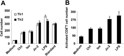
The capacity of Fas-ligated DC supernatant to chemoattract activated T cells (Th1, Th2, and activated CD8+ T cells) was assayed in vitro. The supernatant of Fas-ligated DCs attracted 60% to 70% more Th1 and Th2 cells than that of control DCs (Figure 4A). Notably, the ratio of the attracted Th1 and Th2 cells by Fas-ligated DCs was not altered, suggesting that Fas-ligated DCs do not influence which type of immune response that is initiated. In contrast, supernatants from LPS-treated DCs attracted more Th1 cells than Th2 cells (ratio change from 1:1 to 1.2:1, P < .05), suggesting that Th1 type immune response may play a critical role in clearing bacterial infections (Figure 4A). As shown in Figure 4B, the supernatants of Fas-ligated DCs also attracted 60% to 80% more activated CD8+ T cells than that of control DCs.
Roles of chemokines in attracting T cells and neutrophils
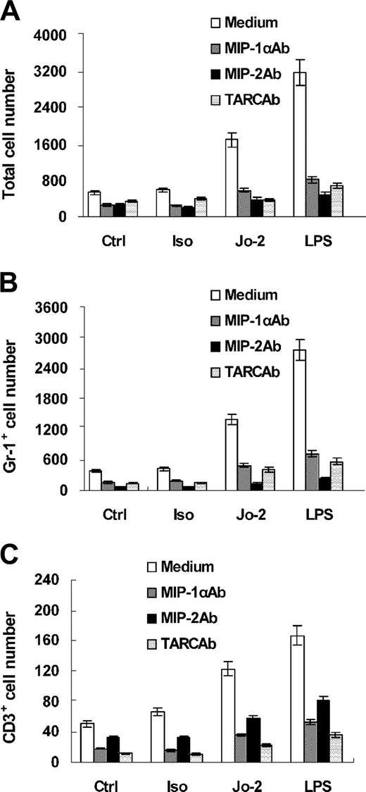
Because our results show Fas ligation induced DCs to produce 2 kinds of chemokines that have different functions and Fas ligation chemoattracts neutrophils and T cells, we wondered whether one type of chemokines is responsible for chemoattracting a defined cell population. We incubated neutralizing antibodies to MIP-1α, TARC, or MIP-2 with conditioned supernatant of Fas-ligated DCs for 30 minutes. We then used 10-fold diluted supernatant to chemoattract mixed splenocytes and bone marrow cells (containing T cells and neutrophils). As shown in Figure 3, neutralizing any one of the chemokines resulted in a significant decrease in the number of chemoattracted cells, indicating that MIP-1α, MIP-2, and TARC in the supernatant were all indispensable in chemoattracting T cells and neutrophils and that there is a substantial synergism among the chemokines. The difference among the 3 chemokines in our function study is that MIP-2 appears to play a dominant role in chemoattracting neutrophils, whereas MIP-1α and TARC appear to be more important in chemoattracting T cells. This might result from the fact that chemokines are known to be promiscuous, and receptors for both the CC and CXC chemokines have been shown to be expressed by both neutrophils and T cells.
CD4+ and CD8+ T cells and phagocytes are chemoattracted by Fas-stimulated DCs in vivo. EGFP+ DCs treated with Jo-2, isotype Ab, or LPS overnight were intraperitoneally injected into mice (n = 5). After 2 hours, PWCs were harvested in 10 mL ice-cold PBS and stained immediately with specific Abs for cell subsets. Total chemoattracted cells (A), T cells, including CD4+ and CD8+ cells (B), and CD11b+ phagocytic cells, including Gr1+ neutrophils (C), were analyzed by flow cytometry, counting the numbers of cells acquired in the same time period. The cell numbers shown are the number we collected on the FACS file. *P < .05.
CD4+ and CD8+ T cells and phagocytes are chemoattracted by Fas-stimulated DCs in vivo. EGFP+ DCs treated with Jo-2, isotype Ab, or LPS overnight were intraperitoneally injected into mice (n = 5). After 2 hours, PWCs were harvested in 10 mL ice-cold PBS and stained immediately with specific Abs for cell subsets. Total chemoattracted cells (A), T cells, including CD4+ and CD8+ cells (B), and CD11b+ phagocytic cells, including Gr1+ neutrophils (C), were analyzed by flow cytometry, counting the numbers of cells acquired in the same time period. The cell numbers shown are the number we collected on the FACS file. *P < .05.
Roles of chemokines in attracting T cells and neutrophils. We incubated the neutralizing antibody to MIP-1α, TARC, or MIP-2 with the conditioned supernatant for 30 minutes. The supernatant was 10-fold diluted and used to chemoattract the mixed cells containing T cells stained with CD3-PE and neutrophils stained with Gr1-FITC in 3.0-μm pore sized transwell insert. After 2 hours of incubation at 37°C, the total cells (top), including Gr1-FITC neutrophils (left bottom) and CD3-PE T cells (right bottom) that had migrated into wells, were harvested and counted by FACSCalibur flow cytometer and analyzed by CellQuest software.
Roles of chemokines in attracting T cells and neutrophils. We incubated the neutralizing antibody to MIP-1α, TARC, or MIP-2 with the conditioned supernatant for 30 minutes. The supernatant was 10-fold diluted and used to chemoattract the mixed cells containing T cells stained with CD3-PE and neutrophils stained with Gr1-FITC in 3.0-μm pore sized transwell insert. After 2 hours of incubation at 37°C, the total cells (top), including Gr1-FITC neutrophils (left bottom) and CD3-PE T cells (right bottom) that had migrated into wells, were harvested and counted by FACSCalibur flow cytometer and analyzed by CellQuest software.
Fas ligation enhanced functions of neutrophils and T cells
Given the fact that Fas ligation on DCs enhances recruitment of neutrophils and T cells, we tested whether such DCs could affect their function. First, we treated DCs as indicated and incubated with purified neutrophils. The endocytosis ability of neutrophils was measured by FACS analysis of Fluosphere uptake by CD11c- neutrophils. As shown in Figure 5A, Fas-ligated DCs enhanced endocytosis by neutrophils dramatically, although the enhancement was lower than that caused by LPS-treated DCs. Meanwhile, we seeded Jo-2–treated DCs on the upper chamber, whereas neutrophils were seeded onto the bottom of a 1.0-μm pore sized transwell, and then detected the endocytosis by neutrophils. The result showed that separation of the 2 cell populations abrogated the enhanced endocytosis by the neutrophils, indicating that DC-neutrophil contact is required for this function.
To examine the effect of Fas-ligated DCs on T-cell function, we treated DCs as indicated and then incubated with purified CD4+ T cells and assayed their activation and proliferation. The supernatant was collected at 24 hours to detect the production of IL-2 and IFN-γ. Fas-ligated DCs displayed a stronger ability to activate naive T cells to undergo proliferation and cytokine production (Figure 5B). Our ELISpot results of IFNγ further confirmed this result (Figure 5C).
Taken together, these data indicate that Fas ligation on DCs promotes endocytosis by neutrophils and activation and proliferation of T cells, therefore enhancing the bridging role of DCs between innate and adaptive immunity.
Th1, Th2, and activated CD8+ T cells are chemoattracted by Fas-ligated DCs in vitro. Th1 and Th2 cells (A) prepared in vitro were stained with CD4-FITC or CD4-PE, respectively, and mixed at a ratio of 1:1, and activated CD8+ T cells (B) prepared in vitro were stained with CD8-PE, and these cells were tested for their chemoattraction to 10-fold diluted DC supernatant by in vitro chemotaxis assay (n = 3). Numbers of migrated cells were analyzed by FACS. Mixed Th1 and Th2 cells were used as a standard, and RPMI 1640 culture medium was used as a negative control. *P < .05.
Th1, Th2, and activated CD8+ T cells are chemoattracted by Fas-ligated DCs in vitro. Th1 and Th2 cells (A) prepared in vitro were stained with CD4-FITC or CD4-PE, respectively, and mixed at a ratio of 1:1, and activated CD8+ T cells (B) prepared in vitro were stained with CD8-PE, and these cells were tested for their chemoattraction to 10-fold diluted DC supernatant by in vitro chemotaxis assay (n = 3). Numbers of migrated cells were analyzed by FACS. Mixed Th1 and Th2 cells were used as a standard, and RPMI 1640 culture medium was used as a negative control. *P < .05.
Fas ligation enhances endocytosis by neutrophils and activation and proliferation of antigen-specific T cells. (A) DCs that were treated with medium, Jo-2, isotype Ab, or LPS for 12 hours were incubated with neutrophils for 20 hours. For separate Jo-2 DC/PMN (polymorphonuclear neutrophil) coculture, we seeded Jo-2–treated DCs on the upper chamber, whereas neutrophils were seeded onto the bottom of a 1.0-μm pore sized transwell. Then, 2 μL Fluospheres per well was added into the coculture system, and 3 hours later the cells were collected and stained with CD11c-PE to gate CD11c- neutrophils. Cells were detected by FACS, and the FL1 fluorescence of the CD11c- cells neutrophils was analyzed. (B) Purified CD4 T cells were cocultured with DCs treated as indicated in the presence of OVA323-339 peptide at a ratio of 1:10 (DC/T) in round-bottom 96-well plates for 5 days. Then cells were collected and double stained with anti-CD4–FITC and 7-AAD and counted by FACS. Supernatants were collected at 24 hours for assay of IL-2 and IFN-γ by ELISA. (C) Purified CD4 T cells were cocultured with DCs treated as indicated in the presence of OVA323-339 peptide at a ratio of 1:10 (DC/T) in the IFN-γ ELISpot plate (3 × 10⁴ T cells/200 μL/well) for 24hours. Then, following the manufacturer's instruction, the microplate was developed and dried in the air. Photos were taken using an Olympus 52X12 dissection microscope with a Cohu high-performance CCD camera (Cohu, San Diego, CA) and Leica QWin imaging software (Hamburg, Germany).
Fas ligation enhances endocytosis by neutrophils and activation and proliferation of antigen-specific T cells. (A) DCs that were treated with medium, Jo-2, isotype Ab, or LPS for 12 hours were incubated with neutrophils for 20 hours. For separate Jo-2 DC/PMN (polymorphonuclear neutrophil) coculture, we seeded Jo-2–treated DCs on the upper chamber, whereas neutrophils were seeded onto the bottom of a 1.0-μm pore sized transwell. Then, 2 μL Fluospheres per well was added into the coculture system, and 3 hours later the cells were collected and stained with CD11c-PE to gate CD11c- neutrophils. Cells were detected by FACS, and the FL1 fluorescence of the CD11c- cells neutrophils was analyzed. (B) Purified CD4 T cells were cocultured with DCs treated as indicated in the presence of OVA323-339 peptide at a ratio of 1:10 (DC/T) in round-bottom 96-well plates for 5 days. Then cells were collected and double stained with anti-CD4–FITC and 7-AAD and counted by FACS. Supernatants were collected at 24 hours for assay of IL-2 and IFN-γ by ELISA. (C) Purified CD4 T cells were cocultured with DCs treated as indicated in the presence of OVA323-339 peptide at a ratio of 1:10 (DC/T) in the IFN-γ ELISpot plate (3 × 10⁴ T cells/200 μL/well) for 24hours. Then, following the manufacturer's instruction, the microplate was developed and dried in the air. Photos were taken using an Olympus 52X12 dissection microscope with a Cohu high-performance CCD camera (Cohu, San Diego, CA) and Leica QWin imaging software (Hamburg, Germany).
Phosphorylation of Ras/Raf/MEK/ERK pathway, but not activation of NF-κB, is responsible for Fas ligation–induced chemokine production in DCs
Our previous report showed that Fas ligation induces phosphorylation of ERK1/2 and nuclear translocation of NF-κB in DCs and that these pathways play different roles in the maturation and survival of DCs.18 To investigate their roles in Fas-induced release of chemokines, we treated Fas-stimulated DCs with PD98059, a selective inhibitor of MEK1, and PDTC, an inhibitor of NF-κB. As shown in Figure 6A, PD98059 significantly inhibited the Fas-induced release of MIP-1α and MIP-2 and partially inhibited LPS-induced chemokine secretion, suggesting that phosphorylation of ERK is important in Fas-induced chemokine production, and that LPS and Fas ligation did not promote chemokine production via the same mechanisms. In contrast, PDTC had no effect on Fas-induced chemokine production (Figure 6B), suggesting that NF-κB activation is not involved in this process. Although previous reports suggest that mitogen-activated protein kinase (MAPK) p38 is involved in Fas-induced chemokine expression in human glioma cells,10,23 it may not be involved in DC chemokine expression after Fas ligation since we did not detect MAPK p38 activation in Fas-ligated DCs, and the p38 inhibitor SB203580 did not affect chemokine production (data not shown).
We have demonstrated that Fas induces activation of ERK1/2 in DCs, but it is unclear which components of Fas downstream signaling cascades lead to activation of ERK1/2. It has been reported that activation of Ras is involved in Fas ligation–induced lymphocyte apoptosis.24,25 We wondered whether Fas induces activation of Ras, Raf, and MEK in DCs. As shown in Figure 6C, Fas ligation on DCs readily activated Ras, Raf, and MEK1/2. Specific inhibitors were used to evaluate their function on Fas-induced chemokine production. As shown in Figure 6D, inhibition of Ras and Raf had similar effects in decreasing the chemokine production in Fas-ligated DCs. Taken together, our data suggest that the poorly defined intracellular portion of the Fas molecule could activate membrane-bound small G proteins such as ras and raf, leading to the activation of MEK1/2 and ERK1/2 pathways and chemokine production upon Fas ligation.
Phosphorylation of Ras/Raf/MEK/ERK pathway, but not activation of NF-κB, is responsible for Fas-induced chemokines production in DCs. (A-B) DCs were pretreated with different doses of PD98059 (A) or PDTC (B) for 30 minutes, then stimulated with Jo-2, isotype Ab (Iso), or LPS for 18 hours. Levels of MIP-1α and MIP-2 in supernatants were determined by ELISA. (C) Western blotting analysis of activation of Ras, Raf, and MEK1/2 in DCs in medium alone as a control (Ctrl) or stimulated with Jo-2 (Jo-2), isotype Ab(Iso), or LPS (LPS) for the indicated times. (D) DCs were pretreated with different doses of FTI-277 or Raf-1 inhibitor for 30 minutes, then stimulated with Jo-2, isotype Ab, or LPS for 18 hours. The levels of MIP-1α or MIP-2 in the supernatants were determined by ELISA.
Phosphorylation of Ras/Raf/MEK/ERK pathway, but not activation of NF-κB, is responsible for Fas-induced chemokines production in DCs. (A-B) DCs were pretreated with different doses of PD98059 (A) or PDTC (B) for 30 minutes, then stimulated with Jo-2, isotype Ab (Iso), or LPS for 18 hours. Levels of MIP-1α and MIP-2 in supernatants were determined by ELISA. (C) Western blotting analysis of activation of Ras, Raf, and MEK1/2 in DCs in medium alone as a control (Ctrl) or stimulated with Jo-2 (Jo-2), isotype Ab(Iso), or LPS (LPS) for the indicated times. (D) DCs were pretreated with different doses of FTI-277 or Raf-1 inhibitor for 30 minutes, then stimulated with Jo-2, isotype Ab, or LPS for 18 hours. The levels of MIP-1α or MIP-2 in the supernatants were determined by ELISA.
Activation of caspases, but not IL-1β autocrine action, is involved in Fas-induced chemokine production
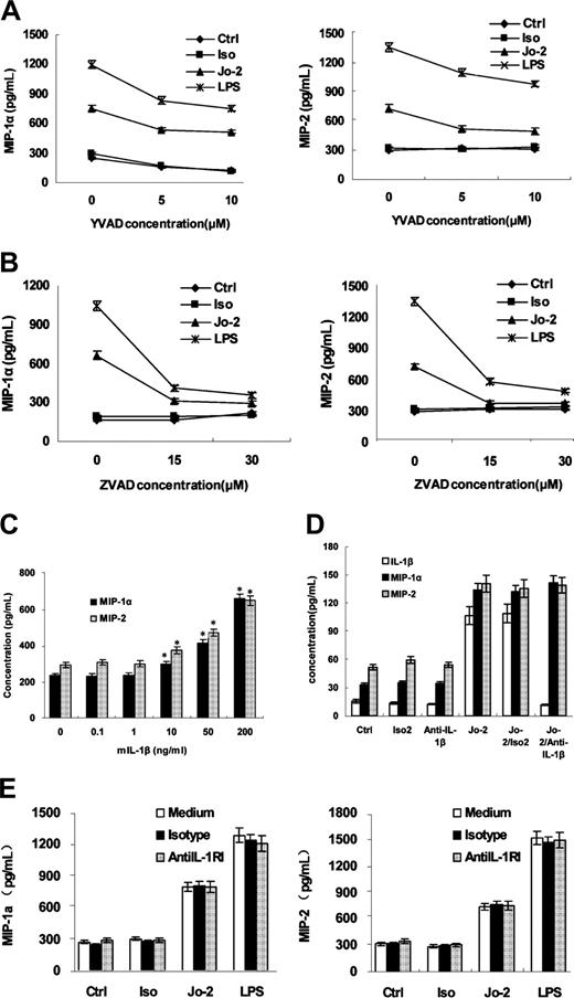
We previously established that caspase 1 is activated in DCs by Fas ligation and plays an important role in IL-1β secretion and the ensuing IL-1β–dependent maturation of DCs.18 To determine whether caspase 1 is involved in chemokine production, we used caspase 1 inhibitor, Ac-YVAD-fmk, in this system. We found that Ac-YVAD-fmk partially inhibited chemokine production (Figure 7A). In contrast, the pan-caspase inhibitor, ZVAD-fmk, almost completely abrogated the effect of Fas ligation on DC (Figure 7B), indicating that caspase 1 only partially contributes to Fas-induced chemokine production. We did not detect activation of caspase 3 or caspase 8, and inhibitors of these 2 caspases did not affect Fas-induced chemokine production in DCs (data not shown), indicating that neither caspase 3 nor caspase 8 is involved in the process. Identification of the caspases involved in Fas-induced chemokine production remains to be done.
Because IL-1β is a proinflammatory cytokine that acts on many cell types to up-regulate expression of chemokines,8 and IL-1β secretion induced by Fas ligation contributes to Fas-induced maturation of DCs,18 we tested whether Fas-induced chemokines production depends on IL-1β secretion. As shown in Figure 7C, only high concentrations of rmIL-1β (10 ng/mL or greater) significantly induced secretion of MIP-1α and MIP-2. The 100-pg/mL dose of IL-1β, equivalent to the quantity secreted by Fas-ligated DCs, did not trigger chemokine production. Both blockade of IL-1β with anti–IL-1β antibody (Figure 7D) or a neutralizing antibody against IL-1R (Figure 7E) did not inhibit chemokine expression, demonstrating that Fas-induced chemokine secretion in DCs is not due to a secondary effect by IL-1β.
Discussion
The Fas-FasL interaction is increasingly regarded as one of the most enigmatic actions in the immune system.26,27 In addition to the generally accepted notion that Fas ligation induces apoptosis of sensitive cells, increasing evidence indicates that it can also elicit inflammation. In our previous report, Fas ligation was found to induce IL-1β secretion in DCs and provoke DC maturation rather than apoptosis.18 Here, we have further investigated the proinflammatory effects of Fas ligation on DCs and demonstrated that Fas-ligated DCs chemoattract naive and activated T cells, as well as neutrophils, by secreting CC and CXC chemokines via an ERK and caspases-dependent mechanism. Fas-ligated DCs also promoted the endocytosis by neutrophils and activation and proliferation of antigen-specific T cells. In this way, Fas ligation appears to enhance the ability of DCs to link innate and adaptive immunity.
FasL is expressed on activated T cells,28 some tumors,29,30 some nonlymphoid cells31 of liver and small intestine, and in sites of immune privilege3,4 such as testis, eye, and placenta, whereas Fas is broadly distributed on many types of cells,28,32 including DCs.33 Under different physiologic or pathologic conditions, these FasL-expressing cells will encounter DCs. As potent antigen-presenting cells, DCs, activated by innate stimuli and loaded with processed foreign antigen, migrate to regional lymph nodes and prime T cells. Activated T cells subsequently find their way back to the site of inflammation and exert effector functions to eliminate pathogens.15,34 During this process, FasL-expressing activated T cells will inevitably interact with Fas on DCs. Our results show that as a result of this interaction Fas-ligated DCs could secret more chemokines and recruit more naive T cells and promote their activation and proliferation, thus enhancing adaptive immunity. Furthermore, at the site of infection, as our results demonstrated, Fas-FasL interaction will recruit more activated T cells and phagocytes and enhance the endocytosis of neutrophils, strengthening the innate immunity.
Activation of caspases, but not IL-1β, is involved in the secretion of MIP-1α and MIP-2 in Fas-ligated DCs. DCs were pretreated with different doses of the relatively specific caspase 1 inhibitor Ac-YVAD-fmk (A) or pan-caspase inhibitor ZVAD-fmk (B) for 30 minutes, then stimulated with Jo-2, isotype Ab (Iso), or LPS for 18 hours. The levels of MIP-1α and MIP-2 in supernatants were determined by ELISA. (C) DCs were stimulated with various doses of rmIL-1β for 18 hours. Then the levels of MIP-1α and MIP-2 in the supernatants were determined by ELISA. (D) DCs were pretreated with anti–mIL-1β Ab or isotype Ab (Iso2) for 30 minutes and then treated with Jo-2 for 18 hours, then levels of IL-1β, MIP-1α, and MIP-2 in supernatants were determined by ELISA. The concentration of MIP-1α and MIP-2 shown here was 5-fold diluted. (E) To confirm the effect of the autocrined IL-1β on Fas-induced production of chemokines in DCs, the blocking antibody to mIL-1R type I (mIL-1RI), by which IL-1β transduces its signal, and its isotype antibody, was added to the system. And then concentration of MIP-1α and MIP-2 was detected by ELISA kits.
Activation of caspases, but not IL-1β, is involved in the secretion of MIP-1α and MIP-2 in Fas-ligated DCs. DCs were pretreated with different doses of the relatively specific caspase 1 inhibitor Ac-YVAD-fmk (A) or pan-caspase inhibitor ZVAD-fmk (B) for 30 minutes, then stimulated with Jo-2, isotype Ab (Iso), or LPS for 18 hours. The levels of MIP-1α and MIP-2 in supernatants were determined by ELISA. (C) DCs were stimulated with various doses of rmIL-1β for 18 hours. Then the levels of MIP-1α and MIP-2 in the supernatants were determined by ELISA. (D) DCs were pretreated with anti–mIL-1β Ab or isotype Ab (Iso2) for 30 minutes and then treated with Jo-2 for 18 hours, then levels of IL-1β, MIP-1α, and MIP-2 in supernatants were determined by ELISA. The concentration of MIP-1α and MIP-2 shown here was 5-fold diluted. (E) To confirm the effect of the autocrined IL-1β on Fas-induced production of chemokines in DCs, the blocking antibody to mIL-1R type I (mIL-1RI), by which IL-1β transduces its signal, and its isotype antibody, was added to the system. And then concentration of MIP-1α and MIP-2 was detected by ELISA kits.
FasL expressed on stromal cells in immune-privileged sites3,4 and tumor cells29,30 was once considered to be the main mechanism for an immune-privileged environment and tumor immune escape. However, in recent years, the role of FasL in immune privilege and tumor escape has been reevaluated.5,26,27 Increasing data support that in many tissues FasL expression induces potent inflammatory responses. Reports regarding FasL-transfected cell–induced inflammation focused mainly on the infiltration of neutrophils,5,7,8 but researchers find now that the early nonspecific inflammatory responses induced by FasL-transfected cells promote tumor-specific T-cell response, which is ultimately responsible for protective immunity.35-37 For this to occur, T-cell subsets, including naive T cells, must be recruited to the site of tumor inoculation following neutrophil infiltration by some chemokines. Which cells produce this panel of chemokines? Most former researches ignored these Fas-expressing DCs. Our experiments indicate that the interaction of widely distributed Fas-expressing DCs with FasL-transfected tumor cells and subsequent secretion of chemokines by DCs may play an important role in this process. Given the established role of neutrophils in T-cell responses,38-40 our data clearly suggest that Fas-ligation on DCs promotes innate and adaptive immunity by enhancing the functions of recruited neutrophils and T cells. Our experiment may provide an explanation for the rejection of FasL-expressing tumor cells by infiltrating neutrophils and the ensuing development of CD8+ T-cell–dependent antitumor immunity.35-37 As to why there is no inflammation in sites of immune privilege, suppressive cytokines such as transforming growth factor β may act to prevent inflammatory responses to FasL.5
The mechanisms underlying Fas-mediated chemokine production still need to be fully elucidated. Our data demonstrate that activation of ERK1/2 and caspases is necessary for Fas-induced chemokine production in DCs, whereas activation of NF-κB and p38 (data not shown) and secretion of IL-1β are not involved. MAPKs are serine-threonine kinases activated by diverse stimuli, and the components include ERK1/2, p38 MAPK, and c-Jun N-terminal kinase/stress-activated protein pathways.41 Combining with our previous study, we demonstrated activation of ERK1/2 determined Fas-induced cytokine and chemokine secretion.18 However, we did not detect p38 activation, despite a report that this molecule is activated following Fas ligation in glioma cells. Some small G proteins such as Rac-1 and Ras have been reported to participate in Fas-induced apoptosis,24,25 Our data demonstrate that Fas signaling could activate membrane-bound small G protein such as ras and raf to ultimately activate ERK1/2 pathways, resulting in DC chemokine production. Because inhibition of Ras and Raf-1 partially abrogates Fas-ligation–induced chemokine production, we speculate that an alternative pathway might exist in Fas-ligation–induced activation of MEK and ERK1/2.
Reports regarding the relationship between caspases and chemokine induction demonstrated the function of caspases varies among different cell types,8,9,12 so it remains important to determine the roles of caspases in Fas-mediated chemokine production in DCs. DCs are resistant to Fas-induced apoptosis. In supporting this, we did not detect activation of caspase-3 or caspase-8 in response to Fas ligation. Furthermore, these caspases are not involved in Fas-ligation–induced chemokine production in DCs because specific inhibition of these caspases did not influence chemokine secretion in DCs. Although caspase-1 inhibitor only partially diminishes chemokine production, a pan-caspase inhibitor almost completely blocks this effect, suggesting that other caspases, in addition to caspase-1, participate in Fas-mediated chemokine production.
Our results also show that Fas-induced chemokine secretion in DCs is independent of NF-κB activation, consistent with results in human hepatocytes.12 Although reports from different groups suggest that Fas ligation induces nuclear translocation of NF-κB and cytokine production,9 none has directly proven that the observed cytokine production is dependent on NF-κB activation.
IL-1β is a proinflammatory cytokine that can up-regulate chemokine expression in many cell types.8 However, the fact that only high concentrations of rmIL-1β induce secretion of chemokines in DCs and neutralizing anti–IL-1β antibody and blocking anti–IL-1R antibody has no effect on chemokine release indicates that Fas ligation–induced chemokine production is a direct function of Fas signaling and independent of concurrent IL-1β secretion triggered by Fas ligation.
Our current and prior findings indicate that Fas ligation on DCs exhibits 2 major functions: (1) induction of DC maturation and survival and (2) recruitment of more neutrophils and naive and activated T cells via increased secretion of CC and CXC chemokines and enhancement of their functions. Activation of ERK1/2, caspases, and NF-κB play different roles in these 2 functions, resulting in amplification of both innate and adaptive immune responses. Owing to the pleiotropic effects of Fas-FasL interaction in immune responses, enhancing or suppressing the various functions of this interaction may serve as potential therapeutics for cancers and allograft transplantation.
Prepublished online as Blood First Edition Paper, June 7, 2005; DOI 10.1182/blood-2004-12-4831.
Supported by grants from the National Key Basic Research Program of China (2001CB510002) and the National Natural Science Foundation of China (30490240, 30121002).
Z.G. and M.Z. contributed equally to this work.
The publication costs of this article were defrayed in part by page charge payment. Therefore, and solely to indicate this fact, this article is hereby marked “advertisement” in accordance with 18 U.S.C. section 1734.
We thank Ms R. Zhang and Ms C. Luo for technical assistance, and Prof You-Wen He, Dr Huazhang An, Dr Jane Rayner, and Dr Yizhi Yu for helpful discussion.


This feature is available to Subscribers Only
Sign In or Create an Account Close Modal