Abstract
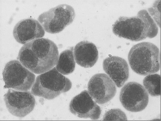
A new human myeloma cell line WuS1 was established from the bone marrow of a 45-year-old Chinese male patient with IgGλ type multiple myeloma (stage IIIB). The growth of WuS1 cells is constitutively independent of exogenous growth factors of feeder cells. The WuS1 cell line proliferated consistently as free-floating single cells in suspension, sometimes in small clusters or slightly adherent on the bottom of the plastic culture flask, without forming clumps. The cell line has been maintained without any external growth factors for over a year, and cells frozen in liquid nitrogen can be revived successfully. The doubling time of the cells was about 11 hours and the colony-forming rate was 55.56±6.33%. WuS1 displayed immature plasma cell features with an obvious heterogeneity in size and a high nuclear-cytoplasmic ratio in Wright-Giemsa staining.
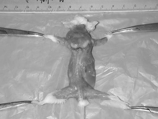
They were positive for ALP, CE, ACP(not inhibited by tartrate) and PAS stainings and negative for POX and NBE. By transmission electron microscopy, the cytoplasm of WuS1 contained abundant mitochondria, and parallel endoplasmic reticulum or Golgi apparatus in some cells. The monoclonal immunoglobulin G and λ light chain were positive in cell lysate and not in cell culture supernatants by immnuoelectrophoresis. The cell surface antigens were positive for CD3, CD59, CD106 and CD138, and negative for CD4, CD5, CD8, CD10, CD13, CD14, CD19, CD20, CD22, CD29, CD31, CD33, CD34, CD38, CD44, CD49d, CD45, CD54, CD56 and HLA-DR by flow cytometry. Chromosomal analysis revealed a hypodiploidy and complex karyotype. WuS1 cells were negative for Epstein-Barr virus by PCR using EBV nuclear antigen-1 specific primers. Twelve SCID mice were injected with WuS1 cells intravenously or subcutaneously, and obvious tumor infiltration in bone marrow, liver, spleen, lung, kidney and injection site (subcutaneously group) were observed by pathologic examination.
The novel WuS1 cell line will be useful in the study of the biology, etiology and treatment of multiple myeloma.
Author notes
Corresponding author


This feature is available to Subscribers Only
Sign In or Create an Account Close Modal