Abstract
Background: Angioimmunoblastic T cell lymphoma (AILT) is a peripheral T- cell lymphoma characterized by systemic disease, a polymorphous infiltrate involving lymph nodes with prominent proliferation of high endothelial venules and follicular dendritic cells. We present a novel technique for the presumptive diagnosis of AILT cases by flow cytometric analysis. The diagnosis of AILT was confirmed by histolopathologic features, immunohistochemical stains and molecular studies.
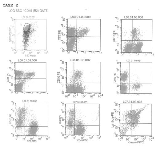
Method: Specimens: Fresh groin excision lymph nodes. Flow cytometric analysis utilizing side scatter versus CD45. Case 1 (figure 1): The gated population consists of 10–12% B cells and 56–61% T cells. CD3, CD4 and CD5 stain 56–61% of the gated cells, while CD7 stains 41% of the gated cells, consistent with aberrant partial loss of CD7 by T cells. A population of abnormal T cells expresses CD10 with CD2, CD3, CD4 and CD7 but not with CD19 (B cells) consistent with aberrant CD10 coexpression by a subset of the T cells (12–19% of the gated cells). Staining for CD34 highlights less than 2% immature cells staining while CD64 stains less than 2% monocytic cells. Surface kappa and lambda staining does not show restriction (kappa: lambda 1–2:1). Cytoplasmic kappa and lambda staining is not interpretable due to non-specific antibody binding. Case 2 (figure 2): The gated population consists of 69% T- cells and 28–33% B- cells. A population of abnormal T cells expresses CD10 with CD2, CD3, CD4 and CD7 but not CD19 consistent with aberrant CD10 expression by a subpopulation of T cells (8–11% of gated cells). Intracellular B cell kappa lambda light chain restriction is not present and there is no aberrant co-expression of CD5, CD10 or CD43 by B cells. The diagnosis of AILT was confirmed by standard morphologic, immunoperoxidase and molecular methods subsequently in both of these cases.
Discussion: Aberrant CD10 expression on neoplastic T cells has been shown with immunohistochemical staining of paraffin embedded tissue in AILT. Our cases show aberrant coexpression of CD10 by CD2, CD3, CD4 and CD7 positive T cells in more than 10% of gated cells by Flow cytometric analysis.
Conclusion: Flow Cytometric analysis is a useful and reproducible tool for immunophenotyping AILT cells and should be considered for presumptive diagnosis of AILT.
Author notes
Corresponding author


This feature is available to Subscribers Only
Sign In or Create an Account Close Modal