Abstract
OX40 (CD134) is a member of the tumor necrosis factor (TNF) receptor family that is transiently expressed on T cells after T-cell receptor (TCR) ligation. Both naive and activated CD4+CD25+ regulatory T cells (T reg's) express OX40 but its functional role has not been determined. Since glucocorticoid-induced tumor necrosis factor receptor (GITR), a related TNF receptor family member, influences T reg function, we tested whether OX40 might have similar effect. Triggering either GITR or OX40 on T reg's using agonist antibodies inhibited their capacity to suppress and restored effector T-cell proliferation, interleukin-2 (IL-2) gene transcription and cytokine production. OX40 abrogation of T reg suppression was confirmed in vivo in a model of graft-versus-host disease (GVHD). In a fully allogeneic C57BL/6>BALB/c bone marrow transplantation, GVHD was lethal unless T reg's were cotransferred with the bone marrow and effector T cells. Strikingly, T reg suppression of GVHD was abrogated either by intraperitoneal injection of anti-OX40 or anti-GITR monoclonal antibodies (mAbs) immediately after transfer, or by in vitro pretreatment of T reg's with the same mAbs before transfer. Cumulatively, the results suggest that in addition to controlling memory T-cell numbers, OX40 directly controls T reg–mediated suppression.
Introduction
Five to 10 percent of naive peripheral CD4+ T cells in normal mice and healthy humans express CD25 and constitute a population of regulatory T cells (T reg's). In multiple in vivo and in vitro situations, these cells have the capacity to suppress immune responses to auto- and alloantigens, tumor antigens, and infections.1 The suppressive effect of T reg's is exerted through inhibition of IL-2 production by effector T cells via an unknown mechanism that requires cell-to-cell contact.2 They themselves are anergic and do not make productive responses to T-cell–receptor (TCR) triggering in vitro unless exogenous interleukin-2 (IL-2) is supplied. The depletion of T reg's in normal mice leads to the development of several autoimmune diseases, whereas reconstitution of this population to mice that lack them prevents autoimmunity. This direct role in controlling peripheral tolerance3,4 has been described in diabetes, experimental encephalomyelitis (EAE)5 and inflammatory bowel disease (IBD).6 T reg's also play a central role in transplantation,7 pathogen clearance,8 and in down-regulation of inflammatory responses.8 In cancer, T reg's increase both in peripheral blood and at the tumor site.9 In mice, the depletion of CD4+CD25+ cells in vivo by means of a monoclonal antibody (mAb) to CD25 (PC61) before tumor challenge enhances the ability of the host to reject several types of immunogenic tumors in different strains of mice.10,11
Phenotypic characterization and microarray analysis of naive T reg's revealed their constitutive expression of several costimulatory molecules including CD28, CD40 ligand (CD40L), cytotoxic T lymphocyte antigen 4 (CTLA-4), glucocorticoid-induced TNF receptor (GITR or TNFRSF18) and OX40 (CD134). CD2812 and CD40L are involved in maintenance of peripheral T reg homeostasis (C.G., B.V., M.P.C. et al, manuscript in preparation), whereas CTLA-4 and GITR regulate T reg–suppressive activity.13,14
Among the members of the tumor necrosis factor (TNF) receptor family expressed on T reg's, GITR is the most studied. Shimizu and colleagues14 have shown that stimulation of GITR abrogates T reg–mediated suppression, while McHugh et al15 demonstrated that such abrogation depends on whether or not T reg's have been preactivated by TCR triggering and IL-2, before GITR engagement. GITR is no longer an exclusive marker of T reg's since it is also expressed at high levels on activated CD4+CD25- T cells and appears to play a general role in the regulation of TCR-driven T-cell activation and cell death.16,17
OX40 is another member of the TNF receptor family that is expressed on naive T reg's.15,18 It was originally identified as an activated T-cell marker in rats, and subsequently was characterized as a costimulatory molecule regulating CD4 and CD8 immunity.19,20 OX40 is transiently expressed upon TCR triggering, peaking at 48 hours and disappearing after 72 to 96 hours.20 OX40 ligand (OX40L or CD134L) is normally expressed on antigen-presenting cells, such as B cells, dendritic cells (DCs), macrophages, and endothelial cells when activated.21-24 OX40-OX40L interaction regulates the production T-helper type 1 (Th1) cytokines and through the up-regulation of antiapoptotic proteins such as Bcl-XL and Bcl-2 it controls T-cell clonal expansion and memory cell development.25-27
Triggering of OX40 in vivo overcomes CD4 T-cell tolerance to peptide antigens and promotes tumor cells rejection and graft-versus-host disease (GVHD).28,29 Both OX40- and OX40L-deficient mice are less susceptible to inflammatory bowel disease (IBD),18 EAE,30 allergic asthma,31 and to virus-induced lung inflammation,32 whereas transgenic mice hyperexpressing OX40L show increased disease severity. The blockade of OX40-OX40L interaction ameliorates wasting disease,33 colitis,34 EAE,35 and GVHD.28
The role of OX40 in autoimmunity and immune response to cancer has been extensively studied; however, the functional significance of its expression on T reg's has not been investigated in detail.36 In this paper we studied the effect of OX40 triggering in response to an agonist mAb, and demonstrated abrogation of T reg–mediated immunosuppression both in vitro and in vivo. We also compared the effect of OX40 triggering with that of GITR and established a system in which T reg's were pretreated with the antibodies to avoid triggering of the same molecules on effector CD4+CD25- T cells during coculture. The results show that GITR-mediated inhibition of T reg function is only apparently stronger than that mediated by OX40 in vitro, while in vivo, both Abs, by blocking T reg's, accelerate GVHD lethality.
Materials and methods
Mice, rats, and treatments
BALB/c and C57BL/6 mice and Wistar rats were purchased from Charles River (Calco, Italy); all mice and rats were used at 8 weeks of age. C57BL/6 OX40-null mice26 were originally generated at the University of California at San Francisco (UCSF) and were maintained under pathogen-free conditions in our animal facility. In vivo treatments with anti-OX40 mAb (OX86 clone; European Collection of Cell Cultures [ECACC], Salisbury, Wiltshire, United Kingdom) and anti-GITR (DTA-1 clone; kindly provided by V. Bronte, University of Padova, Italy) was performed using a single injection of purified mAbs as indicated.
Antibodies and flow cytometric analysis
Fluorescein isothiocyanate (FITC)–conjugated anti-CD4 (L3T4), purified anti–mouse OX40 (OX86), biotin anti–mouse OX40 (OX86), biotin anti–rat OX40 (OX-40), FITC-conjugated anti-rat, phycoerythrin (PE)–conjugated CD25 and purified rat immunoglobulin (Ig) G1 and rat IgG2a isotype controls were all purchased from BD Bioscience (San Diego, CA). Antibodies were used at 5 μg/mL and staining was performed in fluorescence-activated cell-sorting (FACS) buffer (10% fetal calf serum [FCS] in phosphate-buffered saline [PBS]) on ice for 45 minutes. FACS analyses were performed on a FACScan (Becton Dickinson, Franklin Lakes, NJ). IL-2 production was measured by a specific enzyme-linked immunosorbent assay (ELISA) using JES6-1A12 as the capture antibody and JES6-5H4 as the detection antibody (BD Bioscience).
Purification of CD4+CD25+ and CD4+CD25- subsets
T cells were first enriched by passing whole spleen through nylon wool columns. CD8+ cells were removed using anti-CD8 MACS Microbeads (Miltenyi Biotec, Bergisch Gladbach, Germany). CD4+CD25+ cells were then separated from CD4+CD25- cells using the CD25+ T-cell isolation kit (Miltenyi Biotec) according to manufacturer instructions. Flow cytometry showed that the separate fractions were more than 90% pure.
In vitro CFSE labeling
Purified CD4+CD25- or CD4+CD25+ cells were labeled with CFSE (carboxyfluorescein succinimidyl ester; Molecular Probes, Eugene, OR) by incubation with 2 μM CFSE in PBS containing 5% fetal bovine serum (FBS) for 5 minutes at 37°C. Cells where then washed twice with PBS. For the in vitro assay, cells were preincubated or not with anti-OX40 or anti-GITR at the concentration indicated and set up as in a standard 96-well proliferation assay in triplicate. At 72 hours, the triplicates were pooled and analyzed by flow cytometry.
In vitro suppression assay
To test T reg–suppressive activity, 5 × 10⁴ CD4+CD25- cells were cultured with 5 × 10⁴ accessory cells (AC; consisting of irradiated spleen cells) with or without regulatory T cells at the ratio indicated, for 72 hours in complete medium containing RPMI 1640 (Sigma, St Louis, MO) supplemented with 5% FCS, 2 mM L-glutamine, 200 U penicillin, and 200 μg/mL streptomycin (Sigma). Anti-CD3 (1 μ/mL; eBioscience, San Diego, CA) was added to each well for stimulation. [3H]thymidine (0.037 MBq/well [1 μCi/well]; Amersham, Piscataway, NJ) was added for the last 10 hours of culture and measured in a microplate scintillator counter (Tomtec; Wallac, Turku, Finland). Where indicated, T reg's were preincubated at a concentration of 6 × 10⁶/mL with 30 μg/mL of anti-OX40 or anti-GITR mAbs or rat IgG1 or rat IgG2a as isotype control.
Semiquantitative IL-2 mRNA analysis
CD4+CD25- cells or CD4+CD25+ cells cocultured with CD4+CD25- were cultured with AC and 1 μ/mL of anti-CD3 in the presence of indicated reagents for 48 hours. Reactions were set up in 96-well plates as for the proliferation assay and the contents of 1 plate (∼6 × 10⁶ cells) were pooled at 48 hours. Total RNA was extracted with TRIZOL Reagent (Life Technology, Carlsbad, CA). cDNA was synthesized from the RNA using oligo-dT (MWG Biotech, Ebersburg, Germany). Primers to detect the IL-2 cDNA by polymerase chain reaction (PCR) were 5′-ATG TAC AGC ATG CAG CAG CTC GCA TC-3′ and 5′-GGC TTG TTG AGA TGA TGC TTT GAC A-3′. For each sample, the results of IL-2 gene expression were normalized relative to their β-actin expression. To the IL-2 gene expression of normalized CD4+CD25- cells was given an arbitrary unit of 100, and the remaining samples were plotted relative to this value. All PCRs were performed in triplicate with a TaqMan Master Mix (Promega, Madison, WI). A GeneAmp PCR System 9700 (Applied Biosystems, Foster City, CA) was used for 30 cycles of PCR.
GVHD experiments
Recipient BALB/c mice were lethally irradiated with 8.0 Gy total body irradiation. Four hours later, 2 × 10⁶ C57BL/6 donor bone marrow (BM) cells were T-cell depleted by incubation with anti-CD5 MACS Microbeads (Miltenyi) and administered intravenously to recipient mice. To induce GVHD, supplemental T cells (5 × 10⁵) consisting of purified CD4+CD25- T cells were co-injected with BM cells. Protection from GVHD was conferred by co-injection of 3 × 10⁵ CD4+CD25+ T cells. The mice were monitored daily for GVHD lethality. In all experiments control recipients receiving only T-cell–depleted BM with no supplemental T cells survived during the 50 days of observation period without any sign of GVHD. Where indicated, mice received a single dose of 300, 600, or 1200 μg of anti-OX40 or anti-GITR mAbs intraperitoneally the same day of BM transplantation. Alternatively, CD4+CD25+ cells were pretreated with 30 μg/mL of anti-OX40 or anti-GITR mAbs or the relative isotype controls for 2 hours at 37°C and extensively washed before being added to donor BM and effector cells. Survival data were analyzed by life-table methods, and actuarial survival rates are shown.
Statistical analysis
Results are expressed as the means ± SD. Data were analyzed using a 2-sided Student t test. All analyses were performed using Prism software (GraphPad Software, San Diego, CA). Differences were considered significant at a P level less than .05.
Results
T reg–mediated inhibition of CD4+CD25- T-cell proliferation is reversed by addition of anti-OX40 mAb
We found that OX40 is constitutively expressed on 50% of T reg's and rapidly up-regulated upon anti-CD3 stimulation and IL-2 administration (data not shown). To test whether OX40 influences the function of T reg's, we performed a T reg inhibition assay in the presence or absence of an agonist rat mAb specific for OX40 (OX86). For comparison, we also employed an agonist antibody specific for GITR (DTA-1) that has a well-documented effect on T reg function.14,16 Anti-OX40 antibody abrogated suppression by T reg's, and like anti-GITR, it restored robust proliferative responses to CD4+CD25- cells when they were cocultured with T reg's in the presence of anti-CD3 (Figure 1A-C). In the presence of anti-CD3 both anti-OX40 and anti-GITR also have a direct effect on proliferation of CD4+CD25- T cells; however, such effect is significantly stronger in response to anti-GITR mAb at any anti-CD3 Ab concentration (Figure 1D).
OX40 inhibits T reg–suppressive activity. Naive CD4+CD25- (5 × 10⁴) and CD4+CD25+ T cells (named as CD25- and CD25+, respectively) were purified from BALB/c mice and cultured at the indicated ratio together or alone. Cells were cultured in the presence of 5 μg/mL of anti-OX40 (A), anti-GITR (B), left untreated (NT), or treated with the respective isotype controls rat IgG1 (A) and rat IgG2a (B) mAbs. In a dose-response assay, CD25- T cells (5 × 10⁴) were cultured (C) or not (D) with CD25+ T cells (2.5 × 10⁴) at different concentrations of anti-CD3 Ab as indicated with or without (NT) anti-OX40 or anti-GITR Ab (a-OX40 or a-GITR, respectively). In another dose-response assay, CD25- (5 × 10⁴) T cells were cultured with CD25+ T cells at the indicated ratio with or without (NT) anti-OX40 (E) or anti-GITR (F) mAbs at variable concentration, as indicated. All samples were stimulated with 1 μg/mL of anti-CD3 and irradiated accessory cells (5 × 10⁴); proliferation was measured after 72 hours. 3[H]TdR pulse was during the last 10 hours. One representative experiment out of 3 performed in triplicate is shown as mean ± standard deviation (SD). The significance of the data was evaluated by Student t test (***P < .005).
OX40 inhibits T reg–suppressive activity. Naive CD4+CD25- (5 × 10⁴) and CD4+CD25+ T cells (named as CD25- and CD25+, respectively) were purified from BALB/c mice and cultured at the indicated ratio together or alone. Cells were cultured in the presence of 5 μg/mL of anti-OX40 (A), anti-GITR (B), left untreated (NT), or treated with the respective isotype controls rat IgG1 (A) and rat IgG2a (B) mAbs. In a dose-response assay, CD25- T cells (5 × 10⁴) were cultured (C) or not (D) with CD25+ T cells (2.5 × 10⁴) at different concentrations of anti-CD3 Ab as indicated with or without (NT) anti-OX40 or anti-GITR Ab (a-OX40 or a-GITR, respectively). In another dose-response assay, CD25- (5 × 10⁴) T cells were cultured with CD25+ T cells at the indicated ratio with or without (NT) anti-OX40 (E) or anti-GITR (F) mAbs at variable concentration, as indicated. All samples were stimulated with 1 μg/mL of anti-CD3 and irradiated accessory cells (5 × 10⁴); proliferation was measured after 72 hours. 3[H]TdR pulse was during the last 10 hours. One representative experiment out of 3 performed in triplicate is shown as mean ± standard deviation (SD). The significance of the data was evaluated by Student t test (***P < .005).
The inhibition of T reg suppression by anti-OX40 was both dose and effector–T reg ratio dependent (Figure 1E). Indeed, antibody at a low concentration was less effective at blocking suppression as the concentration of T reg's in the cultures increased. By contrast, in the range of concentrations examined, anti-GITR remained potent in relieving suppression regardless of the cell ratios that were used (Figure 1F). The usefulness of this comparison is weakened, however, by the complicating direct, stronger effect of anti-GITR on the proliferative responses of CD4+CD25- T cells.
OX40 triggering directly inhibits T reg function
CD4+CD25- cells acquire OX40 expression upon activation.15,18 Since the results presented in Figure 1 showed evidence of a direct effect of anti-OX40 on the proliferation of CD4+CD25- T cells we evaluated which cells between T reg's and CD4+CD25- T cells were the main target of anti-OX40 mAb. OX86, being the anti-OX40 mAb of rat origin, cannot bind to rat CD4+ lymphocytes (Figure 2A). Thus, in a mixture of mouse T reg's and rat T cells, the antibody can only act on the T reg's. Mouse or rat T cells activated with concanavalin A in the presence of rat accessory cells (ACs) are sensitive to suppression by mouse T reg's (Figure 2B). In both cases, this suppression was completely abrogated by the addition of anti-OX40 to the cultures, showing that the relevant target of the anti-OX40 effect on suppression is the T reg and not the responding T cells.
OX40 specifically triggers T reg's. (A) CD4+ lymphocytes from normal BALB/c mice or Wistar rats were stained with biotin anti–mouse OX40, biotin anti–rat OX40 (thin lines) or the relative biotin isotype control (filled histograms) and FITC-streptavidin. (B) CD4+CD25- (5 × 10⁴) or CD25+ (2.5 × 10⁴) T cells from BALB/c mice were cultured with irradiated rat accessory cells (ACs) in the presence or absence of anti-OX40 mAb (5 μg/mL; left panel). Rat CD4+ T cells (5 × 10⁴) and mouse CD4+CD25+ (2.5 × 10⁴) T cells were cultured with irradiated rat ACs in the presence or absence of anti-OX40 mAb as indicated (right panel). (C) CD25- T cells (5 × 10⁴) cultured with CD25+ T cells at the indicated ratio were stimulated with ACs in the presence or absence (NT) of anti-OX40 antibody (5 μg/mL; ▪); where indicated, CD25+ T cells were preincubated for 2 hours with 30 μg/mL of anti-OX40 mAb (dark gray bars) or their respective isotype control rat IgG1 or rat IgG2a (light gray bars), washed 2 times with PBS and added to CD25- T cells. (D) Cells were prepared and treated as in panel C, except that anti-GITR mAb (5 μg/mL; ▪) was added to coculture or preincubated (30 μg/mL; dark gray bars) or with purified T reg's. All samples were stimulated with 1 μg/mL of anti-CD3 in the presence of ACs. Proliferation was measured after 72 hours and pulsed with 3[H]TdR for the last 10 hours. Data (mean ± SD) are from 1 of 3 independent experiments with similar results. The significance of the data was evaluated by Student t test (**P < .01).
OX40 specifically triggers T reg's. (A) CD4+ lymphocytes from normal BALB/c mice or Wistar rats were stained with biotin anti–mouse OX40, biotin anti–rat OX40 (thin lines) or the relative biotin isotype control (filled histograms) and FITC-streptavidin. (B) CD4+CD25- (5 × 10⁴) or CD25+ (2.5 × 10⁴) T cells from BALB/c mice were cultured with irradiated rat accessory cells (ACs) in the presence or absence of anti-OX40 mAb (5 μg/mL; left panel). Rat CD4+ T cells (5 × 10⁴) and mouse CD4+CD25+ (2.5 × 10⁴) T cells were cultured with irradiated rat ACs in the presence or absence of anti-OX40 mAb as indicated (right panel). (C) CD25- T cells (5 × 10⁴) cultured with CD25+ T cells at the indicated ratio were stimulated with ACs in the presence or absence (NT) of anti-OX40 antibody (5 μg/mL; ▪); where indicated, CD25+ T cells were preincubated for 2 hours with 30 μg/mL of anti-OX40 mAb (dark gray bars) or their respective isotype control rat IgG1 or rat IgG2a (light gray bars), washed 2 times with PBS and added to CD25- T cells. (D) Cells were prepared and treated as in panel C, except that anti-GITR mAb (5 μg/mL; ▪) was added to coculture or preincubated (30 μg/mL; dark gray bars) or with purified T reg's. All samples were stimulated with 1 μg/mL of anti-CD3 in the presence of ACs. Proliferation was measured after 72 hours and pulsed with 3[H]TdR for the last 10 hours. Data (mean ± SD) are from 1 of 3 independent experiments with similar results. The significance of the data was evaluated by Student t test (**P < .01).
In other experiments using mouse CD4+CD25- responder cells, we preincubated T reg's for 2 hours with anti-OX40 or anti-GITR and then washed them free of unbound antibody before adding effector CD4+CD25- cells. Here again, we found that both anti-OX40 and anti-GITR could relieve T reg–mediated suppression of T-cell proliferation (Figure 2C-D). Despite preincubation the mAb to GITR was still able to induce a higher proliferation of the culture mixture than OX86 (Figure 2C-D), raising the possibility that it might also induce proliferation of T reg's. Cumulatively, the data point to a direct effect of anti-OX40 on T reg's that interferes with the capacity of the cells to suppress.
OX40 triggering does not induce CD4+CD25+ cell proliferation, whereas GITR does
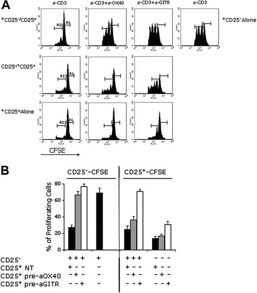
Since T reg's are able to proliferate in response to anti-CD3 and IL-2 (Thornton et al37 and data not shown) we felt that it was important to clarify which T-cell subsets were proliferating in our inhibition assay. T reg's were pretreated with anti-OX40 or anti-GITR mAbs and mixed with effector T cells. To follow the proliferation of the 2 populations of cells in the culture we alternatively labeled them with CFSE and then monitored the dilution of cell-associated fluorescence by flow cytometry. The results of a representative experiment and the cumulative data from four different experiments are shown in Figure 3. The effect of anti-OX40 or anti-GITR on the proliferation of CD4+CD25- cells could be readily observed in these experiments. Whereas anti-GITR also induced a modest proliferative response in CD4+CD25+ cells, no such response was induced by anti-OX40. However, both molecules, by inhibiting T reg–suppressive function, restore the ability of effector T cells to respond to TCR triggering.
Anti-GITR mAb but not anti-OX40 mAb induces the proliferation of CD4+CD25+ T reg's. (A) CD4+CD25- cells (1 × 10⁵) cultured with CD4+CD25+ (5 × 10⁴) cells were stimulated with 1 μg/mL of anti-CD3 and ACs (1 × 10⁵) and seeded in a 96-well plate in triplicates. After 66 hours, the triplicates were pooled and analyzed by flow cytometry. Where indicated, CD4+CD25+ cells were preincubated with anti-OX40 or anti-GITR mAbs (both at 30 μg/mL), washed twice, and added to the CD4+CD25- cells. *CFSE-labeled cells in the coculture. R1 indicates nondividing cells, whereas R2 indicates proliferating cells. The same experiment in panel A was repeated 4 times and the results, expressed as percentage of proliferating cells (R2), were pooled and reported in panel B. In the left part of panel B is reported the percentage (±SD) of CFSE-labeled CD25- proliferating T cells in the presence or absence of CD25+ T cells left untreated or pretreated with anti-OX40 or anti-GITR mAbs, as indicated. In the right part of panel B is reported the percentage of proliferating CFSE-labeled CD25+ T cells. After labeling with CFSE, CD25+ T cells were left untreated (NT) or were preincubated with anti-OX40 or anti-GITR mAbs, and were washed twice before seeding in the presence or absence of CD25- T cells, as indicated.
Anti-GITR mAb but not anti-OX40 mAb induces the proliferation of CD4+CD25+ T reg's. (A) CD4+CD25- cells (1 × 10⁵) cultured with CD4+CD25+ (5 × 10⁴) cells were stimulated with 1 μg/mL of anti-CD3 and ACs (1 × 10⁵) and seeded in a 96-well plate in triplicates. After 66 hours, the triplicates were pooled and analyzed by flow cytometry. Where indicated, CD4+CD25+ cells were preincubated with anti-OX40 or anti-GITR mAbs (both at 30 μg/mL), washed twice, and added to the CD4+CD25- cells. *CFSE-labeled cells in the coculture. R1 indicates nondividing cells, whereas R2 indicates proliferating cells. The same experiment in panel A was repeated 4 times and the results, expressed as percentage of proliferating cells (R2), were pooled and reported in panel B. In the left part of panel B is reported the percentage (±SD) of CFSE-labeled CD25- proliferating T cells in the presence or absence of CD25+ T cells left untreated or pretreated with anti-OX40 or anti-GITR mAbs, as indicated. In the right part of panel B is reported the percentage of proliferating CFSE-labeled CD25+ T cells. After labeling with CFSE, CD25+ T cells were left untreated (NT) or were preincubated with anti-OX40 or anti-GITR mAbs, and were washed twice before seeding in the presence or absence of CD25- T cells, as indicated.
Inhibition of T reg suppression by anti-OX40 and anti-GITR mAbs restores IL-2 production by effector T cells
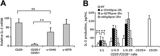
The molecular mechanism explaining T reg–mediated suppression is the blockage of IL-2 gene transcription in CD4+CD25- effector T cells.2 Accordingly, exogenous IL-2 inhibits T reg suppression without inducing IL-2 gene transcription in CD4+CD25- T cells, and allows proliferation of both CD4+CD25- and CD4+CD25+ cells,38 underlying the critical role of IL-2 in effector–T reg function and interaction. We measured IL-2 production during the classic inhibition assay using T reg's that had been preincubated or not with anti-OX40 or anti-GITR mAbs. Reverse transcription (RT)–PCR and ELISA data (Figure 4) show that inhibition of T reg suppression induced by anti-OX40 or anti-GITR mAbs completely restores IL-2 gene transcription and secretion by CD4+CD25-. These data therefore confirm that OX40 triggering on T reg's, like triggering through GITR, inhibits the mechanism that T reg's use to mediate suppression.
Inhibition of T reg's by anti-OX40 and anti-GITR mAbs restores IL-2 production by effector T cells. (A) CD4+CD25+ (CD25+; 5 × 10⁴ cells) were preincubated or not with anti-OX40 (a-OX40) or anti-GITR mAb (a-GITR), both at 30 μg/mL, and washed twice before adding CD4+CD25- (CD25-; 1 × 10⁵ cells) and 1 μg/mL of anti-CD3 plus ACs (1 × 10⁵ cells) in 30 wells of a 96-well plate for every experimental condition. After 72 hours, cells were pooled to isolate mRNA and to perform semiquantitative PCR. Relative IL-2 mRNA levels are reported in percentages of the amount of mRNA produced by CD4+CD25- cells stimulated with anti-CD3. Data shown are the mean (±SD) of 2 independent experiments (**P < .05; ns indicates nonstatistically different). (B) CD4+CD25- (5 × 10⁴) cells were cultured at different ratio with CD4+CD25+ cells preincubated or not with anti-OX40 (a-OX40), anti-GITR mAb (a-GITR), or the relative isotype control (all at 30 μg/mL), in the presence of anti-CD3 (1 μg/mL) and ACs (5 × 10⁴). After 60 hours, supernatants were collected and IL-2 content was measured by ELISA. Results are from 1 representative out of 3 independent experiments. The significance of the data was evaluated by Student t test (**P < .01).
Inhibition of T reg's by anti-OX40 and anti-GITR mAbs restores IL-2 production by effector T cells. (A) CD4+CD25+ (CD25+; 5 × 10⁴ cells) were preincubated or not with anti-OX40 (a-OX40) or anti-GITR mAb (a-GITR), both at 30 μg/mL, and washed twice before adding CD4+CD25- (CD25-; 1 × 10⁵ cells) and 1 μg/mL of anti-CD3 plus ACs (1 × 10⁵ cells) in 30 wells of a 96-well plate for every experimental condition. After 72 hours, cells were pooled to isolate mRNA and to perform semiquantitative PCR. Relative IL-2 mRNA levels are reported in percentages of the amount of mRNA produced by CD4+CD25- cells stimulated with anti-CD3. Data shown are the mean (±SD) of 2 independent experiments (**P < .05; ns indicates nonstatistically different). (B) CD4+CD25- (5 × 10⁴) cells were cultured at different ratio with CD4+CD25+ cells preincubated or not with anti-OX40 (a-OX40), anti-GITR mAb (a-GITR), or the relative isotype control (all at 30 μg/mL), in the presence of anti-CD3 (1 μg/mL) and ACs (5 × 10⁴). After 60 hours, supernatants were collected and IL-2 content was measured by ELISA. Results are from 1 representative out of 3 independent experiments. The significance of the data was evaluated by Student t test (**P < .01).
Contrary to GITR, OX40 triggering does not revert suppression when T reg's are activated
Naive T reg's such as those in all of the experiments described can be expanded in vitro with anti-CD3 plus IL-2.39 This type of expansion increases the expression of costimulatory molecules on T reg's15 and enhances their capacity to suppress.37 Nonetheless, expanded T reg's can still be inhibited by GITR triggering,15 prompting the question of whether they also retain sensitivity to inhibition by anti-OX40. To answer this question, we treated T reg's for 3 days with anti-CD3 plus IL-2 before monitoring their capacity to suppress. mAbs to OX40 or GITR were added in these experiments as follows: (1) during the 3-day activation phase (referred to as Act+Ab 3 days); (2) for 2 hours to washed preactivated T reg's before they were added to effector T cells (Act+Ab; 2 hours); (3) to washed pre-activated T reg's at the time of mixing with CD4+CD25- effector cells (Act then Ab). In all 3 of the conditions anti-OX40 was unable to block T reg–mediated suppression (Figure 5A). Similarly, the anti-GITR mAb was unable to inhibit T reg suppression when it was given during the preactivation period (condition 1). When it was given after withdrawal of IL-2, either transiently (condition 2) or for the duration of the inhibition assay (condition 3), anti-GITR could block suppression mediated by the expanded T reg cells (Figure 5B). These data therefore reveal a clear difference in the effect of anti-GITR and anti-OX40 with the former being effective at inhibiting suppression by either naive or expanded T reg's, while the latter blocks only naive T reg's.
GITR but not OX40 triggering is able to revert suppression of activated T reg's. (A) CD4+CD25+ cells were purified and cultured in the presence of anti-CD3 (1 μg/mL), IL-2 (20 U/mL), and irradiated splenocytes for 3 days. mAbs to OX40 were added during the 3-day culture (Act+Ab 3 days; ▪) to washed, preactivated T reg's for 2 hours and then rewashed before adding effector T cells (Act+Ab 2hr; ▦) to washed, preactivated T reg's together with CD4+CD25- effector cells (Act then Ab; ▨). The CD25-/CD25+ ratio is indicated. (B) The same experiment as in panel A was performed using anti-GITR mAb in place of anti-OX40 mAb. Proliferations were measured after 72 hours and pulsed with 3[H]TdR for the last 10 hours. Shown are the means (±SD) from 1 representative out of 3 independent experiments. The significance of the data was evaluated by Student t test (**P < .01).
GITR but not OX40 triggering is able to revert suppression of activated T reg's. (A) CD4+CD25+ cells were purified and cultured in the presence of anti-CD3 (1 μg/mL), IL-2 (20 U/mL), and irradiated splenocytes for 3 days. mAbs to OX40 were added during the 3-day culture (Act+Ab 3 days; ▪) to washed, preactivated T reg's for 2 hours and then rewashed before adding effector T cells (Act+Ab 2hr; ▦) to washed, preactivated T reg's together with CD4+CD25- effector cells (Act then Ab; ▨). The CD25-/CD25+ ratio is indicated. (B) The same experiment as in panel A was performed using anti-GITR mAb in place of anti-OX40 mAb. Proliferations were measured after 72 hours and pulsed with 3[H]TdR for the last 10 hours. Shown are the means (±SD) from 1 representative out of 3 independent experiments. The significance of the data was evaluated by Student t test (**P < .01).
OX40 triggering on T reg's reverts their suppressive activity in vivo
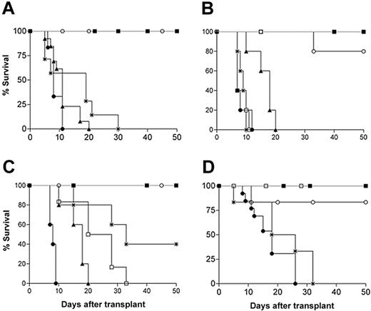
To explore the influence of OX40 on T reg function in additional detail, we turned to an in vivo setting. The depletion of CD4+CD25+ from donor T cells exacerbates GVHD induced by allogeneic bone marrow transplantation.40 If anti-OX40 interferes with T reg suppression, then it too should aggravate GVHD. To test this hypothesis, we transferred T-cell–depleted bone marrow cells together with freshly purified CD4+CD25- and CD4+CD25+ T cells from C57BL/6 mice into irradiated BALB/c mice that also received an intraperitoneal injection of 600 μg of anti-OX40 or anti-GITR mAbs. Whereas the transfer of T reg's together with donor bone marrow cells protected the recipient mice from lethal GVHD, this protection was lost when the mice were treated with anti-OX40 or anti-GITR mAbs immediately after transplantation. Strikingly, treatment with anti-OX40 seemed to be more effective than anti-GITR based on the complete absence of any evidence of T reg protection in the anti-OX40–treated mice. A dose-response experiment not only confirmed that anti-OX40 mAb was more effective then anti-GITR in worsening GVHD lethality but also showed an apparent paradoxical effect of anti-GITR that ameliorates GVHD effects at the highest dose of 1200 μg (Figure 6B-C).
OX40 or GITR triggering on T reg's impedes T reg–mediated inhibition of GVHD. (A) Lethally irradiated BALB/c mice were reconstituted with 2 × 10⁵ C57BL/6 T-cell–depleted BM cells in the absence (▪) or presence (▴) of 5 × 10⁵ CD4+CD25- cells. Some mice also received 3 × 10⁵ CD4+CD25+ cells and were left untreated (○) or injected intraperitonally with 600 μg of anti-OX40 (•) or anti-GITR (*) mAb immediately after transplantation (6 mice/group). The same experiment was repeated as in panel A, treating the mice intraperitonally with 300 μg(•), 600 μg(□), or 1200 μg (*) of anti-OX40 (B) or anti-GITR antibody (B-C). Results are shown in 2 separate panels (B-C) for clarity. Additional experiments were performed (D) in which mAbs were given to T reg's in vitro with anti-OX40 (•) or anti-GITR (*) mAbs (30 μg/mL) and washed before their injection with BM and effector T cells: 2 groups of T reg's pretreated with control isotype rat IgG1 (□) or rat IgG2a (○) were added (6 mice/group). All mice were monitored daily for signs of GVHD and lethality was recorded. One representative experiment, for both in vivo and in vitro treatment with mAbs, out of 2 with similar results is shown.
OX40 or GITR triggering on T reg's impedes T reg–mediated inhibition of GVHD. (A) Lethally irradiated BALB/c mice were reconstituted with 2 × 10⁵ C57BL/6 T-cell–depleted BM cells in the absence (▪) or presence (▴) of 5 × 10⁵ CD4+CD25- cells. Some mice also received 3 × 10⁵ CD4+CD25+ cells and were left untreated (○) or injected intraperitonally with 600 μg of anti-OX40 (•) or anti-GITR (*) mAb immediately after transplantation (6 mice/group). The same experiment was repeated as in panel A, treating the mice intraperitonally with 300 μg(•), 600 μg(□), or 1200 μg (*) of anti-OX40 (B) or anti-GITR antibody (B-C). Results are shown in 2 separate panels (B-C) for clarity. Additional experiments were performed (D) in which mAbs were given to T reg's in vitro with anti-OX40 (•) or anti-GITR (*) mAbs (30 μg/mL) and washed before their injection with BM and effector T cells: 2 groups of T reg's pretreated with control isotype rat IgG1 (□) or rat IgG2a (○) were added (6 mice/group). All mice were monitored daily for signs of GVHD and lethality was recorded. One representative experiment, for both in vivo and in vitro treatment with mAbs, out of 2 with similar results is shown.
As for in vitro experiments, to distinguish Ab's effect on T reg's from effector T cells, we pretreated T reg's for 2 hours with anti-OX40 or anti-GITR mAbs before cotransferring them with mismatched bone marrow cells into irradiated recipients that received no antibody injections. Despite the short length of this pretreatment, both antibodies blocked the suppressive activity of the T reg's as shown by the failure of the treated cells to prevent GVHD (Figure 6D). Cumulatively, these in vivo experiments substantiate the importance of both OX40 and GITR in the control of T reg activity.
Discussion
In this paper we show that the engagement of OX40 on naive T reg's confers a similar effect to that of GITR engagement in limiting the capacity of these cells to mediate suppression. A previous study18 describing OX40 expression on T reg's pointed to its possible functional dual role: one to mask OX40L expressed on DCs, rendering OX40L unavailable for effector T-cell costimulation; the other to control T reg homeostasis, because mice lacking OX40 were found to have reduced numbers of CD4+CD25+ T cells early in life. In that study, however, the effect of OX40 triggering on T reg's was missed because authors used T reg's from OX40-/- mice. In addition, the reciprocal combination in which wild-type T reg's were mixed with OX40 null–CD4+CD25- T cells, which do not respond to anti-CD3 stimulation,26 was not testable. Not conflicting with our results, the wild-type CD4+CD25- T-cell proliferation in the presence of an excess of anti-OX40 mAb was likely due to a direct costimulation of CD4+CD25- T cells as we show in Figure 1. Besides differences in Ab concentration or effector–T reg ratio, all titrated in our study, through the use of rat effector T cells that could not be engaged by anti-OX40 mAb, and through a T reg pretreatment regimen, in which free anti-OX40 was removed before exposing the cells to CD4+CD25- effectors, we have shown that T reg's can be directly targeted by the Ab. Cumulatively, the data presented here show that OX40 signaling in T reg's can have a profound impact on T reg suppression and tolerance. Moreover, differences in T reg numbers observed by Takeda et al18 was not readily apparent in the OX40-/- mice that we and others have examined (Tang et al12 and data not shown), but it is possible that genetic background differences and/or environmental factors may have obscured a subtle effect. We note, however, that OX40-/- mice do not develop spontaneous autoimmune disease, suggesting that T reg homeostasis and function are not grossly disturbed by the deficiency.12,18 Additional experiments involving the transfer of OX40-null T cells into T-cell–deficient mice are warranted as a further test of how the absence of OX40 might compromise either the homeostasis or the suppressive functions of T reg's. Transfer experiments of this sort have previously proven useful in revealing defects in the suppressive activity of T reg's isolated from CD28-/- or CD40-/- mice.12,41
We observed a marked difference between T reg's collected from naive mice and T reg's that had been activated in vitro with anti-CD3 plus IL-2. Whereas suppression by the former was inhibited by anti-OX40, the antibody had no detectable effect on T reg's expanded in vitro. During culture with anti-CD3 plus IL-2, T reg's are unresponsive to inhibition by either OX40 or GITR triggering; however, GITR but not OX40 inhibition could be restored by IL-2 withdrawal. At this point, just as it is unclear how triggering through these 2 molecules inhibits suppression by naive T reg's, it is also unclear why preculture in IL-2 would eliminate their sensitivity to anti-OX40. However, T reg's that are activated in vivo are unlikely to be exposed to the high concentrations of IL-2 that we and others have used in preactivation protocols.42 The differences between the 2 molecules, however, are independent from their surface expression, since activated T reg's up-regulate both OX40 and GITR at the same level (McHugh et al15 and data not shown). A clue to test whether T reg's retain their sensitivity to inhibition by OX40 triggering in physiologic conditions was indeed through the GVHD experiments. In these experiments, irradiated BALB/c mice developed lethal GVHD when reconstituted with a mixture of T-cell–depleted bone marrow and purified CD4+CD25- from C57BL/6 donors. While the cotransfer of CD4+CD25+ T reg's prevented lethal GVHD, the mice succumbed to disease if the T reg's were pretreated with anti-OX40 mAb for 2 hours before transfer, or if they received a single intraperitoneal injection of anti-OX40 mAb at the time of transplantation. The results of pretreatment experiments in particular show that triggering OX40 on T reg's rather than effector cells blocks the capacity of these cells to suppress the disease.
We have completely excluded a possible involvement of complement or antibody-mediated cell cytotoxicity (ADCC) in killing in vivo T reg's precoated in vitro, since CFSE-labeled T reg's either pretreated with OX86 or not can be recovered at the same number from recipient syngenic mice 15 days after inoculation (data not shown). We also performed a GVHD experiment using the F(ab′)2 fragment of the anti-OX40 mAb and as for the whole Ab we observed the same accelerated GVHD lethality (data not shown). Several publications regarding in vivo injection of Ab to OX40 or GITR speaks against the possibility of Ab-mediated killing of cells expressing such molecules.14,27,29,43,44
Although anti-OX40 mAb inhibited T reg's at any tested dose, anti-GITR mAb was effective at the lower doses, whereas at the highest it had the opposite effect on GVHD. This contrasting dose-effect is explained by a recent demonstration that GITR-triggers of allostimulated CD4+CD25- effector T cells has such a strong costimulatory activity to induce activation-induced cell death (AICD) via Fas (CD95).44 We hypothesized that low doses (300-600 μg) of anti-GITR mAb are insufficient to induce AICD of CD4+CD25- T cells but enough to trigger CD4+CD25+ T reg's that already express high levels of GITR at the naive state.
Faster up-regulation of GITR molecules than OX40 molecules on CD4+CD25- T cells upon TCR activation might explain why the former induces stronger cell proliferation than the latter. This explanation is unlikely to be germane, however, in the case of CD4+CD25+ T reg's because significant numbers of these cells express both molecules prior to intentional activation in vitro. Thus, it seems likely that the signals delivered by GITR and OX40 to T reg's are not equivalent even though both types of signals can apparently induce T reg inactivation. In search of evidence for differences in the 2 signals, we have examined CTLA-4 and winged helix/forkhead transcription factor (Foxp3) expression levels in T reg's exposed to anti-OX40 or anti-GITR mAbs, but have not discovered any differences (data not shown).
Recently, it has been demonstrated that conditions of low costimulation and low antigen concentrations (eg, as might typically be the case for antigen presentation by immature nonactivated DCs) are favorable for suppression by T reg's.45,46 OX40L and GITRL are mainly expressed by activated DCs,20,47 and the expression of OX40L in particular seems to be dependent on prior CD40 triggering.48 Our results suggest that the low levels of OX40L and GITRL on immature DCs would be conducive to suppression by T reg's, while activation of the DCs leading to up-regulation of these ligands would allow for inhibition of suppression through the OX40 and GITR molecules expressed on the T reg's. Consistent with this, blockade of CD40-CD40L may induce tolerance in alloreactive T cells,49 perhaps because it inhibits OX40L acquisition by DCs and therefore indirectly potentiates T reg activity. Our data suggest that Treg–mediated suppression in the absence of OX40 triggering is highly active, and offer additional explanation of several papers showing that blockade of OX40-OX40L interaction ameliorates the symptoms of autoimmune disease such as IBD and EAE.33,35 In tumors, OX40 ligation by means of OX40L-Ig fusion protein,50 agonist antibody,50 or OX40L gene transduction51 has been found to stimulate antitumor immunity; we speculate that such an effect might also result from inhibition of T reg function.
In conclusion, the data presented here support a crucial role for OX40 signaling in the control of tolerance induced by T reg's. Although OX40 signaling has been shown to enhance T-cell survival and memory T-cell numbers,25 the new data show that it also has a striking capacity to block the suppressive functions of T reg's through a direct mechanism. Understanding the details of how OX40 signaling abrogates T reg suppression may provide a useful avenue for exploring the broader problem of how suppression itself is manifest. Moreover, it is possible that additional work on OX40 in T reg's may lead to the development of new strategies for augmenting or decreasing suppression in situations where such manipulations might afford therapeutic benefit.
Prepublished online as Blood First Edition Paper, December 9, 2004; DOI 10.1182/blood-2004-07-2959.
Supported by Associazione Italiana ricerca sul Cancro (AIRC), and by Fondo per gli Investimenti della Ricerca di Base (FIRB) code RBNE017B4C. B.V. is supported by a fellowship from Federazione Italiana Ricerca sul Cancro (FIRC).
B.V. and C.G. equally contributed to this work.
The publication costs of this article were defrayed in part by page charge payment. Therefore, and solely to indicate this fact, this article is hereby marked “advertisement” in accordance with 18 U.S.C. section 1734.
We thank Mariella Parenza and Ivano Arioli for technical assistance. We gratefully acknowledge Elena Luison and Silvana Canevari for helping in F(ab′)2 fragment preparation.

![Figure 1. OX40 inhibits T reg–suppressive activity. Naive CD4+CD25- (5 × 10⁴) and CD4+CD25+ T cells (named as CD25- and CD25+, respectively) were purified from BALB/c mice and cultured at the indicated ratio together or alone. Cells were cultured in the presence of 5 μg/mL of anti-OX40 (A), anti-GITR (B), left untreated (NT), or treated with the respective isotype controls rat IgG1 (A) and rat IgG2a (B) mAbs. In a dose-response assay, CD25- T cells (5 × 10⁴) were cultured (C) or not (D) with CD25+ T cells (2.5 × 10⁴) at different concentrations of anti-CD3 Ab as indicated with or without (NT) anti-OX40 or anti-GITR Ab (a-OX40 or a-GITR, respectively). In another dose-response assay, CD25- (5 × 10⁴) T cells were cultured with CD25+ T cells at the indicated ratio with or without (NT) anti-OX40 (E) or anti-GITR (F) mAbs at variable concentration, as indicated. All samples were stimulated with 1 μg/mL of anti-CD3 and irradiated accessory cells (5 × 10⁴); proliferation was measured after 72 hours. 3[H]TdR pulse was during the last 10 hours. One representative experiment out of 3 performed in triplicate is shown as mean ± standard deviation (SD). The significance of the data was evaluated by Student t test (***P < .005).](https://ash.silverchair-cdn.com/ash/content_public/journal/blood/105/7/10.1182_blood-2004-07-2959/6/m_zh80070576200001.jpeg?Expires=1767696364&Signature=hXqobxJUTmQbvZIOVypVz1VbwLuy4B97x4MroZO-BPl3-VoGMCE3P6hRbn0cYg7nwodCYnTcA-f3QvgthYO6gQGiBhUvANDps-Q2VupUn-Qgy10lEL0NmpcoSsqyTV7GKlXLEoLJk3JV-bffPociHTPvCz5yuVPrD3PW43Pp5O76pVvJssYOtxU0x0Itbok8s06aPH8FLHdY1uJrufoHLZ6jntU7audh9Ry6EgBgmiYm6MMqXThww6ziU3VHaDjnO3JqbEg8hFFv6hN0AyJfZ7ORtB7~2QvIL-BhB-TAYizXAH0TdVa3uVrm2dTPLKi9GgJ9WWwNO5Uw6g3wLB4j6Q__&Key-Pair-Id=APKAIE5G5CRDK6RD3PGA)
![Figure 2. OX40 specifically triggers T reg's. (A) CD4+ lymphocytes from normal BALB/c mice or Wistar rats were stained with biotin anti–mouse OX40, biotin anti–rat OX40 (thin lines) or the relative biotin isotype control (filled histograms) and FITC-streptavidin. (B) CD4+CD25- (5 × 10⁴) or CD25+ (2.5 × 10⁴) T cells from BALB/c mice were cultured with irradiated rat accessory cells (ACs) in the presence or absence of anti-OX40 mAb (5 μg/mL; left panel). Rat CD4+ T cells (5 × 10⁴) and mouse CD4+CD25+ (2.5 × 10⁴) T cells were cultured with irradiated rat ACs in the presence or absence of anti-OX40 mAb as indicated (right panel). (C) CD25- T cells (5 × 10⁴) cultured with CD25+ T cells at the indicated ratio were stimulated with ACs in the presence or absence (NT) of anti-OX40 antibody (5 μg/mL; ▪); where indicated, CD25+ T cells were preincubated for 2 hours with 30 μg/mL of anti-OX40 mAb (dark gray bars) or their respective isotype control rat IgG1 or rat IgG2a (light gray bars), washed 2 times with PBS and added to CD25- T cells. (D) Cells were prepared and treated as in panel C, except that anti-GITR mAb (5 μg/mL; ▪) was added to coculture or preincubated (30 μg/mL; dark gray bars) or with purified T reg's. All samples were stimulated with 1 μg/mL of anti-CD3 in the presence of ACs. Proliferation was measured after 72 hours and pulsed with 3[H]TdR for the last 10 hours. Data (mean ± SD) are from 1 of 3 independent experiments with similar results. The significance of the data was evaluated by Student t test (**P < .01).](https://ash.silverchair-cdn.com/ash/content_public/journal/blood/105/7/10.1182_blood-2004-07-2959/6/m_zh80070576200002.jpeg?Expires=1767696364&Signature=uv3D7UbZ4TlrF15GjWY2esoqycHaOUclvklKaWD6U6aZFfzBJmhir8j0WGA5fAzLV7Mnoy28myGPfEtGaH8glKA3yJoHeLzQn5-9TQaT4NghFES79BGo~Ld56FS86Vd1D1xDDOqyHU5rXKy~02fMWHGXGMTXmGp5EJpZLbpXDcr74yhii9p8agi9glIImZZtgSmT1pt29VjLwP9eWoDDA0uFq6IebD12UzmgePmzF4~DUz~7puLChsJ-mUG1RyjrEIVweKES8YHxB7qeZjrqBGkVd4ciBA47AEhiw~wbNcN9M3zfyRm~VuZXVV7aqPuS1QdUe7R8bmzGUQ3Rx7d69Q__&Key-Pair-Id=APKAIE5G5CRDK6RD3PGA)


![Figure 5. GITR but not OX40 triggering is able to revert suppression of activated T reg's. (A) CD4+CD25+ cells were purified and cultured in the presence of anti-CD3 (1 μg/mL), IL-2 (20 U/mL), and irradiated splenocytes for 3 days. mAbs to OX40 were added during the 3-day culture (Act+Ab 3 days; ▪) to washed, preactivated T reg's for 2 hours and then rewashed before adding effector T cells (Act+Ab 2hr; ▦) to washed, preactivated T reg's together with CD4+CD25- effector cells (Act then Ab; ▨). The CD25-/CD25+ ratio is indicated. (B) The same experiment as in panel A was performed using anti-GITR mAb in place of anti-OX40 mAb. Proliferations were measured after 72 hours and pulsed with 3[H]TdR for the last 10 hours. Shown are the means (±SD) from 1 representative out of 3 independent experiments. The significance of the data was evaluated by Student t test (**P < .01).](https://ash.silverchair-cdn.com/ash/content_public/journal/blood/105/7/10.1182_blood-2004-07-2959/6/m_zh80070576200005.jpeg?Expires=1767696364&Signature=HM~JjZPi1cvR2EL7cVixluZoHi6GiEwkijHzL0r45DYLsaX3xChexhxZwJUAQjzQnPBGnM32gSK9N1Nu7izLzB9Ob2qYNv6bmPN4Y60cLP2r0nz-qt6EYh4lUeox0sqS6GQ2Oxax9I6eFXHBLb2Dhh3YDJR6RZrC8OV0HKAcxySigAr-esNxgSZgCc-gfW3pSk0RBfx3iT7FOLse5-GVmRlf1K9tQd-1if~ZEGPPlpyp1skmIcX0xluIw6AkBZiuWZTMGKCZiNVbvrfW-wXFwMyFJsbv1XVpO0dOZztYauFC~VN5ljA8YVdxBLeAZgVJOqA-xMNbbnJRwjjHQF0jig__&Key-Pair-Id=APKAIE5G5CRDK6RD3PGA)

This feature is available to Subscribers Only
Sign In or Create an Account Close Modal