Abstract
B-cell activation and differentiation is regulated through the coordinated function of a dynamic array of cell surface receptors. At different stages in their differentiation, human B cells may express one or more members of a large family of immunoglobulin Fc receptor homologs (FcRH) with regulatory potential. Among these newly identified transmembrane molecules, FcRH1 is unique in having 2 immunoreceptor tyrosine-based activation motif (ITAM)–like motifs in its intracellular domain. Here we used the Fab fragments of new monoclonal anti-FcRH1 antibodies and mRNA analysis to evaluate FcRH1 expression and function during B-cell differentiation. FcRH1 expression begins in pre-B cells, reaches peak levels on naive B cells, and is down-regulated after B cells are activated to begin to form germinal centers. This FcRH1 down-regulation coincides with dramatic enlargement of the pre-germinal center cells, cell cycle entry, and other overt signs of activation that include CD80 and CD86 up-regulation and immunoglobulin D (IgD) down-regulation. In vitro analysis indicates that ligation of FcRH1 leads to its tyrosine phosphorylation and to modest B-cell activation and proliferation. Concomitant FcRH1 ligation enhances B-cell antigen receptor (BCR)–induced Ca2+ mobilization and proliferation. FcRH1 thus has the potential to serve as an activating coreceptor on B cells.
Introduction
Receptors for the Fc portion of immunoglobulins (FcRs) may differentially modulate both cellular and humoral immune responses depending upon their immunoglobulin isotype-binding specificity and the type of cells that bear them.1,2 The FcRs often pair with adaptor transmembrane proteins that possess immunoreceptor tyrosine-based activation motifs (ITAMs) or, alternatively, may possess either ITAMs or immunoreceptor tyrosine-based inhibitory motifs (ITIMs) in their cytoplasmic domain. After FcR ligation by antibodies complexed with antigen, tyrosines in the ITAM or ITIM are phosphorylated by Src family kinases to engage Src homology region 2 (SH2)–containing molecules and other downstream signaling components in the cellular response cascade.3-6
A recently identified family of Fc receptor homolog (FcRH) genes are located within the FcR locus on chromosome 1 in humans.7-14 At the transcript level, the FcRH genes are differentially expressed by B-lineage cells and overexpressed in some B-cell malignancies.7-14 The predicted FcRH1-5 transmembrane glycoproteins contain consensus ITIM, ITAM-like, or both types of motifs. FcRH1, the subject of this study, has 2 ITAM-like motifs, an acidic glutamic acid residue in its transmembrane region, and 3 extracellular immunoglobulin-like domains. Ligands for FcRH1 have not been identified, but the presence of ITAM-like motifs suggests its potential for B-cell activation. To begin examining the function of FcRH1, we have produced anti-FcRH1–specific monoclonal antibodies and used Fab fragments of these in conjunction with mRNA analysis to determine when FcRH1 is expressed during B-cell differentiation. After determining that FcRH1 is preferentially expressed on naive and memory B cells, we examined the functional consequences of FcRH1 ligation in B-cell differentiation. The findings obtained in these studies indicate that FcRH1 has the potential to serve as an activating coreceptor on B cells.
Materials and methods
Cells
Human and mouse cell lines were cultured in RPMI-1640 medium containing 100 U/mL penicillin, 100 mg/mL streptomycin, 2 mM l-glutamine, and 10% fetal calf serum (Life Technologies, Grand Island, NY). Human blood samples, tonsils, and rib sections were obtained in accordance with policies established by the University of Alabama at Birmingham (UAB) Institutional Review Board and with informed consent according to the Declaration of Helsinki. Mononuclear cells in these tissues were isolated by Ficoll-Hypaque gradient centrifugation. Naive B cells in tonsil samples were purified to more than 90% purity by depletion of CD10+, CD27+, CD38+, CD3+, and CD14+ cells using monoclonal antibodies, antibody conjugated microbeads, or goat anti–mouse immunoglobulin G (IgG)–conjugated microbeads (Miltenyi Biotec, Auburn, CA). Stained cells were analyzed on a FACSCalibur flow cytometer (BD Biosciences, Mountain View, CA) and plotted using WINMDI software (Scripps Institute, La Jolla, CA).
Production of monoclonal anti-FcRH1 antibodies
Balb/c mice hyperimmunized with baculovirus-derived recombinant FcRH1 extracellular region protein (10 μg/injection) were boosted with Daudi, Ramos, and Raji cells on the day before fusion of regional lymph node cells with the Ag8.653 plasmacytoma cell line.15 Hybridoma supernatants were screened by enzyme-linked immunosorbent assay (ELISA) for anti-FcRH1 antibody activity before testing for immunofluorescence reactivity with B-cell lines and Western blotting of recombinant FcRH1 to FcRH5 proteins. Hybridomas producing anti-FcRH1–specific antibodies were subcloned by limiting dilution, and the antibody isotype was determined by an indirect capture ELISA (Zymed, San Francisco, CA).
Western blot analysis
Baculoviral-derived recombinant FcRH1 to FcRH5 ectodomains were made by Dr Peter Snow of the Protein Expression Facility, California Institute of Technology, PaloAlto, CA. Recombinant FcγR proteins were the gifts of Drs Katsumi Maenaka (Kyushu University, Fukuoko, Japan) and Peter Sondermann (GLYCART Biotechnology Ag, Schlieren, Switzerland).16 Recombinant FcRH and control proteins (1 μg each) were resolved by sodium dodecyl sulfate–polyacrylamide gel electrophoresis (SDS-PAGE) and transferred onto nitrocellulose membranes. Antibody reactivity was assessed by incubation of these protein-loaded membranes with test antibodies (3 μg/mL) and horseradish peroxidase–labeled goat anti-mouse immunoglobulin antibody (1:5000 dilution; Dako, Glostrup, Denmark). Antigen-antibody reactivity was visualized by enhanced chemiluminescence (Amersham Life Science, Braunschweig, Germany). A HA-FcRH1 chimeric receptor overexpressing IIA1.6 cell line was established by methods previously described.17 Before and after FcRH1 ligation by anti-FcRH1 antibody, cell lysate (200 μg) was immunoprecipitated with anti-hemagglutinin (HA) antibody (Roche Diagnostics, Mannhein, Germany), and the immunoprecipitates were immunoblotted with either anti-phosphotyrosine antibody (4G10; Upstate Biotechnologies, Lake Placid, NY) or anti-FcRH1 antibody.
Immunofluorescence cell sorting and real-time PCR analysis of FcRH1 transcript expression
Tonsillar B-cell subpopulations were purified by immunofluorescent cell sorting with a MoFlow instrument (Cytomation, Fort Collins, CO) as follows: naive cells (CD27–CD38–IgD+CD19+), pre-GC cells (CD38+IgD+CD19+), centroblasts (CD77+CD38+CD19+), centrocytes (CD77–CD38+CD19+), memory B cells (CD27+CD38–CD19+), and plasma cells (CD38++IgD–CD19+). Sorted cells were lysed in TRIzol reagent (Gibco, Grand Island, NY) before preparation of total RNA and first-stand cDNA synthesis using Superscript II system (Invitrogen, Carlsbad, CA). After inactivating the reactions at 50°C for 2 minutes, real-time polymerase chain reaction (PCR) was performed by using SYBR Green PCR Master Mix (Applied Biosystems, Foster City, CA) denaturing at 95°C for 10 minutes, amplification for 40 cycles at 95°C for 15 seconds, annealing and extension at 60°C for 1 minute using an ABI Prism 7900 HT sequence detection system (Applied Biosystems). FcRH1 gene-specific primers used for PCR amplification were 5′-AGGAGATCCCAGATAAATGTG-3′ and 5′-CTGTGCCCATAGCAACTGAG-3′.
Transient transfectants
Full-length FcRH1 to FcRH5 cDNAs were ligated into the pEGFP-N1 mammalian expression vector (Clontech, Palo Alto, CA). Purified plasmid (5 μg) was transfected into the human 293T cell line using Lipofectamine reagent (Invitrogen). Transfectants were harvested at 48 hours and stained for reactivity with FcRH1 antibodies. FcRH2 to FcRH5 surface expression was confirmed by reactivity with FcRH-specific antibodies (data not shown).
Antibodies, immunofluorescence reagents, and chemicals
Fluorescein isothiocyanate (FITC)–conjugated anti-human CD3, CD27, and CD34 antibodies; phycoerythrin (PE)–conjugated anti–human CD19, CD3, CD14, CD56, CD38, and IgD antibodies; and allophycoreythrin (APC)–conjugated anti–human CD34 and CD19 antibodies were purchased from Becton Dickinson (Mountain View, CA). Streptavidin-PE, streptavidin-APC, streptavidin, FITC-conjugated anti–human IgM, and FITC-conjugated anti–rat immunoglobulin antibodies were from Southern Biotechnology Associates (Birmingham, AL). Monoclonal anti–human CD77 antibody was from Coulter/Immunotech (Marseille Cedex, France). Immobilized pepsin and sulfo-NHS-LC-biotin were obtained from Pierce (Rockford, IL).
B-cell activation, cell cycle status, and proliferation assays
B cells purified from tonsils were incubated in 96-well plates (105/well in 200 μL RPMI supplemented with 10% fetal calf serum [FCS]) for 72 hours in the presence or absence of biotinylated Fab fragments of anti-FcRH1 mAbs with 20 μg/mL streptavidin. Cells pulsed for an additional 16 hours with 3H-thymidine (1 μCi/well [0.037 MBq/well]) were then harvested, and 3H-thymidine incorporation was assessed with a liquid scintillation counter. Cell surface expression of IgD, IgM, CD69, CD80, and CD86 assessed before and after naive B cells (5 × 10⁵/24 wells) were incubated for 48 hours in the presence or absence of biotinylated Fab fragments of anti-FcRH1 monoclonal antibodies (mAbs) with 20 μg/mL streptavidin. To evaluate the cell cycle status, sorted cells were fixed with 100% ethanol, treated with RNAase A, and stained with 40 μg/mL propidium iodide. DNA content was assessed using a FACSCaliber flow cytometer (Becton Dickinson).
Calcium mobilization and apoptosis assays
Cells (5 × 10⁶) were washed twice with Hanks balanced salt solution (HBSS) before resuspension in 1 mL HBSS containing the indication dye, Fluo-4-am (2 μM), and reference dye, SNARF-1 (4 μM) (Molecular Probes, Eugene, OR).After incubation for 30 minutes at 37°C, cells were washed twice with HBSS, and the Ca2+ levels were measured before and after receptor ligation using a FACSCaliber flow cytometer. Calcium concentrations detected by Fluo-4 am were normalized to the readouts by SNARF-1 and illustrated as the Fluo-4/SNARF-1 ratio. Apoptotic cells were identified with an In Situ Cell Detection kit (Roche Diagnostic, Mannheim, Germany) using terminal deoxynucleotidyl transferase-mediated dUTP (deoxyuridine triphosphate) nick end labeling (TUNEL; Roche Diagnostics). Cells fixed with 2% paraformaldehyde for 15 minutes were permeabilized with 0.1% Triton-100 in 0.1% sodium citrate for 3 minutes and incubated with deoxynucleotidyl transferase in labeling buffer for 1 hour at 37°C. The percentage of TUNEL-positive cells was determined by flow cytometric analysis.
Statistics analysis
The Student t test was used to evaluate the significance of differences in experimental results.
Results
Generation of monoclonal anti-FcRH1 antibodies
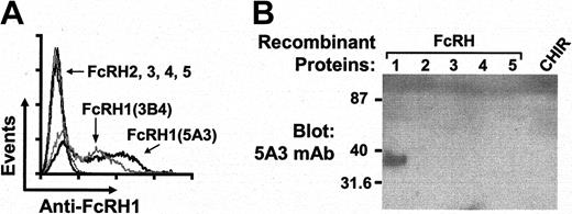
Lymph node cells from mice hyperimmunized with recombinant protein corresponding to the 3 extracellular immunoglobulin domains of FcRH1 and boosted with FcRH1 transcript-positive human B cell lines7 were fused with a non–immunoglobulin-producing plasmacytoma cell line. Antibodies produced by 12 hybridoma clones were found to be reactive with an FcRH1-transfected cell line when assessed by cell surface immunofluorescence. The fine specificity of 2 of these monoclonal antibodies, 5A3 (γ2bκ) and 3B4 (γ1κ), were shown by their lack of reactivity with other FcRH family members by immunofluorescence analysis of transfected cells (Figure 1A). The specificity was confirmed by Western blot assays demonstrating that the 5A3 antibody reacted with the recombinant FcRH1 protein, but not with other FcRH family members (FcRH2 to FcRH5), chicken Ig-like receptor (CHIR), FcγRIIa, FcγRIIb, and FcγRIII (Figure 1B; data not shown). Likewise, the 3B4 antibody was found to be specific for FcRH1, except that it did not react with recombinant FcRH1 protein in Western blots. These data indicate that both the 5A3 and 3B4 mAbs specifically recognize native FcRH1 molecules. Biotinylated Fab monomers of these antibodies were therefore prepared for use in identification and ligation of the FcRH1 molecules on B-lineage cells.
Specificity of 2 anti-FcRH1 monoclonal antibodies. (A) Human 293T cells were transiently transfected with FcRH1 to FcRH5 expression vectors and stained with 3B4 (thick gray line) or 5A3 (thick black line) monoclonal antibodies for immunofluorescence analysis. Cell surface expression of FcRH2 to FcRH5 was confirmed by the use of specific monoclonal antibodies against each molecule. (B) Recombinant FcRH proteins and the chicken Ig-like receptor (CHIR) were immunoblotted with anti-FcRH1 (5A3).
Specificity of 2 anti-FcRH1 monoclonal antibodies. (A) Human 293T cells were transiently transfected with FcRH1 to FcRH5 expression vectors and stained with 3B4 (thick gray line) or 5A3 (thick black line) monoclonal antibodies for immunofluorescence analysis. Cell surface expression of FcRH2 to FcRH5 was confirmed by the use of specific monoclonal antibodies against each molecule. (B) Recombinant FcRH proteins and the chicken Ig-like receptor (CHIR) were immunoblotted with anti-FcRH1 (5A3).
FcRH1 is variably expressed as a function of B-cell differentiation
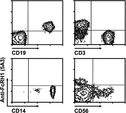
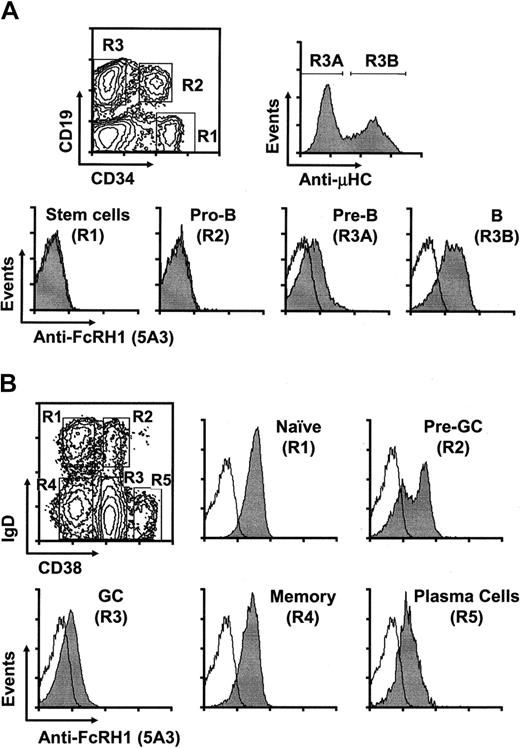
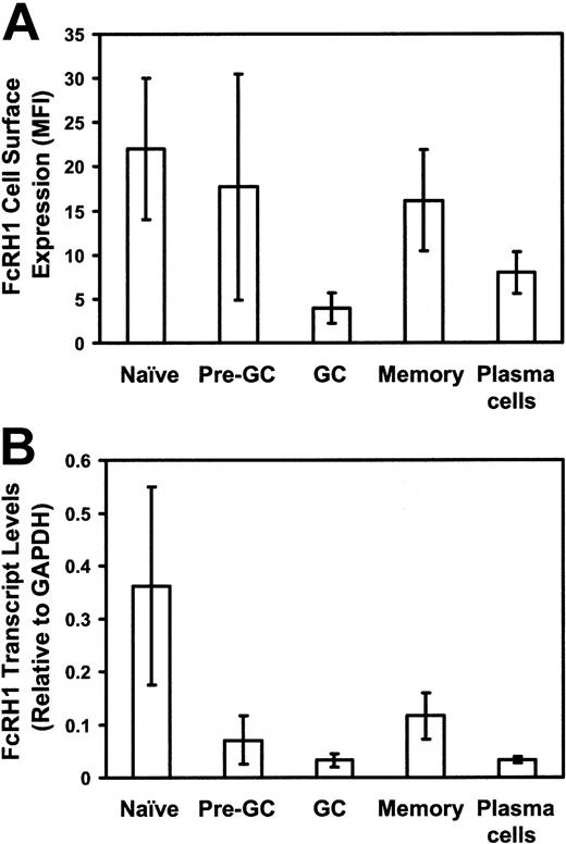
Previous analysis of FcRH1 transcripts in blood and tonsillar cells has suggested that FcRH1 expression is B lineage specific.7,9 To determine which cells express the FcRH1 protein, blood mononuclear cells were stained with biotinylated Fab fragments of the 5A3 anti-FcRH1 mAb and streptavidin-APC in conjunction with PE-labeled mAbs against lineage-specific markers. FcRH1 was found on all of the circulating B cells, and not on T cells, natural killer (NK) cells, monocyte/macrophages, granulocytes, and platelets (Figure 2; data not shown). Reverse transcription (RT)–PCR analysis of FcRH1 transcripts in B cells, T cells, NK cells, and monocyte/macrophages confirmed the exclusive expression of FcRH1 by B cells (data not shown). FcRH1 protein expression was correspondingly found on human B-cell lines and not on T, monocytoid, or erythroid cell lines (Table 1). Notably, FcRH1 was also undetectable on pro/pre-B cell lines and, correspondingly, was found on only a subpopulation of the CD19+ B-lineage cells in the bone marrow. The hematopoietic stem cells (CD34+CD19–) and pro-B cells (CD34+CD19+) did not express detectable FcRH1 (Figure 3A, R1 and R2), while most cells (60%-89%) in a mixed pre-B and B-cell population (CD34–CD19+) expressed FcRH1 (data not shown). When this population was divided into pre-B and immature B-cell subpopulations according to their μ heavy chain expression levels (R3A and R3B subpopulations in Figure 3A), relatively low levels of FcRH1 expression were found on pre-B cells and higher levels were found on B cells. (IgD–CD38–), and plasma cells (CD38++). Relatively high levels of cell surface FcRH1 expression were found on the naive (follicular mantle) B cells (Figure 3B, R1). Whereas GC B cells and plasma cells (Figure 3B, R3 and R5) expressed FcRH1 at very low levels, memory B cells were found to express FcRH1 in levels almost as high as those found on naive B cells (Figure 3B, R4). For the tonsillar pre-GC population the levels of FcRH1 expression were remarkably variable in different donors, presumably reflecting differences in their antigenic activation status. In 2 of 7 tonsils, FcRH1 surface expression levels divided the pre-GC cells into relatively high- and low-expressing subpopulations (Figure 3B, R2). The pre-GC cells in 2 donor samples expressed higher FcRH1 levels, while those in the 3 remaining samples had medium-to-low FcRH1 levels. When FcRH1 transcript levels were examined by real-time RT-PCR analysis of the different subpopulations of purified tonsillar B cells, the naive B cells were found to express the highest levels of FcRH1 transcripts, while the pre-GC and memory B cells expressed intermediate levels. The lowest B-cell levels of FcRH1 transcripts were found for the GC B cells and plasma cells (Figure 4A). This analysis indicates that FcRH1 transcription begins in pre-B cells, increases as B cells mature, is down-regulated as B cells are activated to form germinal centers and later to undergo plasma cell differentiation. Memory B cells regain FcRH1 expression, although at lower levels than for naive B cells.
FcRH1 expression by peripheral blood mononuclear cells. Human blood mononuclear cells purified by Ficoll centrifugation were stained with biotinylated Fab fragments of 5A3 followed by streptavidin-APC and PE-conjugated antibodies to lineage specific markers: CD19+ B lineage cells, CD3+ T cells, CD14+ myeloid lineage cells, and CD56+ NK cells. Cells in the lymphocyte light-scatter gate were analyzed for CD19, CD3, and CD56, and cells in the myeloid gate are shown here for CD14 and FcRH1 staining. The same FcRH1 expression pattern was found for 10 donors of European, African, or Asian ancestry. Similar results were obtained with the 3B4 antibody.
FcRH1 expression by peripheral blood mononuclear cells. Human blood mononuclear cells purified by Ficoll centrifugation were stained with biotinylated Fab fragments of 5A3 followed by streptavidin-APC and PE-conjugated antibodies to lineage specific markers: CD19+ B lineage cells, CD3+ T cells, CD14+ myeloid lineage cells, and CD56+ NK cells. Cells in the lymphocyte light-scatter gate were analyzed for CD19, CD3, and CD56, and cells in the myeloid gate are shown here for CD14 and FcRH1 staining. The same FcRH1 expression pattern was found for 10 donors of European, African, or Asian ancestry. Similar results were obtained with the 3B4 antibody.
Expression of FcRH1 on cell lines
Cell type . | Cell line . | Cell surface staining by anti-FcRH1 . |
|---|---|---|
| Pro-B | Nalm16 | - |
| Pre-B | ||
| 697 | - | |
| 207 | - | |
| B | ||
| Daudi | +++ | |
| Raji | + | |
| Ramos | +++ | |
| BJAB | + | |
| T | Jurkat | - |
| Monocytic | THP-1 | - |
| Myelomonocytic | U937 | - |
| Erythroid | K562 | - |
- indicates lack of staining, and + symbols indicate level of positive anti-FcRH1 staining.
FcRH1 expression by B-lineage cells in bone marrow and tonsils. (A) Bone marrow mononuclear cells from adult ribs were purified and 3-color immunofluorescent staining was performed. This pattern of FcRH1 expression by pre-B and B cells was confirmed for 3 additional bone marrow samples. (B) Tonsillar B cells purified using CD19 microbeads were stained with antibodies against CD38, IgD, and biotin-5A3 followed by streptavidin-APC. The different B-cell subpopulations were gated for FcRH1 analysis based on their CD38 and IgD expression. Note the biphasic pattern of FcRH1 expression by the pre-germinal center subpopulation. FcRH1 stained populations are shaded, and Ig isotype controls are unshaded.
FcRH1 expression by B-lineage cells in bone marrow and tonsils. (A) Bone marrow mononuclear cells from adult ribs were purified and 3-color immunofluorescent staining was performed. This pattern of FcRH1 expression by pre-B and B cells was confirmed for 3 additional bone marrow samples. (B) Tonsillar B cells purified using CD19 microbeads were stained with antibodies against CD38, IgD, and biotin-5A3 followed by streptavidin-APC. The different B-cell subpopulations were gated for FcRH1 analysis based on their CD38 and IgD expression. Note the biphasic pattern of FcRH1 expression by the pre-germinal center subpopulation. FcRH1 stained populations are shaded, and Ig isotype controls are unshaded.
Comparative analysis of FcRH1 expression by different subpopulations of tonsillar B lineage cells. (A) Mean fluorescent intensity (MFI) of cell surface FcRH1 (± 1 SD) is shown for 7 individuals. (B) Tonsillar subpopulations were sorted and FcRH1 mRNA levels were examined by real-time PCR and normalized to glyceraldehyde-3-phosphate dehydrogenase (GAPDH) expression. Mean levels (± SDs) are shown for each subpopulation in 3 tonsillar samples.
Comparative analysis of FcRH1 expression by different subpopulations of tonsillar B lineage cells. (A) Mean fluorescent intensity (MFI) of cell surface FcRH1 (± 1 SD) is shown for 7 individuals. (B) Tonsillar subpopulations were sorted and FcRH1 mRNA levels were examined by real-time PCR and normalized to glyceraldehyde-3-phosphate dehydrogenase (GAPDH) expression. Mean levels (± SDs) are shown for each subpopulation in 3 tonsillar samples.
FcRH1 down-regulation occurs during pre-germinal center activation
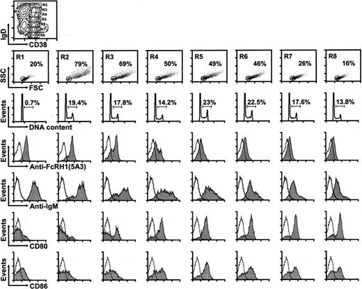
To examine more precisely the FcRH1 down-regulation that occurs during the naive to GC B-cell transition, we divided the naive, pre-GC, and GC cells into multiple subsets on the basis of their cell surface IgD and CD38 levels (Figure 5). When these subsets were examined for expression of FcRH1 and other cell surface molecules, FcRH1 levels were found to be uniformly high on naive B cell subsets (not shown). In contrast, coinciding with the onset of CD38 expression by pre-GC cells, progressive FcRH1 down-regulation occurred in concert with diminishing levels of cell surface IgD and IgM, and this pattern was maintained for GC B cells (Figure 5). A dramatic increase in cell size was also observed with the onset of CD38 expression, presumably reflecting activation of the B cells entering the pre-GC compartment. The size of pre-GC cells then progressively decreased in concert with the decline in their IgD and IgM expression levels (Figure 5). Cell cycle entry also coincided with the initiation of CD38 expression. Whereas more than 99% of the naive B cells were in the G0 or G1 phases, 14% to 23% of B cells were found to be in the S and G2/M phases throughout the pre-GC and GC stages (Figure 5). Expression of the CD80 and CD86 costimulatory molecules was also up-regulated after the B cells entered the pre-GC stage (Figure 5). This constellation of findings suggests that FcRH1 is well positioned to potentially influence the activation of naive B cells.
Correlation between FcRH1 levels with cell size, cell cycle status, and surface IgD, IgM, CD80, and CD86 expression in tonsillar B cells. Tonsillar B cells were purified as described in “Materials and methods” for 4-color immunofluorescent analysis. Naive, pre-GC, and GC populations were subdivided into the indicated R1 to R8 subsets for analysis. DNA content analysis was conducted after cell fixation in 100% ethanol, treatment with RNase A, and staining with propidium iodide (40 μg/mL). Subsequent analysis of the naive subpopulations was conducted but is not shown here because of insignificant variation. Shaded areas indicate staining with the indicated antibodies, while the unshaded areas show Ig isotope control staining.
Correlation between FcRH1 levels with cell size, cell cycle status, and surface IgD, IgM, CD80, and CD86 expression in tonsillar B cells. Tonsillar B cells were purified as described in “Materials and methods” for 4-color immunofluorescent analysis. Naive, pre-GC, and GC populations were subdivided into the indicated R1 to R8 subsets for analysis. DNA content analysis was conducted after cell fixation in 100% ethanol, treatment with RNase A, and staining with propidium iodide (40 μg/mL). Subsequent analysis of the naive subpopulations was conducted but is not shown here because of insignificant variation. Shaded areas indicate staining with the indicated antibodies, while the unshaded areas show Ig isotope control staining.
B cells are activated by FcRH1 ligation
To examine the activation potential for FcRH1, B cells of the Daudi cell line were treated with biotinylated anti-FcRH1 Fab fragments plus streptavidin. FcRH1 ligation alone had no demonstrable effect on intracellular calcium levels, whereas concomitant FcRH1 ligation enhanced the Ca2+ flux induced by B-cell antigen receptor (BCR) ligation (Figure 6A). Also consistent with its possession of ITAM-like motifs, transient FcRH1 tyrosine phosphorylation was observed after its ligation on an FcRH1-transfected cell line (Figure 6B). When the FcRH1 activation potential was examined for native B cells in tonsillar samples, FcRH1 ligation was found to induce a significant increase in the proportion of relatively large B cells, 38.2 ± 1.0 versus 24.7 ± 1.3 for unstimulated control cells (P = .01), and of CD69+ cells (59.0 ± 0.9 versus 33.4 ± 1.9 for controls, P = .037) after 48 hours in culture. Conversely, surface IgD levels were reduced by FcRH1 cross-linkage (mean fluorescent intensity of 25.9 ± 0.1 for control cells versus 18.8 ± 0.5 for stimulated cells, P = .04), whereas the proportion of CD86+ cells was enhanced, 40.0 ± 1.1 versus 15.6 ± 0.4 for unstimulated cells (P = .02). The ligation of FcRH1 on tonsillar B cells also induced an increase in 3H-thymidine uptake (Figure 6C), and FcRH1 and BCR coligation resulted in an additive effect on B-cell proliferation when suboptimal doses of anti-IgM were used (Figure 6D; data not shown). The ability of FcRH1 to induce B cell proliferation was confirmed by the finding of an anti-FcRH1 dose-related increase in cell numbers without an accompanying alteration of cell survival (42.6 ± 2.0 TUNEL-positive cells versus 43.7 ± 1.9 for unstimulated control cells at 24 hours, P = .898). FcRH1 ligation also had no effect on the expression levels of antiapoptotic proteins (Bcl-2, Bcl-xL, and Mcl-1) or a proapoptotic protein (Bax) (data not shown).
Analysis of B-cell activation by FcRH1 ligation. (A) Concomitant FcRH1 ligation enhances BCR-induced calcium flux. Daudi B cells were labeled with the calcium indicator dye Fluo-4 and, a reference dye, SNARF-1, and calcium levels were evaluated by flow cytometry before and after stimulation. Thin black line indicates streptavidin crosslinker alone (20 μg/mL). Thick black line indicates biotinylated F(ab′)2 fragments of goat anti–human μ HC (2 μg/mL) plus streptavidin. Thick gray line indicates biotinylated F(ab′)2 fragments of goat anti–human μ HC (2 μg/mL), biotinylated Fab fragments of anti-FcRH1 (5A3; 1 μg/mL), and streptavidin. (B) Ligation induced FcRH1 tyrosine phosphorylation. HA-tagged FcRH1 overexpressing mouse IIA1.6 cells were serum-starved for 2 hours before incubation with biotinylated Fab fragments of anti-FcRH1 mAb plus streptavidin (20 μg/mL). Cell lysate (200 μg) was immunoprecipitated with anti-HA antibody, and the immunoprecipitates were immunoblotted with either anti-phosphotyrosine antibody (anti-pTyr) or anti-FcRH1 antibody. (C) FcRH1 ligation induces DNA synthesis. Purified tonsillar B cells were incubated in 96-well plates (105/well) for 72 hours in the presence or absence of varying concentrations of biotinylated Fab fragments of anti-FcRH1 or control mAbs plus streptavidin (20 μg/mL). Cells were pulsed for an additional 16 hours with 3H-thymidine (1 μCi/well [0.037 MBq/well]) before measuring 3H-thymidine incorporation. (D) FcRH1 coligation enhances BCR-induced B-cell proliferation. Tonsillar B cells were incubated in 96-well plates (105/well) for 72 hours in the presence or absence of anti-μ HC antibody (DA4.4; 1 μg/mL), biotinylated Fab fragments of anti-FcRH1 mAbs (3 μg/mL) plus streptavidin (20 μg/mL), or the combination of both antibodies. Cells were analyzed as in panel C.
Analysis of B-cell activation by FcRH1 ligation. (A) Concomitant FcRH1 ligation enhances BCR-induced calcium flux. Daudi B cells were labeled with the calcium indicator dye Fluo-4 and, a reference dye, SNARF-1, and calcium levels were evaluated by flow cytometry before and after stimulation. Thin black line indicates streptavidin crosslinker alone (20 μg/mL). Thick black line indicates biotinylated F(ab′)2 fragments of goat anti–human μ HC (2 μg/mL) plus streptavidin. Thick gray line indicates biotinylated F(ab′)2 fragments of goat anti–human μ HC (2 μg/mL), biotinylated Fab fragments of anti-FcRH1 (5A3; 1 μg/mL), and streptavidin. (B) Ligation induced FcRH1 tyrosine phosphorylation. HA-tagged FcRH1 overexpressing mouse IIA1.6 cells were serum-starved for 2 hours before incubation with biotinylated Fab fragments of anti-FcRH1 mAb plus streptavidin (20 μg/mL). Cell lysate (200 μg) was immunoprecipitated with anti-HA antibody, and the immunoprecipitates were immunoblotted with either anti-phosphotyrosine antibody (anti-pTyr) or anti-FcRH1 antibody. (C) FcRH1 ligation induces DNA synthesis. Purified tonsillar B cells were incubated in 96-well plates (105/well) for 72 hours in the presence or absence of varying concentrations of biotinylated Fab fragments of anti-FcRH1 or control mAbs plus streptavidin (20 μg/mL). Cells were pulsed for an additional 16 hours with 3H-thymidine (1 μCi/well [0.037 MBq/well]) before measuring 3H-thymidine incorporation. (D) FcRH1 coligation enhances BCR-induced B-cell proliferation. Tonsillar B cells were incubated in 96-well plates (105/well) for 72 hours in the presence or absence of anti-μ HC antibody (DA4.4; 1 μg/mL), biotinylated Fab fragments of anti-FcRH1 mAbs (3 μg/mL) plus streptavidin (20 μg/mL), or the combination of both antibodies. Cells were analyzed as in panel C.
The possibility that FcRH1 could be an Fc receptor was examined, because of the close relationship between FcR and FcRH gene families. These experiments were conducted by incubating FcRH1-transfected cell lines with either soluble or aggregated forms of human IgM, IgA, IgG1, IgG2, IgG3, and IgG4 and subsequent immunofluorescence assessment of binding. The results of these analyses did not reveal evidence of FcRH1 binding for any of these human Ig isotypes (Figure S1, available at the Blood website; see the Supplemental Figure link at the top of the online article). These results indicate that FcRH1 does not serve as an Fc receptor for IgM, IgG, or IgA antibodies.
Discussion
The findings in this study indicate FcRH1 is an intrinsic activation molecule that has the potential to augment BCR-induced activation. Two new monoclonal anti-FcRH1 antibodies were used to show that FcRH1 is expressed by mature B-lineage cells, naive and memory B cells in particular. Naive B cells were found to express the highest FcRH1 levels, pre-GC and memory cells express intermediate levels, while germinal center B cells and plasma cells express the lowest levels. Correspondingly, FcRH1 transcript levels were highest in naive B cells; reduced to low levels in pre-GC, GC, and plasma cells; and restored to an intermediate level in memory cells. These results complement and extend the in situ hybridization analysis of Miller et al9 in which IRTA5 (FcRH1) transcripts were found in tonsillar mantle zones wherein naive B cells predominate. Those investigators failed to detect FcRH1 transcripts outside this region, while we found FcRH1 transcripts in memory cells as well, albeit at lower levels than in the naive B cells. This difference in distribution is likely attributable to the higher threshold of detection required by the in situ hybridization assay.
FcRH1 is unique among molecules of the FcRH family in having an acidic residue (glutamic acid) in the transmembrane domain and 2 ITAM-like motifs in the cytoplasmic region. The presence of a negative-charged residue in the FcRH1 transmembrane region suggests that it could associate with another transmembrane protein, although FcRH1-associated proteins have not yet been identified. The sequences of the 2 tyrosine-based ITAM-like motifs in FcRH1, E-F-T-Y-L-N-S-X9-Y-E-N-V and D-I-Y-S-R-L-X9-Y-E-D-A, differ from the canonical ITAM sequence D/E-X-X-Y-X-X-L/I-X6-8-Y-X-X-L/I found in the BCR subunits, Igα and Igβ. The failure of FcRH1 ligation alone to induce a calcium flux is a further indication that FcRH1 differs from the BCR complex in its B-cell activation potential. However, several lines of evidence support the conclusion that FcRH1 has significant activation potential and may serve as an activation coreceptor on B cells. (1) Cross-linkage of FcRH1 alone stimulates its tyrosine phosphorylation. (2) FcRH1 ligation also induces a modest increase in B-cell size, enhances CD69 and CD86 expression, and decreases cell surface IgD levels. (3) Although FcRH1 ligation alone has no effect on intracellular Ca2+ levels, concomitant FcRH1 ligation can enhance BCR-triggered Ca2+ mobilization. (4) FcRH1 ligation induces a modest proliferative response and can enhance the proliferative response induced by suboptimal BCR ligation. Notably, the FcRH1 potentiation of BCR-mediated signaling does not require coligation of the 2 receptors. Although the B-cell responses triggered by FcRH1 ligation indicate its potential to modify the activation status of B cells, FcRH1 differs from the previously defined activation enhancing coreceptors, CD19 and CD40, in its expression pattern and biologic activity. Whereas CD19 and CD40 are expressed throughout B-cell differentiation, FcRH1 is preferentially expressed on naive B cells and on memory cells to a lesser extent. FcRH1 has the potential to deliver activation signals on its own, while CD19 mediates its effects through physical and functional association with the surface immunoglobulin receptor complex.19-22 CD40, in contrast to FcRH1, serves as an activation coreceptor on mature B cells and specifically enhances the survival of germinal center B cells.23-27
Although sequence homology with Fc receptors led to its identification,7 the extracellular domains of FcRH1 lack consensus sequences shown to be important for Fc binding. Our failure to detect binding of human IgM, IgA, or IgGs by FcRH1-transfected cell lines provides further evidence that FcRH1 is not an Fc receptor. The future identification of an FcRH1 ligand could shed new insight into the regulation of B-cell activation. For example, if an FcRH1 ligand were expressed on activated T cells, its interaction with FcRH1 could play a role in B-cell activation. While other equally plausible scenarios can be envisioned, further study is needed for elucidation of the physiologic role of the FcRH1 activation potential in B-cell responses.
Prepublished online as Blood First Edition Paper, October 12, 2004; DOI 10.1182/blood-2004-06-2344.
Supported in part by National Institutes of Health (NIH) (grant AI 39816) and by NIH K08 award (AI55638) (R.S.D.).
The online version of the article contains a data supplement.
The publication costs of this article were defrayed in part by page charge payment. Therefore, and solely to indicate this fact, this article is hereby marked “advertisement” in accordance with 18 U.S.C. section 1734.
We thank Drs Peter D. Burrows and Louis B. Justement for helpful discussion, Dr Guzide Aksu for help with monoclonal antibody preparation, Dr Larry Gartland for help with flow cytometry, and Ann Brookshire and Marsha Flurry for help in manuscript preparation.

![Figure 6. Analysis of B-cell activation by FcRH1 ligation. (A) Concomitant FcRH1 ligation enhances BCR-induced calcium flux. Daudi B cells were labeled with the calcium indicator dye Fluo-4 and, a reference dye, SNARF-1, and calcium levels were evaluated by flow cytometry before and after stimulation. Thin black line indicates streptavidin crosslinker alone (20 μg/mL). Thick black line indicates biotinylated F(ab′)2 fragments of goat anti–human μ HC (2 μg/mL) plus streptavidin. Thick gray line indicates biotinylated F(ab′)2 fragments of goat anti–human μ HC (2 μg/mL), biotinylated Fab fragments of anti-FcRH1 (5A3; 1 μg/mL), and streptavidin. (B) Ligation induced FcRH1 tyrosine phosphorylation. HA-tagged FcRH1 overexpressing mouse IIA1.6 cells were serum-starved for 2 hours before incubation with biotinylated Fab fragments of anti-FcRH1 mAb plus streptavidin (20 μg/mL). Cell lysate (200 μg) was immunoprecipitated with anti-HA antibody, and the immunoprecipitates were immunoblotted with either anti-phosphotyrosine antibody (anti-pTyr) or anti-FcRH1 antibody. (C) FcRH1 ligation induces DNA synthesis. Purified tonsillar B cells were incubated in 96-well plates (105/well) for 72 hours in the presence or absence of varying concentrations of biotinylated Fab fragments of anti-FcRH1 or control mAbs plus streptavidin (20 μg/mL). Cells were pulsed for an additional 16 hours with 3H-thymidine (1 μCi/well [0.037 MBq/well]) before measuring 3H-thymidine incorporation. (D) FcRH1 coligation enhances BCR-induced B-cell proliferation. Tonsillar B cells were incubated in 96-well plates (105/well) for 72 hours in the presence or absence of anti-μ HC antibody (DA4.4; 1 μg/mL), biotinylated Fab fragments of anti-FcRH1 mAbs (3 μg/mL) plus streptavidin (20 μg/mL), or the combination of both antibodies. Cells were analyzed as in panel C.](https://ash.silverchair-cdn.com/ash/content_public/journal/blood/105/3/10.1182_blood-2004-06-2344/6/m_zh80030573360006.jpeg?Expires=1767798855&Signature=YpPvw5923Fn1eb5-zxOnCb-y~~fUHSfNmjKWE47CC1pNTafqUdsZlhZfP4cL2e4NHRNEEFWBQxQruEa~7DvFJFOX7DRO3fxofZUYeLdXtJyBR4qhKZsSarUeqylkIyn-V6RCWGNSk0LtWdeHHvgybe~CFzd4QV4E-hpzegqrugQ1OCZabWe~Qel259UzXnyoMYTpI7fMgRVpoNRn4IxvdQH1oJMhkwIxaPFXTzh-uKIuLZW-FFTnph0EeiUFG5Wd50nfmqzIzIKrZpM0XZWd8PqqOlenA3HORZOLcvlcc9PdwhBI9xY6KM1G3QWs5vuQGLLSgGYRZiIWn6x0f08eLQ__&Key-Pair-Id=APKAIE5G5CRDK6RD3PGA)
This feature is available to Subscribers Only
Sign In or Create an Account Close Modal