Thrombosis can be initiated when activated platelets adhere to injured blood vessels via the interaction of subendothelial collagen with its platelet receptor, glycoprotein (GP) VI. Here we observed that incubation of platelets with convulxin, collagen, or collagen-related peptide (CRP) resulted in GPVI signaling-dependent loss of surface GPVI and the appearance of an approximately 55-kDa soluble fragment of GPVI as revealed by immunoblotting. Ethylenediaminetetraacetic acid (EDTA) or GM6001 (a metalloproteinase inhibitor with broad specificity) prevented this loss. In other receptor systems, calmodulin binding to membrane-proximal cytoplasmic sequences regulates metalloproteinase-mediated ectodomain shedding. In this regard, we have previously shown that calmodulin binds to a positively charged, membrane-proximal sequence within the cytoplasmic tail of GPVI. Incubation of platelets with calmodulin inhibitor W7 (150 μM) resulted in a time-dependent loss of GPVI from the platelet surface. Both EDTA and GM6001 prevented this loss. Surface plasmon resonance demonstrated that W7 specifically blocked the association of calmodulin with an immobilized synthetic peptide corresponding to the calmodulin-binding sequence of GPVI. These findings suggest that disruption of calmodulin binding to receptor cytoplasmic tails by agonist binding to the receptor triggers metalloproteinase-mediated loss of GPVI from the platelet surface. This process may represent a potential mechanism to regulate GPVI-dependent platelet adhesion.
Introduction
Platelets adhere to sites of endothelial cell damage in a process requiring rapid activation of multiple signaling events. Under conditions of low shear stress, the platelet membrane receptor glycoprotein (GP) VI is able to bind collagen and mediates intracellular signaling events that lead to an up-regulation of platelet adhesion receptors including platelet P-selectin and the fibrinogen receptor on platelets, αIIbβ3 integrin.1,2 Platelet adhesion to exposed collagen is then stabilized by an additional collagen receptor, α2β1 integrin.
GPVI is a 60- to 65-kDa protein containing 2 extracellular immunoglobulin (Ig)–like domains, a single transmembrane domain and a cytoplasmic tail of about 51 amino acids.3,4 Along with collagen, other ligands such as collagen-related peptide (CRP)5 and the snake venom protein convulxin6 can bind to the extracellular region of GPVI, rapidly inducing intracellular tyrosine phosphorylation of numerous proteins, leading to the activation of αIIbβ3 integrin and formation of platelet aggregates. The rate and strength of an observed intracellular signal appears to be proportional to the potency of each GPVI agonist to cross-link the receptor and induce clustering of GPVI molecules.1,7,8 Although the precise mechanism by which GPVI signals remains unclear, the receptor is homologous to immune receptors and associates with the Fc receptor (FcR) γ subunit of FcR9,10 via a salt bridge within the platelet membrane. This association allows ligand-bound GPVI to use a single immunoreceptor tyrosine-based activation motif (ITAM) within the cytoplasmic tail of FcRγ and signal via Syk tyrosine kinase.11 More recently, the Src tyrosine kinase, Lyn,12 and calmodulin13 have been demonstrated to bind directly to the cytoplasmic tail of GPVI, suggesting that the tail of GPVI also plays a direct role in GPVI-mediated signaling. Indeed, a requirement for the entire cytoplasmic tail of GPVI as well as associated FcRγ for complete convulxin-mediated signaling has been confirmed.14
Loss of GPVI from the surface of mouse platelets is protective against thrombosis in a mouse carotid artery model of thromboembolism.15 The loss of GPVI in this study15 was induced by treatment of mouse platelets with the anti-GPVI rat monoclonal antibody JAQ116 and was proposed to be mediated primarily through receptor internalization mechanisms, although proteolytic shedding of GPVI was not excluded. Regulation of activated adhesion receptors through proteolysis is well recognized as a mechanism for modulation of cell surface proteins17,18 and has been identified in a number of systems including proteolytic cleavage of Notch,19 and adhesion proteins,20-22 as well as numerous cytokines, growth factors, and their receptors.17,23-25 In the majority of cases, one or more membrane-bound metalloproteinases appear to be responsible for the extracellular processing observed.26,27 A role for calmodulin in the metalloproteinase-dependent release of L-selectin from leukocytes has also been described.28 This ectodomain proteolysis of L-selectin was dependent on loss of binding of calmodulin to a membrane proximal amphipathic helix within the cytoplasmic tail of L-selectin.29
In the course of our studies on the association of calmodulin with the GPVI cytoplasmic tail,13 we investigated whether GPVI was lost from the platelet surface under both resting conditions and conditions of activation. On ligation of GPVI, or when calmodulin was dissociated from the cytoplasmic tail of GPVI, we observed a loss of up to 90% of GPVI from the surface of platelets that could be blocked by inclusion of a metalloproteinase inhibitor. These results show for the first time that levels of GPVI receptor can be down-regulated on human platelets and support the hypothesis that calmodulin binding to cytoplasmic sequences of membrane-expressed receptors is a specialized control mechanism for proteolytic shedding common to several different cell and receptor types.
Materials and methods
Materials
Soybean trypsin inhibitor, thrombin, and pepstatin A were purchased from Sigma (St Louis, MO). Leupeptin and thrombin-related agonist peptide (TRAP) were purchased from Auspep (Parkville, Australia). Horseradish peroxidase (HRP)–conjugated or fluorescein isothiocyanate (FITC)–conjugated antimouse and antirabbit immunoglobulin (Ig) antibodies raised in sheep were from Chemicon (Melbourne, Australia). GM6001 (a hydroxamic acid-based metalloproteinase inhibitor with broad specificity)30 and the calmodulin inhibitor W7 (N-(6-aminohexyl)-5-chloro-1-naphthalenesulfonamide) as well as inhibitors of intracellular signaling molecules, PP1 and PP2 (Src kinase inhibitors), piceatannol (Syk inhibitor), and wortmannin (phosphatidylinositol 3 kinase [PI3K] inhibitor) were obtained from Calbiochem (La Jolla, CA). Purified convulxin from the venom of the South American rattlesnake Crotalus durissus terrificus was a kind gift from Dr Kenneth Clemetson (Berne, Switzerland). von Willebrand factor from human factor VIII concentrates and botrocetin from Bothrops jararaca were purified as previously described.31,32 A monoclonal antibody, 6B12-3, against human platelet GPVI was raised essentially as described elsewhere.7 6B12-3 recognized GPVI on intact platelets by flow cytometry (not shown) and recognized a single platelet protein of approximately 60 kDa by Western blot analysis at an immunoglobulin protein concentration of approximately 2 μg/mL (100-fold dilution of hybridoma medium).
Synthetic peptides with sequence corresponding to the cytoplasmic calmodulin-binding sequence of human GPVI or the C-terminus of human GPIbα were purified by reverse-phase high-pressure liquid chromatography and characterized by mass spectrometry (Mimotopes, Clayton, Victoria, Australia). The GPVI-specific agonist, CRP with amino acid sequence GCO(GPO)10GCOG-NH2 where O represents hydroxyproline, was a kind gift of Dr Su Kulkarni (Monash University). Cysteinyl residues at the N- and C-termini of CRP were cross-linked using the chemical cross-linker (3-(2-pyridyldithio)propionic acid N-hydroxysuccinimide (Sigma) as described.33
Shedding of GPVI from platelets
Human platelets were isolated from venous blood using acid-citratedextrose as anticoagulant and washed 3 times by centrifugation in 0.15 M NaCl containing 37.5 mM sodium citrate and 5 mM glucose, pH 7.0, as previously described.34 Platelets were counted and resuspended in Tyrode buffer (5 × 10⁸ platelets/mL). To examine the effect of GPVI ligands on platelet GPVI levels, aliquots (0.1 mL) of platelets were mixed with 0.1 mL Tyrode buffer containing (final concentration) convulxin (0.5 μg/mL), collagen (20 μg/mL), CRP (20 μg/mL), W7 (0.1 mM), thrombin (1 U/mL), or botrocetin (2.5 μg/mL) plus von Willebrand factor (10 μg/mL) and incubated at room temperature for the indicated times. Some platelet suspensions also contained 1 to 10 mM leupeptin, pepstatin A, soybean trypsin inhibitor, GM6001, or ethylenediaminetetraacetic acid (EDTA). In experiments investigating a role for intracellular signaling events in GPVI shedding, platelet suspensions were treated with 10 μM PP1, 10 μM PP2, 0.1 μM wortmannin, or 30 μg/mL piceatannol for 10 minutes prior to addition of convulxin. Where appropriate, an equivalent volume of dimethyl sulfoxide (DMSO; vehicle) was added to samples. Incubations were halted by addition of 0.1 mL of 10 mM EDTA and platelets were pelleted by centrifugation at 15 000g for 1 minute. The supernatant fraction was isolated and mixed with 0.1 mL sodium dodecyl sulfate (SDS) sample loading buffer. The platelet pellet was treated with 0.3 mL lysis buffer (0.01 M Tris [tris(hydroxymethyl)aminomethane], pH 7.5, containing 0.15 M NaCl, 5 mM EDTA, 1% Triton X-100, 10 μg/mL each of soybean trypsin inhibitor, leupeptin, and pepstatin, and 1 mM sodium fluoride, sodium orthovanadate, and 5 mM phenylmethylsulfonyl fluoride) on ice for 30 minutes. Platelet lysates were then mixed with 0.1 mL SDS sample loading buffer. All aliquots of platelet lysates or supernatants were electrophoresed on 5% to 20% polyacrylamide-SDS gels, electrotransferred to nitrocellulose, and immunoblotted with anti-GPVI monoclonal antibody 6B12-3. Alternatively, some nitrocellulose filters were probed with the anti-β3 integrin monoclonal antibody CRC54,35 the antiplatelet endothelial cell adhesion molecule 1 (PECAM-1) monoclonal antibody, WM59,36 or the anti-GPIbα monoclonal antibody WM23.37 All blots were probed with HRP-labeled antimouse IgG secondary antibody (Silenus, Hawthorn, Australia) and visualized using the enhanced chemiluminescence (ECL) detection method (Amersham, Buckinghamshire, United Kingdom).
Convulxin-dependent association of GPVI with GPIb by immunoprecipitation
For immunoprecipitation experiments, washed platelets (5 × 10⁸/mL) were resuspended in 0.01 M HEPES (N-2-hydroxyethylpiperazine-N′-2-ethanesulfonic acid) buffer, pH 7.6, containing 5 mM EDTA and 0.15 M NaCl, and left untreated or treated with GPVI agonists for 60 minutes. Platelets were then lysed by the addition of ice-cold lysis buffer (final concentration: 20 mM Tris-HCl, pH 7.4, 5 mM EGTA [ethylene glycol tetraacetic acid], 1% Triton X-100, Complete protease inhibitor cocktail [Roche Diagnostics, Basel, Switzerland]). Lysates were centrifuged at 15 000g for 10 minutes at 4°C to pellet insoluble material. Immunoprecipitation of GPIbα from the Triton-soluble supernatant was performed by addition of 10 μg purified polyclonal antiglycocalicin (or nonimmune rabbit immunoglobulin) to precleared platelet lysates incubated overnight at 4°C as previously described.34,38 Immunoprecipitated proteins were resolved by 5% to 20% SDS-polyacrylamide gel electrophoresis (PAGE) and transferred to nitrocellulose membrane for Western blotting/ECL. Membranes were initially probed with anti-GPVI monoclonal antibody 6B12-3, followed by an HRP-labeled antimouse IgG secondary antibody (Silenus), and visualized using ECL. Membranes were stripped and reprobed with a monoclonal anti-GPIbα antibody (WM23).
Effect of W7 on binding of calmodulin to its GPVI interaction sequence
A cDNA encoding full-length human calmodulin was cloned into the pGEX-2T vector (Pharmacia, Uppsala, Sweden) using BamH1 and Xho1 restriction sites and the sequence was verified by DNA sequencing. For expression of the GST-calmodulin fusion protein, the construct was transfected into the BL21 strain of Escherichia coli and grown in the presence of 1 mM isopropylthiogalactoside (IPTG) for 4 hours at 37°C with constant shaking. The cells were lysed and expressed protein was isolated by affinity chromatography on glutathione-Sepharose, then GST was removed by treating the isolated protein with thrombin according to the manufacturer's instructions (Pharmacia). A second affinity chromatography step on a column of W7 immobilized on Affigel 10/15 resin39 was used to isolate calmodulin.
The association of calmodulin with cytoplasmic sequences of GPVI was measured by surface plasmon resonance using a BIAcore 3000 (Pharmacia Biosensor AB, Uppsala, Sweden) maintained at 25°C. Aliquots (5-10 μL) of biotinylated peptides (10 μM in 10 mM HEPES-saline, pH 7.0) with sequences matching either the calmodulin-binding sequence of GPVI, RRKRLRHRGRAVQRPL, or the final 10 residues of the cytoplasmic tail of GPIbα, VSIRYSGHSL, were passed over streptavidin-coated sensor chips (SA5; Pharmacia) to an equivalent of 200 to 250 resonance units (RU) at a flow rate of 20 μL/min. A preparation of calmodulin (analyte) was dialyzed and diluted in 0.01 M HEPES-saline, pH 7.0, containing 1 mM CaCl2, to protein concentrations ranging from 10 nM to 30 μM and volumes of 10 to 50 μL injected at a constant flow rate of 10 μL/min. To regenerate the surface after each sample, 10 μL 0.5% SDS in HEPES-saline was injected to dissociate the calmodulin/peptide complex without disturbing the biotinylated peptide/streptavidin interaction. The capacity of the peptide-coated sensor chip to bind calmodulin alone was identical at the start and at the end of each experiment.
Results
Treatment of platelets with GPVI agonists induces loss of GPVI from the platelet surface
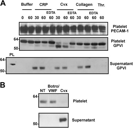
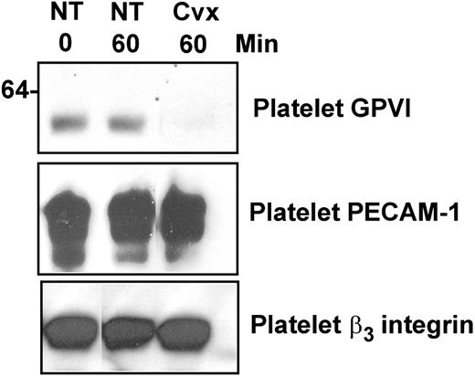
Initially we investigated whether the level of GPVI was regulated on platelets under conditions where GPVI was bound by a ligand. Washed platelets were resuspended in Tyrode buffer and incubated at room temperature for various times with convulxin, CRP, or collagen, all of which bind GPVI and activate platelets. Platelet and supernatant samples were analyzed by SDS-PAGE, transferred to nitrocellulose, and immunoblotted with an anti-GPVI monoclonal antibody 6B12-3. Figure 1A shows that 6B12-3 recognizes a single band of molecular weight about 60 kDa in untreated platelets. In contrast, in platelets treated with either CRP, convulxin, or collagen, the 60-kDa band was lost with time, and a band of molecular weight about 55 kDa appeared in the corresponding supernatant fraction probed with 6B12-3. Although convulxin has been reported to also bind GPIbα,40,41 the release of GPVI fragment was not detected after treatment for 1 hour with either thrombin (Figure 1A) or a combination of von Willebrand factor and botrocetin (Figure 1B). Loss of more than 80% of GPVI protein from platelets isolated from 5 different donors was observed with convulxin after a 1-hour incubation (data not shown). By comparison, removal of GPVI from platelets treated with the weaker GPVI agonists, CRP or collagen, was less evident; however, after 30 minutes, the 55-kDa GPVI fragment could be detected in the platelet supernatant (Figure 1A). Levels of PECAM-1 on platelets remained stable throughout the experiment (Figure 1A). Inclusion of 10 mM EDTA in the platelet incubation medium prior to the addition of convulxin, collagen, or CRP abrogated the loss of GPVI from the platelet surface suggesting a role for one or more metalloproteinases in GPVI shedding. Surface levels of other platelet membrane proteins such as PECAM-1 and αIIbβ3 integrin (Figure 2) or GPIbα (data not shown) remained constant throughout the 1-hour incubation with GPVI agonists. We did not analyze supernatants for release of receptors other than GPVI; however, platelet-associated levels of these proteins are essentially maintained under conditions where virtually all of the GPVI is shed (Figures 1, 2).
Treatment of platelets with GPVI agonists induces loss of GPVI from the platelet surface. (A) Washed human platelets (5 × 10⁸/mL) were resuspended in Tyrode buffer and treated with either 20 μg/mL CRP or collagen or 0.5 μg/mL convulxin (Cvx) for up to 60 minutes, or 1 U thrombin (Thr) for 60 minutes. EDTA (10 mM), where indicated, was included in some incubations. Platelet suspensions were incubated at room temperature, then mixed with 10 mM EDTA (final concentration) and centrifuged to isolate platelets from incubation medium supernatants. Platelet pellets were lysed in 1% Triton X-100 and aliquots of platelet extracts or supernatants were electrophoresed on SDS/acrylamide gels, transferred to nitrocellulose, then probed with anti-PECAM-1 antibody WM59 or anti-GPVI monoclonal antibody 6B12-3. An aliquot of resting platelet lysate is included on the left hand side of supernatant fractions for molecular weight comparison. Data are representative of at least 3 experiments with different donors. Two separate preparations each of collagen and CRP were tested and no difference was observed between each preparation of GPVI agonist for production of GPVI fragment. (B) Washed platelets were treated with 0.5 μg/mL convulxin or a combination of 2.5 μg/mL botrocetin (Botr0) plus 10 μg/mL von Willebrand factor (VWF) for 60 minutes. Incubations were then treated and analyzed as described for panel A.
Treatment of platelets with GPVI agonists induces loss of GPVI from the platelet surface. (A) Washed human platelets (5 × 10⁸/mL) were resuspended in Tyrode buffer and treated with either 20 μg/mL CRP or collagen or 0.5 μg/mL convulxin (Cvx) for up to 60 minutes, or 1 U thrombin (Thr) for 60 minutes. EDTA (10 mM), where indicated, was included in some incubations. Platelet suspensions were incubated at room temperature, then mixed with 10 mM EDTA (final concentration) and centrifuged to isolate platelets from incubation medium supernatants. Platelet pellets were lysed in 1% Triton X-100 and aliquots of platelet extracts or supernatants were electrophoresed on SDS/acrylamide gels, transferred to nitrocellulose, then probed with anti-PECAM-1 antibody WM59 or anti-GPVI monoclonal antibody 6B12-3. An aliquot of resting platelet lysate is included on the left hand side of supernatant fractions for molecular weight comparison. Data are representative of at least 3 experiments with different donors. Two separate preparations each of collagen and CRP were tested and no difference was observed between each preparation of GPVI agonist for production of GPVI fragment. (B) Washed platelets were treated with 0.5 μg/mL convulxin or a combination of 2.5 μg/mL botrocetin (Botr0) plus 10 μg/mL von Willebrand factor (VWF) for 60 minutes. Incubations were then treated and analyzed as described for panel A.
Treatment of platelets with GPVI agonists does not induce loss of PECAM-1 or β3 integrin from platelets. Samples of resting and convulxin-treated platelet lysates were prepared as described in Figure 1 and were analyzed for levels of GPVI, PECAM-1, or β3 integrin by SDS-PAGE and immunoblotting with 6B12-3, WM59, or CRC54, respectively.
Treatment of platelets with GPVI agonists does not induce loss of PECAM-1 or β3 integrin from platelets. Samples of resting and convulxin-treated platelet lysates were prepared as described in Figure 1 and were analyzed for levels of GPVI, PECAM-1, or β3 integrin by SDS-PAGE and immunoblotting with 6B12-3, WM59, or CRC54, respectively.
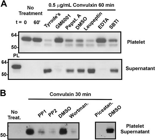
To further characterize the type of protease responsible for the shedding of GPVI, washed platelets were treated with 0.5 μg/mL convulxin in the presence of various proteolytic inhibitors (Figure 3A). Treatment of platelets with either EDTA or the hydroxamic acid-based, broad-range metalloproteinase inhibitor, GM6001, but not pepstatin A, leupeptin, or soybean trypsin inhibitor blocked the loss of GPVI from the platelet surface and the liberation of the 55-kDa GPVI fragment into the supernatant (Figure 3A). This concentration of GM6001 (100 μM) has been used previously to demonstrate the involvement of metalloproteinases in loss of receptors from the surface of aging platelets.42 Those studies demonstrated that GM6001 did not inhibit platelet receptor/agonist binding or act via a nonspecific interaction with the platelet membrane.42 Our results thus suggest that the observed loss of GPVI from platelets treated with convulxin, CRP, or collagen, is also mediated by metalloproteinase-derived proteolysis.
Release of GPVI from platelets is blocked by a broad-spectrum metalloproteinase inhibitor. Washed human platelets were resuspended in Tyrode buffer and treated with 0.5 μg/mL convulxin in the presence of (A) 100 μM GM6001 or DMSO, 5 mM EDTA, 0.1 mM pepstatin A, 0.1 mM leupeptin, or 0.1 mM soybean trypsin inhibitor (SBTI) or (B) 10 μM PP1, 10 μM PP2, 0.1 μM wortmannin, 30 μg/mL piceatannol, or DMSO. Platelets were incubated at room temperature for 1 hour, then pelleted and lysed in buffer containing proteinase inhibitors and Triton X-100 as described in “Materials and methods.” Aliquots of supernatants and platelet lysates were electrophoresed on SDS/acrylamide gels, transferred to nitrocellulose, and probed with anti-GPVI monoclonal antibody. An aliquot of resting platelet lysate is included on the left hand side of supernatant fractions for molecular weight comparison in panel A. Data are representative of at least 3 experiments with different donors.
Release of GPVI from platelets is blocked by a broad-spectrum metalloproteinase inhibitor. Washed human platelets were resuspended in Tyrode buffer and treated with 0.5 μg/mL convulxin in the presence of (A) 100 μM GM6001 or DMSO, 5 mM EDTA, 0.1 mM pepstatin A, 0.1 mM leupeptin, or 0.1 mM soybean trypsin inhibitor (SBTI) or (B) 10 μM PP1, 10 μM PP2, 0.1 μM wortmannin, 30 μg/mL piceatannol, or DMSO. Platelets were incubated at room temperature for 1 hour, then pelleted and lysed in buffer containing proteinase inhibitors and Triton X-100 as described in “Materials and methods.” Aliquots of supernatants and platelet lysates were electrophoresed on SDS/acrylamide gels, transferred to nitrocellulose, and probed with anti-GPVI monoclonal antibody. An aliquot of resting platelet lysate is included on the left hand side of supernatant fractions for molecular weight comparison in panel A. Data are representative of at least 3 experiments with different donors.
To further characterize the mechanism of loss of GPVI from platelets, platelets were preincubated with inhibitors of intracellular tyrosine kinases including Src tyrosine kinases (PP1, PP2), PI3K (wortmannin), and Syk kinase (piceatannol) at concentrations known to interfere with GPVI-related signaling events.6,13,43 Shedding of GPVI was induced by addition of convulxin and levels of GPVI fragment in samples of platelet supernatant were assessed by Western blot as described in “Materials and methods.” Figure 3B shows that inhibitors of Src, PI3K, and Syk blocked the release of GPVI fragment from convulxin-treated platelets, suggesting a role for these signaling proteins in the platelet GPVI-shedding mechanism.
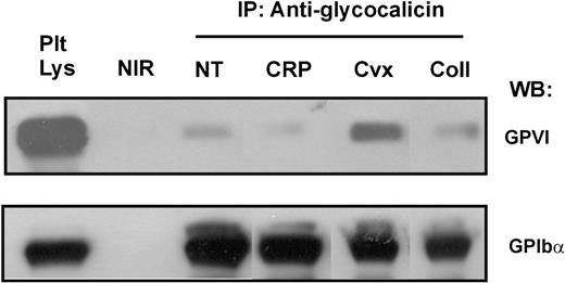
A GPVI+ fragment of approximately 55 kDa was also evident in increased amount in the pellet of convulxin-treated platelets as well as in the supernatant (Figures 1A and 3A). Convulxin is a disulfide-linked heterodimer consisting of homologous α and β chains further linked by disulfide bridges to form cyclic α4β4 heterotetramers.44 Because both GPIb and GPVI bind convulxin,40 it is likely that the increased association of shed GPVI with platelets treated with convulxin is due to convulxin-bridging association of the shed GPVI with membrane-bound GPIb. To investigate whether this is the case, platelets were treated with convulxin in the presence of excess EDTA to prevent GPVI shedding, lysed, immunoprecipitated with anti-GPIb antibody, and Western blotted for coassociated GPVI. Figure 4 shows that the relative levels of GPIb-associated GPVI are increased in the presence of convulxin as compared with untreated platelets or platelets treated with CRP or collagen, although some GPVI was detectable in these lanes. In this regard, we have observed that GPVI is associated with GPIb-IX-V in both resting and activated platelets (J.F.A. et al, manuscript in preparation).
Immunoprecipitation of GPIb-GPVI complexes is increased by treatment of platelets with convulxin. Washed human platelets (5 × 10⁸/mL) were resuspended in HEPES-saline buffer, pH 7.4, containing 10 mM EDTA, and treated with either 20 μg/mL CRP or collagen or 0.5 μg/mL convulxin for 60 minutes, then lysed in the same buffer containing Triton X-100 and proteinase inhibitors as described in “Materials and methods.” Platelet lysates were precleared, mixed with 10 μg/mL of either antiglycocalicin antibody or a nonimmune rabbit control antibody and immune complexes precipitated by mixing with protein A-Sepharose. After washing the beads, captured proteins were eluted in SDS sample loading buffer and analyzed for levels of GPVI by SDS-PAGE and immunoblotting using the anti-GPVI monoclonal antibody, 6B12-3. Blots were stripped and reprobed with the anti-GPIbα monoclonal antibody, WM23. Data are representative of at least 3 experiments with different donors. IP indicates immunoprecipitation; WB, Western blotting; NT, not treated; and NIR, nonimmune rabbit IgG.
Immunoprecipitation of GPIb-GPVI complexes is increased by treatment of platelets with convulxin. Washed human platelets (5 × 10⁸/mL) were resuspended in HEPES-saline buffer, pH 7.4, containing 10 mM EDTA, and treated with either 20 μg/mL CRP or collagen or 0.5 μg/mL convulxin for 60 minutes, then lysed in the same buffer containing Triton X-100 and proteinase inhibitors as described in “Materials and methods.” Platelet lysates were precleared, mixed with 10 μg/mL of either antiglycocalicin antibody or a nonimmune rabbit control antibody and immune complexes precipitated by mixing with protein A-Sepharose. After washing the beads, captured proteins were eluted in SDS sample loading buffer and analyzed for levels of GPVI by SDS-PAGE and immunoblotting using the anti-GPVI monoclonal antibody, 6B12-3. Blots were stripped and reprobed with the anti-GPIbα monoclonal antibody, WM23. Data are representative of at least 3 experiments with different donors. IP indicates immunoprecipitation; WB, Western blotting; NT, not treated; and NIR, nonimmune rabbit IgG.
Inhibition of calmodulin induces loss of GPVI from platelets
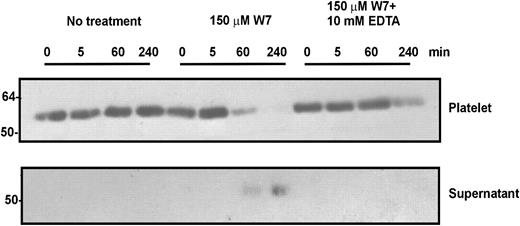
We have previously identified that calmodulin binds to a positively charged membrane-proximal sequence within the cytoplasmic tail of GPVI. This association of calmodulin with GPVI was reduced in platelets treated with GPVI agonists but not other platelet receptor agonists.13 Several other receptors including L-selectin and the epidermal growth factor (EGF) receptor are known to be associated with calmodulin via positively charged cytoplasmic sequences.29,45 Interestingly, these receptors undergo rapid extracellular domain shedding either on activation or after treatment with calmodulin inhibitors such as W7.28,46 In both instances, shedding has been proposed to be a mechanism for autoregulation of each receptor's biologic activity. We therefore investigated whether inhibition of calmodulin in platelets also resulted in a loss of GPVI from the platelet surface. Figure 5 shows that the amount of surface GPVI in suspensions of washed platelets in Tyrode buffer was stable for up to 4 hours, with no detectable levels of the 55-kDa fragment in the supernatant. However, after treatment of washed platelets with 150 μM W7 for 1 hour, more than 80% of GPVI was lost from the platelet surface coinciding with the appearance of a 55-kDa fragment in the supernatant (Figure 5). Loss of GPVI was observed at W7 concentrations from 25 to 300 μM (data not shown). This loss of GPVI from platelets was also observed with comparable concentrations of a different calmodulin inhibitor, trifluoperazine (data not shown). Similar to treatment of platelets with GPVI agonists, convulxin, CRP, and collagen, the loss of GPVI induced by calmodulin inhibition was blocked by both EDTA (Figure 5) and GM 6001 (data not shown).
Treatment of platelets with a calmodulin inhibitor induces loss of GPVI from the platelet surface. Washed human platelets were resuspended in Tyrode buffer and treated with 150 μM W7. EDTA (10 mM) was included in some incubations. Platelet suspensions were incubated at room temperature for up to 4 hours, then mixed with 10 mM EDTA (final concentration), and centrifuged to isolate platelets from the incubation medium supernatant. Platelet pellets were lysed in buffer containing Triton X-100 and proteinase inhibitors and aliquots of platelet extracts or supernatants were electrophoresed on SDS/acrylamide gels, transferred to nitrocellulose, and probed with anti-GPVI monoclonal antibody, 6B12-3. Data are representative of at least 3 experiments with different donors.
Treatment of platelets with a calmodulin inhibitor induces loss of GPVI from the platelet surface. Washed human platelets were resuspended in Tyrode buffer and treated with 150 μM W7. EDTA (10 mM) was included in some incubations. Platelet suspensions were incubated at room temperature for up to 4 hours, then mixed with 10 mM EDTA (final concentration), and centrifuged to isolate platelets from the incubation medium supernatant. Platelet pellets were lysed in buffer containing Triton X-100 and proteinase inhibitors and aliquots of platelet extracts or supernatants were electrophoresed on SDS/acrylamide gels, transferred to nitrocellulose, and probed with anti-GPVI monoclonal antibody, 6B12-3. Data are representative of at least 3 experiments with different donors.
W7 causes the dissociation of calmodulin from its GPVI interaction site
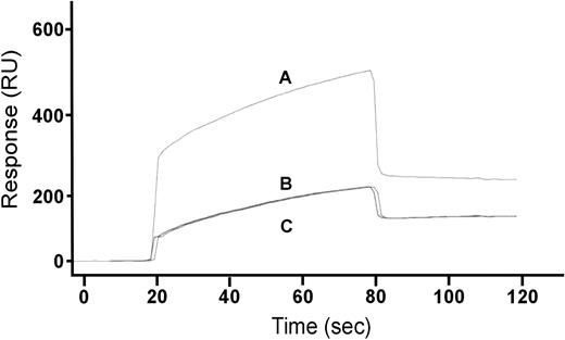
W7 is known to bind calmodulin and thus is thought to compete with other binding partners such as amphipathic positively charged peptide sequences as occur in L-selectin, the EGF receptor, and GPVI.13,29,45 To directly test whether W7 is able to disrupt calmodulin binding, surface plasmon resonance was performed using immobilized biotinylated synthetic peptides either with sequence matching the known calmodulin binding sequence of GPVI or with control peptides. Peptides were immobilized on streptavidin-coated biosensor chips and unoccupied binding sites were blocked with excess biotin. Preparations of purified calmodulin (analyte) were passed over the peptide surface and the interaction of calmodulin determined. Figure 6 shows sensorgrams depicting the interaction over time of calmodulin with immobilized GPVI peptide, but not a control GPIbα peptide (data not shown). The association of calmodulin with the immobilized peptide could be disrupted by mixing the fusion protein with 150 μM W7 or 10 mM EDTA prior to passing the analyte solution across the sensor surface.
W7 causes the dissociation of calmodulin from its GPVI interaction site. Aliquots of purified calmodulin (10 μM) were passed over a surface plasmon resonance sensor chip containing immobilized biotinylated peptide with sequence matching the calmodulin-binding region of GPVI cytoplasmic tail, as described in “Materials and methods.” The association of calmodulin in HEPES-buffered saline containing 1 mM CaCl2 (trace A) or the same buffer containing 10 mM EDTA (trace B) or calmodulin that had been mixed with 150 μM W-7 prior to passage across the chip (trace C) was measured by surface plasmon resonance.
W7 causes the dissociation of calmodulin from its GPVI interaction site. Aliquots of purified calmodulin (10 μM) were passed over a surface plasmon resonance sensor chip containing immobilized biotinylated peptide with sequence matching the calmodulin-binding region of GPVI cytoplasmic tail, as described in “Materials and methods.” The association of calmodulin in HEPES-buffered saline containing 1 mM CaCl2 (trace A) or the same buffer containing 10 mM EDTA (trace B) or calmodulin that had been mixed with 150 μM W-7 prior to passage across the chip (trace C) was measured by surface plasmon resonance.
The mechanism of release of GPVI from platelets does not require active αIIbβ3 integrin
At least one other protein, CD40 ligand (CD40L), can be proteolytically shed from the surface of activated platelets.47 Although this shedding results from stimulation of platelets with a wide variety of agonists and is thus distinct from the GPVI agonist-dependent shedding observed in the present study, additional experiments were performed to further evaluate whether a similar mechanism applied for both events. CD40L shedding from activated platelets is almost completely inhibited by blockade of αIIbβ3 integrin with RGD mimetics.48,49 In contrast, blockade of αIIbβ3-integrin function by RGD peptide had no effect on overall GPVI-dependent shedding induced by convulxin or by W7 (Figure 7). The available data therefore suggest that a specific metalloproteinase-dependent mechanism exists for the selective cleavage of GPVI on activated platelets.
Treatment of platelets with RGD peptide does not block ligand-induced loss of GPVI from platelets. Washed human platelets (5 × 10⁸/mL) were resuspended in Tyrode buffer alone or containing 100 μM RGD peptide and treated with either 0.5 μg/mL convulxin, 150 μM W7, or 1 U thrombin (Thr) for 90 minutes at room temperature. The treated platelets were then mixed with 10 mM EDTA and the platelets isolated by centrifugation. The platelet pellets were lysed in buffer containing Triton X-100 and proteinase inhibitors, and aliquots of the platelet extracts were electrophoresed on SDS/acrylamide gels, transferred to nitrocellulose, then probed with the anti-GPVI monoclonal antibody 6B12-3. This experiment is representative of at least 3 experiments with different donors.
Treatment of platelets with RGD peptide does not block ligand-induced loss of GPVI from platelets. Washed human platelets (5 × 10⁸/mL) were resuspended in Tyrode buffer alone or containing 100 μM RGD peptide and treated with either 0.5 μg/mL convulxin, 150 μM W7, or 1 U thrombin (Thr) for 90 minutes at room temperature. The treated platelets were then mixed with 10 mM EDTA and the platelets isolated by centrifugation. The platelet pellets were lysed in buffer containing Triton X-100 and proteinase inhibitors, and aliquots of the platelet extracts were electrophoresed on SDS/acrylamide gels, transferred to nitrocellulose, then probed with the anti-GPVI monoclonal antibody 6B12-3. This experiment is representative of at least 3 experiments with different donors.
Discussion
In the present study, we have made the novel finding that treatment of platelets with specific GPVI agonists such as collagen, convulxin, and CRP not only results in platelet activation but also subsequent proteolytic shedding of GPVI. Concomitant with loss of GPVI from the platelet surface, a soluble 55-kDa fragment of GPVI was detected in the platelet supernatant. Convulxin was the most powerful inducer of GPVI proteolysis, with more than 90% of the platelet GPVI shed within 30 minutes (Figure 1A). The relative abilities of different GPVI agonists to induce GPVI shedding correlates with their ability to induce tyrosine phosphorylation, platelet aggregation, and other readouts of platelet activation.6,11 The latter are likely to depend on the multimeric nature of each GPVI ligand and the relative ability of the GPVI agonist to induce clustering or cross-linking (or both) of the receptor.1,2,8 Our results imply that cross-linking may also facilitate shedding although further studies are required to confirm this possibility. The ligand-induced loss of GPVI could be abrogated by both EDTA and a broad range metalloproteinase inhibitor, but not by inhibitors of calpain, pepsin, and cathepsins (leupeptin and pepstatin) or trypsin (soybean trypsin inhibitor; Figure 3A), suggesting that release of the GPVI ectodomain was mediated by a platelet-derived metalloproteinase.
Three lines of evidence suggest GPVI shedding involves a specific mechanism regulated by GPVI-associated calmodulin. First, only GPVI-binding ligands, but not other platelet agonists (thrombin, von Willebrand factor), induced loss of GPVI from platelets and appearance of the soluble GPVI fragment in the supernatant (Figure 1). In contrast, collagen, convulxin, and CRP did not cause depletion of other platelet membrane glycoproteins under conditions where GPVI was completely lost (Figures 1, 2). Second, the metalloproteinase-dependent shedding of GPVI could be induced in the absence of ligand by treating platelets with the calmodulin inhibitor, W7 (Figure 5). Again, loss of GPVI was specific, and W7 blocked binding of calmodulin to a peptide based on the calmodulin-binding sequence of GPVI in vitro (Figure 6). Convulxin-induced dissociation of calmodulin from GPVI is at least partly inhibitable by PP1,13 implying an activation-dependent mechanism of calmodulin release and is consistent with the present study, where inhibition of GPVI-related signaling molecules Src, PI3, and Syk kinases also inhibits shedding (Figure 3B). Third, the mechanism for shedding is apparently different from that reported for the other platelet receptors, such as GPIb-IX-V42 or CD40L.48 In the case of CD40L, shedding was dependent on αIIbβ3 integrin and inhibitable by RGD mimetics. In contrast, GPVI shedding was not affected by RGD-dependent blockade of αIIbβ3 integrin.
The proteolytic shedding of adhesion receptors is a well-documented autocrine mechanism to control receptor activity21,22,27,50 and, for platelets, functional autocrine loops mediated by secreted factors such as adenosine diphosphate (ADP) and CD40L are well established.47,51-53 Recent work by Bergmeier and coworkers examining a platelet model of mitochondrial injury has linked the profound proteolytic shedding of GPIbα observed in this system to a platelet-derived metalloproteinase.42 The authors proposed that levels of soluble GPIbα ectodomain in plasma could be used as an indicator of platelet hemostatic status and function. More recently, Boylan and colleagues54 reported the detection of a 52-kDa fragment of GPVI from the plasma of healthy individuals, indicating that a soluble form of GPVI occurs in normal human plasma. In other systems, the release of soluble adhesion proteins, particularly the ligand-binding domains of proteins including vascular cell adhesion molecule 1 (VCAM-1), intercellular adhesion molecule 1 (ICAM-1), L-selectin, and P-selectin have been used as clinical markers of vascular disorders.55 Much less is known about the extent to which the shedding of specific adhesion proteins modulates cell adhesion.
The loss of GPVI observed here, together with the work of Nieswandt and colleagues15 demonstrating lack of responsiveness to collagen in vivo by murine platelets depleted in GPVI by treatment with the rat monoclonal antibody, JAQ1, implies that removal of GPVI from platelet membranes would significantly attenuate platelet interactions with collagen. Chen and colleagues7 also suggested human platelets may respond to collagen in a graded fashion depending on GPVI receptor density. The GPVI-calmodulin interaction provides a potential mechanism for liberation of the 55-kDa ectodomain fragment of GPVI, and therefore modulation of GPVI function. Calmodulin is a ubiquitous intracellular regulatory protein.56,57 Within platelets, calmodulin has been identified as a cytoplasmic binding partner for GPVI13 and dissociates from the receptor by ligand binding to GPVI.13,14 Data presented here (Figure 6) suggest that W7 or EDTA can also cause dissociation of calmodulin from a calmodulin-binding peptide based on GPVI. W7 binds specifically to calmodulin (Kd ∼11 μM) and competes directly with the binding of calmodulin-dependent proteins.58,59 The dissociation of calmodulin from cytoplasmic sequences of membrane-expressed receptors could represent a specialized control mechanism for proteolytic shedding.28 We used W7 treatment as an approach to investigate potential mechanisms underlying GPVI shedding, rather than trying to study the effect of W7 on platelet function. W7 has been reported to have other inhibitory actions on platelets, for example, blocking collagen-dependent platelet aggregation.60
The identity of the metalloproteinases on platelets responsible for GPVI ectodomain shedding, as well as the mechanism by which the metalloproteinase influences levels of GPVI, remain to be established. Four members of the matrix metalloproteinase (MMP) family, MMP-1, MMP-2, MMP-4, and MMP-9, have been identified in platelets.61-64 Platelets also express at least one disintegrin and metalloprotease (ADAM) family member, ADAM10.65 Interestingly, a role for platelet-derived MMP-161 and MMP-262,66 has recently been described in modulating platelet intracellular signaling and aggregation induced by several platelet agonists, although the precise mechanism remains to be elucidated. Although several other platelet membrane proteins in addition to GPVI are released either via proteolysis or within platelet microparticles,42,67-71 additional studies are clearly necessary to evaluate the physiologic significance of this in regulating platelet function.
During submission of this manuscript, a paper by Bergmeier and colleagues72 describing loss of GPVI from murine platelets in a mitochondrial injury model was published. This also involved a metalloproteinase-dependent mechanism generating a soluble 55-kDa fragment of GPVI.
Prepublished online as Blood First Edition Paper, August 12, 2004; DOI 10.1182/blood-2004-04-1549.
Supported by the National Health and Medical Research Council of Australia, Monash University, and the National Heart Foundation of Australia.
The publication costs of this article were defrayed in part by page charge payment. Therefore, and solely to indicate this fact, this article is hereby marked “advertisement” in accordance with 18 U.S.C. section 1734.
We thank Dr Yang Shen for providing the GST-calmodulin construct and Ms Andrea Aprico for outstanding technical assistance.


This feature is available to Subscribers Only
Sign In or Create an Account Close Modal