Abstract
Background: Folate receptor (FR) is a recently recognized tumor marker that is being investigated for targeted delivery of drugs, imaging of cancer cells, and tumor immunotherapy. The small size of the molecule, simple conjugation chemistry and lack of immunogenicity make folate an ideal candidate for targeted delivery to tumors bearing the FR (Lu Y, Low PS. Folate-mediated delivery of macromolecular anticancer therapeutic agents. Adv Drug Deliv Rev. 2002 Sep 13;54(5):675–93). FR is overexpressed in 90% of ovarian cancers and in other human tumors including endometrial, colorectal, breast, and neuroendocrine carcinomas, it is negatively or only weakly expressed in most normal tissues including some myeloid cells. In view of the above we wished to learn if overexpression of FR by marrow plasma cells (PC) might differentiate MM from MGUS and SMM.
Methods: We obtained marrow biopsy sections from 20 patients each with MM, MGUS, or SMM, and 11 normals and stained them by immunohistochemistry using a FR-beta antibody. Expression was graded 0 (negative), 1 (equivocal), 2 (positive) or 3 (strong positive) based on immunohistochemical staining of marrow PC. We recorded the percent PC, percent positive PC, and the staining of other cell types including myeloid, erythroid, lymphocytes, macrophages, megakaryocytes and control tissues.
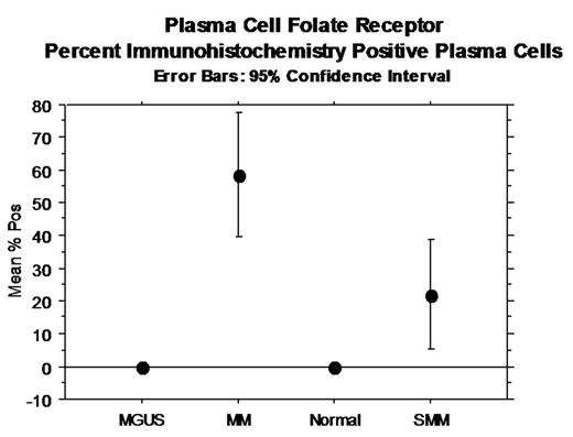
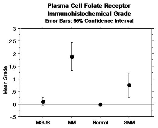
Results: PC of patients with MM positively expressed FR compared to MGUS, SMM and normal PC (p<0.0001) (Figure 1 and 2). Of interest, 6 of 20 SMM patients showed positive expression. Maturing myeloid cells were also positive, acting as internal positive controls. Macrophages were often positive; some endothelial cells were positive; early erythroid precursors were rarely positive; megakaryocytes, maturing erythroid precursors and lymphocytes did not express FR. The chromosomal location of the FOLR gene cluster is 11q13. Gene expression arrays of purified PC failed to show evidence of overexpression of FOLR1, FOLR2, or FOLR3. We are currently performing Western blots to examine levels of protein product of purified PC.
Conclusion: Immunohistochemistal studies show differential expression of FR in MM PC compared to absent or equivocal expression in MGUS and normal PC and variable expression in SMM. The data suggest that FR may be used as a diagnostic tumor marker and has implications for development of folate-based tumor imaging, drug delivery, and immunotherapy.
Author notes
Corresponding author


This feature is available to Subscribers Only
Sign In or Create an Account Close Modal