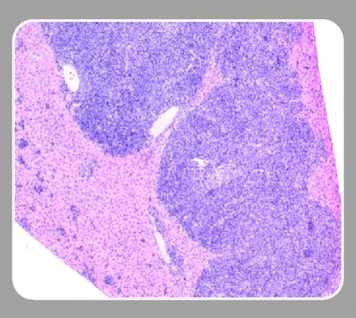
Common B-cell non-Hodgkin lymphomas (B-NHLs) arise by malignant transformation of defective germinal center (GC)–stage B cells.1 These lymphomas include follicular lymphoma (FL) and more aggressive Burkitt (or Burkitt-variant) and diffuse large B-cell lymphomas. A characteristic t(14;18) is detectedin over 80% of FL, which causes overexpression of the BCL2 cell survival gene by rearrangements with immunoglobulin (Ig) heavy chain locus control elements. Initially indolent, FL may transform to a more aggressive tumor with increasingly complex cytogenetics over time.
FL is also the most common B-cell lymphoma naturally arising in old mice and in Eμ-Pim-1 transgenic mice. White pulp expansions of sIgM+/B220+/CD19+ GC B cells usually begin in the spleen and may contain large (centroblasts, immunoblasts), small (centrocytes), or a mixture of large and small tumor cells that lose the usual GC spatial relationships. Unlike human FL, naturally occurring mouse FLs are not associated with Bcl2 gene overexpression and do not display a typical follicular pattern. Despite several attempts, a BCL2-based model of FL has not been generated in mice, until now.
In this issue of Blood, Egle and colleagues (page 2276) describe a new model of FL by Bcl2 overexpression using VavP control sequences. About 15% to 25% of mice developed a syndrome resembling autoimmune glomerulonephritis that was strain dependent. However, 37% to 50% of mice developed FL by 18 months of age following a florid GC hyperplasia.2 Other hematologic tumors occurred at lower frequencies, including plasma cell tumors, lymphoblastic or large B-cell lymphoma, thymic lymphoma, and histiocytic sarcoma. Interestingly, levels of the Bcl2 transgene expression were independent of lymphomagenesis; rather, CD4+ T-cell help appeared essential for FL.
There are several exciting features in this valuable genetic model of human FL. The key seems to be panlymphoid Bcl2 expression and time, which yield increased numbers of CD4+ T cells that support robust GC B-cell expansions. Antigenic stimulation through surface Ig (sIg), with associated somatic hypermutation (SHM), has been described previously in human FL but not in mouse FL, which usually lacks SHM and GC-signature Bcl6 protein expression.3 Egle and colleagues show that antigenic selection is ongoing in this model and that the FL cells express the proliferating cell nuclear antigen (PCNA) GC-signature marker, features similar to human FL. Furthermore, Egle et al suggest that increased CD4+ T cells in VavP-Bcl2, but not Eμ-Bcl2, transgenic mice support the premalignant GC expansion required to generate enough apoptosis-resistant B cells for a secondary transforming mutation. If this hypothesis is correct, why are T-cell expansions not part of human follicular lymphomagenesis? Is there a T-cell help mechanism in human GCs that is so powerful that it obviates the need for excess CD4+ T cells beyond those usually present? And what are the additional genetic/epigenetic mistakes that complement Bcl2 overexpression to cause mouse FL? Do they have similar counterparts in human FL? This model, and one other that causes a spectrum of GC-based B-cell malignancies by overexpression of the TCL1 oncogene in both B and T cells,4 provide systems for determining some of the most difficult mechanisms in early GC B-cell transformation.


This feature is available to Subscribers Only
Sign In or Create an Account Close Modal