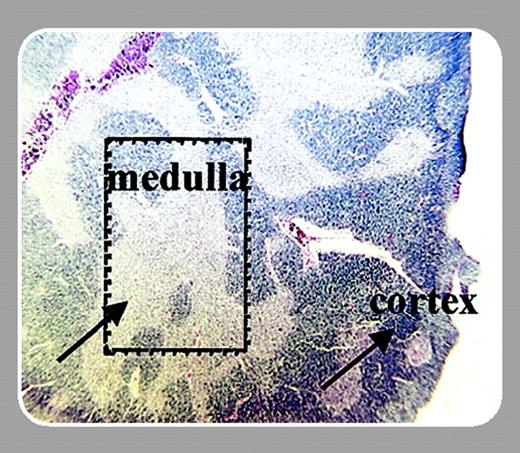
Models of leukemic transformation have focused on binary phenomena: either loss of function of tumor suppressors or gain of function via high-level, dysregulated expression of a dominant transforming oncogene or an abnormal oncogenic fusion protein. The AKR/J mouse is a model of susceptibility to T-lymphoid malignancies. AKR mice develop thymic lymphomas spontaneously or rapidly after neonatal challenge with transforming murine retroviruses, such as the T-lymphomagenic strain SL3-3.1 The mechanisms of increased susceptibility to transformation of AKR lymphohematopoietic progenitors are unknown.
On page 1985 of the present issue of Blood, Laouar and colleagues describe differences in expression of a cytokine receptor, the interleukin 7 receptor (IL-7R), between thymocytes from AKR mice and other strains not prone to lymphoma. The IL-7R is composed of the IL-7Rα and common gamma chain (γc) subunits. Signal transduction through the IL-7R, like other γc-containing receptors (IL-2R, IL-4R, IL-9R, IL-15R, IL-21R), involves activation of the Janus kinase–signal transducer and activator of transcription (Jak-STAT) and other signaling pathways. Engagement of the IL-7R with IL-7 produced by thymic epithelial cells is critical for multiple processes in thymocyte development, including induction of both proliferation and survival signals, and differentiation events such as V(D)J recombination of the T-cell receptor genes.
Laouar et al demonstrated that AKR thymocytes express IL-7Rα mRNA and surface protein at significantly higher levels than do other strains of mice. The overexpression of IL-7Rα was specific, as expression of γc was unaltered. Overexpression of IL-7Rα altered the proportions of immature thymocytes and led to increased survival of AKR thymocytes in vitro. Importantly, bone marrow chimeras containing lymphohematopoietic progenitors from both AKR and non-susceptible strains selectively produced thymocytes derived from the AKR population, while maintaining fixed proportions of non-lymphoid cells. Thus, the AKR-derived thymocytes have a selective advantage in vivo and in vitro over normal cells.
With some caveats, Laouar et al's data strongly suggest that high-level IL-7R expression is a causal event in leukemogenesis. An autocrine or paracrine signaling loop is unlikely since thymocytes do not themselves produce IL-7. A signaling mutation of the IL-7Rα that also affects expression was not formally excluded. The high-level expression of IL-7Rα could be an epiphenomenon downstream of an event that leads to increased expression of other survival signals, as well as IL-7Rα.
Type I cytokine receptors like IL-7R are not thought to be oncogenic, because they lack intrinsic receptor tyrosine kinase activity and need ligand engagement for signal transduction. However, graded differences in signal intensity may be oncogenic. For example, the increased risk of leukemia observed in granulocyte colony-stimulating factor (G-CSF)–treated patients with severe congenital neutropenias may be related to the effects of coding sequence mutations of the G-CSF receptor, leading to enhanced susceptibility to the growth-promoting effects of high-dose G-CSF.2 Recently, T-cell leukemias were described in 2 patients who received gene therapy for γc-deficient severe combined immune deficiency (X-SCID).3 The X-SCID gene therapy patients were successfully reconstituted with CD34+ hematopoietic progenitors transduced with a constitutively expressed wild-type γc cDNA under the control of a murine leukemia virus promoter. Elegant analyses of the leukemia cells demonstrated that proviral insertion had occurred near the LMO-2 proto-oncogene locus. What remains unexplained is why leukemia has been observed only in an X-SCID gene therapy trial, and not in other diseases treated with similar vectors carrying different transgenes. Is it possible that high-level expression of γc, which by itself is not thought to be a transforming gene, led to increased growth factor stimulation via IL-7R or another receptor, thereby promoting the outgrowth of cells containing these insertions?
The relevance of the results of Laouar et al to human leukemogenesis is unknown. The authors' observations indicate that naturally occurring quantitative differences in signal transduction can promote abnormal cell growth and survival. Analyses of single nucleotide polymorphism (SNP) and associated quantitative trait loci (QTL) for human leukemia susceptibility may uncover quantitative differences in gene expression of the sort described by Laouar et al. It will be fascinating to see whether variations in the genome that promote intensity of cytokine receptor signaling, as suggested in the inbred AKR mouse strain, will be associated with increased susceptibility to cancers in humans.


This feature is available to Subscribers Only
Sign In or Create an Account Close Modal