Abstract
Factor V (FV), a central regulatory protein in hemostasis, is distributed into distinct plasma and platelet compartments. Although platelet FV is highly concentrated within the platelet α-granule, previous analysis of human bone marrow and liver transplant recipients has demonstrated that platelet FV in these individuals originates entirely from the uptake of plasma FV. In order to examine further the biosynthetic origins of the platelet and plasma FV pools, we performed bone marrow transplantations of Fv-null (Fv–/–) fetal liver cells (FLCs) into wild-type mice. Fractionation of whole blood from control mice demonstrated that approximately 14% of total blood FV activity is platelet-associated. Mice that received transplants of Fv-null FLCs displayed a high degree of engraftment and appeared grossly normal, with no evidence for spontaneous hemorrhage. Although total FV levels in Fv-null FLC recipients were only mildly decreased, the FV activity within the platelet compartment was reduced to less than 1% of that in normal mice. We conclude that the murine platelet FV compartment is derived exclusively from primary biosynthesis within cells of marrow origin, presumably megakaryocytes, and that an intact platelet FV pool is not required for protection from spontaneous hemorrhage or bleeding following minor trauma.
Introduction
Coagulation factor V (FV) is an essential protein required for hemostasis. Plasma FV circulates as a 330-kDa single-chain procofactor and undergoes site-specific proteolytic activation to FVa by thrombin cleavage. Activated FV (FVa) serves as a cofactor for activated factor X (FXa). In the absence of FVa, the catalytic efficiency of FXa is reduced by at least 3 orders of magnitude. Bound together with calcium on a phospholipid surface, these 2 proteins form the prothombinase complex responsible for converting prothrombin to activated thrombin (reviewed in Mann and Kalafatis1 ). Patients deficient in FV suffer a moderate to severe bleeding disorder,2 although most patients have low but detectable residual levels of FV. Absolute deficiency of FV in mice results in a lethal phenotype with approximately 50% dying in utero and approximately 50% dying at birth.3 FVa also is central to a physiologic anticoagulation pathway, serving as the primary substrate target for proteolytic inactivation by activated protein C (APC).1,4 FV Leiden, a common human polymorphism substituting Gln at the initial site of APC cleavage within FV (Arg506), results in slowed inactivation of FVa by APC and is associated with a predisposition to thrombosis.4
Whole human blood contains 7 μg/mL FV, with approximately 80% located in the plasma and the remaining 20% contained within platelet α-granules (4:1 ratio).5 The platelet FV pool is released upon platelet activation, along with other α-granule contents,6 potentially providing a high local concentration of FV on the platelet surface at sites of vascular injury. Although the level of plasma FV appears to correlate poorly with severity of bleeding symptoms in FV-deficient patients,2 platelet FV levels may be a better predictor.7 The absence of significant bleeding in a patient with markedly reduced plasma FV due to a circulating anti-FV autoantibody8 and the particular effectiveness of platelet transfusion in the treatment of hemorrhage associated with FV deficiency9 further supports the critical role of platelet FV in normal hemostasis. Also consistent with a distinct role for platelet FV, patients with FV Quebec10 demonstrate degradation of their platelet FV associated with significant bleeding, while their plasma FV appears relatively normal. However, this disorder recently has been shown to be associated with generalized proteolysis of platelet α-granule contents, likely due to mislocalization of a urokinase-like protease.11-13
Although the liver appears to be the primary source of plasma FV,14 FV biosynthesis also has been detected in human lymphocytes,15 cultured bovine endothelial cells,16 and vascular smooth muscle cells.17 Several lines of evidence had previously suggested that platelet FV is synthesized primarily within the megakaryocytes. FV is highly concentrated within the platelet pool relative to plasma,5 similar to several proteins known to be synthesized within the megakaryocyte, including von Willebrand factor and platelet factor 4.18,19 In addition, FV mRNA has been detected within megakaryocytes,20,21 and biosynthesis of FV protein has been demonstrated in isolated megakaryocytes.21-23 However, the work of Camire and colleagues24 has changed this view. Using FV Leiden as a marker in a human bone marrow transplant recipient and a second patient who received a liver transplant, platelet FV was found to reflect the liver genotype, suggesting that platelet FV is derived entirely through uptake from plasma FV. Although these results imply a highly efficient mechanism for the specific endocytosis and targeting of FV to the α-granule, no uptake of labeled FV by resting platelets could be demonstrated,24 suggesting that transfer from the plasma compartment must occur in the megakaryocyte.
In order to further explore the cellular origin and functional significance of platelet FV, we generated mice deficient in megakaryocyte/platelet FV synthesis by bone marrow transplantations of fetal liver cells (FLCs) from Fv-null mice into irradiated wild-type C57BL/6J mice. Our results demonstrate that murine platelet FV is derived entirely from primary biosynthesis in the megakaryocyte, with no evidence for significant uptake of FV from plasma.
Materials and methods
Isolation of FLCs
Fetuses from a Fv+/–X Fv+/–intercross3 were collected at 17.5 days after coitus. FLC suspensions from each individual fetus were prepared according to a previously described procedure.25 Briefly, livers from each fetus were separated from other tissues and placed in individual Petri dishes containing 2 mL RPMI media supplemented with 2% fetal calf serum (FCS) (GIBCO-BRL, Gaithersburg, MD). Unless otherwise noted, all manipulations with the FLCs were performed at 4°C. Liver tissue was disrupted into a suspension, transferred into individual 2.0 mL cryogenic storage vials (Corning, Corning, NY), washed twice with 1 mL cold RPMI/2% FCS, and resuspended in cryopreservation media (65% RPMI, 25% FCS, 10% dimethyl sulfoxide [DMSO]). Vials were frozen at –70°C for at least 3 hours before transfer to storage at –135°C. The genetic background of the donor FLCs was 74.2% C57BL/6C, 25% SJL, and 0.8% 129Sv.
Genomic DNA was prepared from tail tissue as previously described26 and Fv genotype determined by polymerase chain reaction (PCR) amplification using oligonucleotide primers: 5′-ACTTGGCAAAACGGGTTAGAATGG-3′ (primer i7B) in intron 7 of the Fv gene and common to both Fv–wild-type and Fv-null alleles; 5′-CGACTGTGGCCGGCTGGGTGTGG-3′ (primer neoA1) in the neomycin resistance gene of the Fv-null allele; and 5′-CAGTGCGTTTGGTGAAGGTCTCT-3′ (primer e8B) in the Fv gene's eighth exon (which is deleted in the Fv-null allele).3 All oligonucleotides were synthesized by the DNA Synthesis Core Service at the University of Michigan. All 3 primers are used simultaneously under conditions of 94°C, 30 seconds; 58°C, 1 minute; 72°C, 1 minute for 40 cycles. Amplification of sequence between i7B and e8B from the Fv–wild-type allele yields a 630–base pair (bp) product, whereas i7B and neoA1 yield a 470-bp product from the Fv-null allele.
Immediately prior to transplantation, cells were thawed rapidly by 2 minutes of incubation at 37°C, centrifuged at 1200 rpm for 10 minutes, and washed in 1 mL RPMI with 2% FCS. After a second centrifugation, cells were resuspended in 1 mL RPMI without FCS, and cell counts determined on a hemacytometer under light microscopy. Individual cell suspensions were diluted to 1.3 × 10⁷ cells/mL in RPMI media.
Bone marrow transplantation
Female 13-week-old specific-pathogen–free (SPF) C57BL/6J mice (Jackson Laboratories, Bar Harbor, ME) received two 650 rad doses separated by 3 hours in a Gammacell 40 Cs-137 irradiator (Nordion International, Kanata, ONT, Canada). Under Metofane (Mallinckrodt Veterinary, Mundelein, IL) anesthesia, mice receiving transplants each received 4 × 10⁶ fetal liver cells in 300 μL RPMI via injection into the retro-orbital sinus with a 25-gauge needle. Mice were housed in the SPF facility at the University of Michigan and fed acidified water (6 mM HCl, pH∼2.3) for 1 week prior and up to 6 weeks after the transplantation procedure.
Assay for engraftment in Fv-null FLC recipient mice
The degree of engraftment was assayed as the percentage of Fv-null alleles in the peripheral blood cell DNA of a recipient mouse as measured by competitive PCR. At 7 weeks after transplantation, blood was collected by tail transection at a 5/128-inch-diameter cross section (measured by passing the tail through a stencil) with drops of blood from the wound blotted onto cards of S&S 903 paper (Schleicher & Schuell; Keene, NH). A second set of blood samples on S&S 903 paper was collected 2 months after transplantation from the inferior vena cava for mice R and S and 5 months after transplantation from a tail wound for mice J through Q. Blood spots were amplified by PCR after processing with the Dry Blood Extraction Kit (Bio-Rad, Hercules, CA) according to the manufacturer's instructions. Cutting instruments were washed with Eliminase (Fisher, Pittsburgh, PA) and rinsed between each sample. PCR primers and conditions are identical to those described in “Isolation of FLCs,” except that primer i7B was 32P end-labeled with polynucleotide kinase (New England Biolabs, Beverly, MA), samples separated on a 4% denaturing polyacrylamide gel, and signals quantitated on a phosphorimager using ImageQuant software (Molecular Dynamics, Sunnyvale, CA). A standard curve was generated from control Fv-wild-type and Fv-null allele PCR products purified by the QiaQuick PCR Purification Kit (Qiagen, Santa Clarita, CA) as templates.
Gel filtration of mouse platelets
Under anesthesia, 0.5 mL to 1.0 mL of blood was drawn from the inferior vena cava via a 25-gauge needle into a syringe containing 0.2 mL acid-citrate dextrose (ACD) and 10 μL 500 μM prostaglandin E1 (Sigma, St Louis, MO). The citrated blood was spun 160g × 10 minutes in a tabletop centrifuge at 20°C to prepare platelet-rich plasma (PRP). Mouse platelets were purified from PRP by gel filtration27 on a column containing a bed volume of 7 mL washed Sepharose 2B beads (Pharmacia Biotech, Piscataway, NJ) between 2 sheets of 30-μm pore Nitex membrane (Tetko, Kansas City, MO) held in place with rubber gaskets constructed from a 10-mL syringe piston. Columns were stored in 0.9% saline and 20% ethanol and washed and equilibrated with degassed solutions of 0.9% saline and platelet buffer (134 mM NaCl, 12 mM NaHCO3, 2.9 mM KCl, 0.34 mM Na2HPO4, 1 mM MgCl2, 10 mM HEPES [N-2-hydroxyethylpiperazine-N′-2-ethanesulfonic acid], 5 mM glucose, 3% bovine serum albumin, pH = 7.4; filter-sterilized before use).
The PRP was loaded and eluted with platelet buffer. Platelet counts were determined on a hemacytometer under phase-contrast microscopy. Fractions were separated into supernatant (plasma) and pellet (platelet) components by centrifugation at 1500g × 10 minutes on an approximately 40% bovine serum albumin cushion.28 Pellets were resuspended in platelet buffer.
Assays for FV activity
FV coagulation activity assays were performed as previously described.20 Standard curves were generated with pooled human plasma (1000 mU/mL) (George King Bio-Medical; Overland Park, KS) for each lot of FV-deficient plasma (George King Bio-Medical) or activated partial thromboplastin time (APTT) reagent (thromboplastin with calcium [Sigma] or Simplastin Excel [Organon-Teknika, Durham, NC]). Platelet-associated FV activity in the platelet peak fraction was divided by the platelet count in that fraction to yield FV activity per platelet. Whole blood platelet-associated FV activity was determined by multiplying the FV activity per platelet times the whole blood platelet count. Whole blood plasma-associated activity was calculated by subtracting the platelet-associated activity of the PRP from total PRP FV activity and correcting for hematocrit and the volume of added anticoagulant.
A modified 2-stage FV activity assay also was performed for plasma and platelet fractions from control C57BL/6J mouse samples B through E (Table 1). Test samples were incubated with 1 National Institutes of Health (NIH) U/mL mouse thrombin (Sigma) for 5 minutes at room temperature. Test samples were then diluted 1:20 in buffer and a 50-μL aliquot mixed 1:1 with human FV–deficient plasma (George King Bio-Medical) and warmed for 3 minutes at 37°C. Thromboplastin (200 μL) with 25 mM calcium (Sigma) was then added and the time to clot formation measured in an Electra 750 fibrinometer (Medical Laboratory Automation, Pleasantville, NY). A standard curve was generated using dilutions of pooled normal C57BL/6 mouse plasma. Comparison of the 1- and 2-stage assays and Western blot data indicated that the 1-stage assay overestimated platelet FV activity by a factor of approximately 3.2 (see “Results”). Insufficient material was available from the transplant recipients (mice F-S in Table 1) to repeat the FV activity measurements using the 2-stage assay.
Factor V activity in platelet pool of control mice and mice that received transplants
. | Whole blood FV activity, mU/mL* . | Platelet FV activity, mU/mL* . | % Platelet FV pool† . | % Platelet FV pool‡ . |
|---|---|---|---|---|
| Wild-type C57BL/6J | ||||
| A | 5015 | 1650 | 33 (10) | — |
| B | 5026 | 3845 | 77 (24) | 16 |
| C | 2788 | 758 | 27 (8) | 26 |
| D | 4797 | 2998 | 63 (19) | 6 |
| E | 5535 | 1212 | 22 (7) | 7 |
| Mean ± SD | 4632 ± 1066 | 2047 ± 1289 | 44 ± 24 (14) | 14 ± 9 |
| Wild-type Fv FLC recipients | ||||
| F | 3648 | 1893 | 52 (16) | — |
| G | 2102 | 1322 | 63 (20) | — |
| H | 5017 | 2187 | 44 (14) | — |
| I | 5104 | 1199 | 24 (7) | — |
| Mean ± SD | 3968 ± 1411 | 1804 ± 469 | 45 ± 17 (14) | — |
| Fv-null FLC recipients | ||||
| J | 4151 | 10 | 0.2 (<0.1) | — |
| K | 1579 | 20 | 1.3 (0.4) | — |
| M | 2265 | 3 | 0.1 (<0.1) | — |
| O | 4628 | 2 | <0.1 (<0.1) | — |
| P | 2192 | 6 | 0.3 (<0.1) | — |
| Q | 5282 | 5 | 0.1 (<0.1) | — |
| R | 3997 | 1 | <0.1 (<0.1) | — |
| S | 4092 | 9 | 0.2 (<0.1) | — |
| Mean ± SD | 3523 ± 1330 | 10 ± 6 | 0.3 ± 0.4 (0.1) | — |
— indicates not done.
FV activity measured with a 1-stage assay in whole blood and in platelets.
Size of the platelet FV pool expressed as percent of the total activity measured in whole blood. Corrected estimates of the platelet FV pool size based on the 2-stage FV assay are shown in parentheses (see “Materials and methods”).
2-stage assay.
Western blotting
Samples were separated by electrophoresis on a 4%-15% sodium dodecyl sulfate (SDS)–polyacrylamide gradient Pharmacia Phastgel (factor V) or a 5%/10% SDS-polyacrylamide minigel (fibrinogen), and Western blotting performed as previously described20 or per manufacturer's instructions. Murine FV was detected with an affinity-purified rabbit anti–mouse FV polyclonal antibody,20 followed by a peroxidase-tagged mouse anti–rabbit IgG antibody (Accurate, Westbury, NY) and developed with the ECL Chemiluminescence Kit (Amersham; Arlington Heights, IL). Fibrinogen was detected with a goat anti–mouse fibrinogen antibody (Accurate), followed by a peroxidase-tagged rabbit anti–goat IgG antibody (Zymed, South San Francisco, CA), and developed with the ECL Chemiluminescence Kit. Signal strength was quantified densitometrically and analyzed using ImageQuant software.
Results
Fetal liver cell transplantation in irradiated mice
Ten control irradiated mice that did not receive transplants succumbed to complications within 8 to 14 days of the procedure. Although one Fv–wild-type FLC recipient died under anesthesia during the transplantation procedure, the remaining 18 mice that received transplants survived (8 receiving Fv-wild-type FLCs, 10 receiving Fv-null FLCs). All mice that received transplants appeared healthy without evidence of spontaneous hemorrhage. One Fv–wild-type FLC recipient and one Fv-null FLC recipient died at 4.5 and 6 months after transplantation, respectively.
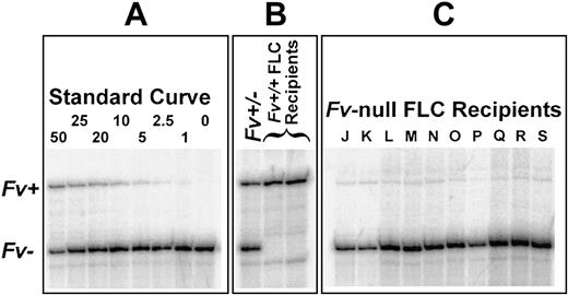
Mice that received transplants were not thrombocytopenic (1.26 × 10⁶ platelets/μL ± 0.12 SE) compared to control mice that did not receive transplants (9.03 × 10⁵ platelets/μL ± 0.51 SE; Student t test, P > .6), and no spontaneous hemorrhage or increased bleeding following routine tail transection was observed. Competitive PCR analysis of peripheral blood from Fv-null FLC recipients demonstrated that 82.5% to 98.9% of circulating leukocytes were of donor origin (Figure 1). Samples obtained at later time points showed higher levels of engraftment (94.3%-98.9%).
PCR quantitation of transplant engraftment. Competitive PCR amplification of Fv-wild-type (Fv+) and null (Fv–) alleles was performed as described in “Materials and methods.” The Fv+ gene product is 630 bp, and the Fv– product is 470 bp. Numbers for the standard curve (A) are the molar percentage of Fv+ template. (B) Approximately equal amounts of Fv+ and Fv– product are seen in a heterozygote (Fv+/–) DNA sample, as expected, with no Fv– allele detected in recipients of Fv-wild-type (Fv+/+) FLCs. (C) Only a faint signal from residual host Fv+ cells (1.1%-5.7%) is evident in each of the 10 Fv-null FLC recipients (J-S).
PCR quantitation of transplant engraftment. Competitive PCR amplification of Fv-wild-type (Fv+) and null (Fv–) alleles was performed as described in “Materials and methods.” The Fv+ gene product is 630 bp, and the Fv– product is 470 bp. Numbers for the standard curve (A) are the molar percentage of Fv+ template. (B) Approximately equal amounts of Fv+ and Fv– product are seen in a heterozygote (Fv+/–) DNA sample, as expected, with no Fv– allele detected in recipients of Fv-wild-type (Fv+/+) FLCs. (C) Only a faint signal from residual host Fv+ cells (1.1%-5.7%) is evident in each of the 10 Fv-null FLC recipients (J-S).
Analysis of murine platelet and plasma FV pools
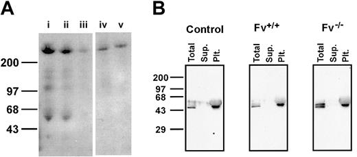
Whole blood FV activity levels and platelet/plasma distribution were measured in 5 unirradiated C57BL/6J mice that did not receive transplants, 4 Fv-wild-type FLC recipients, and 8 Fv-null FLC recipients (Figure 2; Table 1). The average FV activity in unirradiated C57BL/6J mice that did not receive transplants was 4632 mU/mL (as measured by a one-stage FV activity assay), with approximately 44% of the activity being platelet-associated. Values obtained from Fv–wild-type FLC recipients were not significantly different from those of unirradiated mice that did not receive transplants (3968 mU/mL [Student t test, P > .4], with approximately 45% of the activity being platelet associated). However, Western blot analysis of plasma and platelet FV indicates that murine plasma contains approximately 6- to 8-fold more FV antigen than the equivalent volume of platelets (Figure 3A). These data suggested that the one-stage FV activity assay was reporting a higher relative activity for platelet-associated FV, perhaps due to a partially cleaved state within the platelet. Therefore, we used a modified 2-stage FV activity assay that incorporated complete activation with murine thrombin prior to assay determination (see “Materials and methods”). The 2-stage assay, performed on 4 wild-type C57BL/6 mice (Table 1), demonstrated that 14% +/– 9% of the PRP FV activity was platelet-associated, in excellent agreement with the relative distribution of FV antigen in the platelet and plasma pools, as detected by Western blotting (Figure 3A).
Distribution of FV activity in mice that received transplants. One mouse each of unirradiated controls (left panel; Table 1, mouse A), Fv-wild-type (Fv+/+) FLC recipients (middle panel; Table 1, mouse F), and Fv-null (Fv–/–) FLC recipients (right panel; Table 1, mouse J) are shown. Total FV activity per fraction (dark gray) and the pellet-associated component of the total FV activity (light gray) both measured with the one-stage assay (“Materials and methods”) are plotted for each fraction. The platelet count for each fraction is also plotted (black line). Little or no platelet-associated FV activity is seen in fractions from the Fv-null FLC recipient.
Distribution of FV activity in mice that received transplants. One mouse each of unirradiated controls (left panel; Table 1, mouse A), Fv-wild-type (Fv+/+) FLC recipients (middle panel; Table 1, mouse F), and Fv-null (Fv–/–) FLC recipients (right panel; Table 1, mouse J) are shown. Total FV activity per fraction (dark gray) and the pellet-associated component of the total FV activity (light gray) both measured with the one-stage assay (“Materials and methods”) are plotted for each fraction. The platelet count for each fraction is also plotted (black line). Little or no platelet-associated FV activity is seen in fractions from the Fv-null FLC recipient.
Western blot analysis of plasma and platelet FV and platelet fibrinogen. (A) Samples prepared from pooled C57BL/6 mice are shown as follows, with concentrations reported as a percentage relative to undiluted plasma: (i) 20% solution of platelet-rich plasma; (ii) 20% platelet-poor plasma (PPP); (iii) 10% PPP; (iv) 100% platelets, centrifuged from PRP; (v) 100% platelets, gel-filtered. Single-chain FV (∼300 kDa) is seen near the top of the gel. Fainter bands below represent nonspecific signals that are also present on Western blots of plasma from mice genetically deficient for FV (not shown). (B) Western blots of the peak platelet fraction prepared from an unirradiated normal C57BL/6 mouse (control), an Fv–wild-type FLC (Fv+/+), and an Fv-null FLC (Fv–/–) recipient are shown. For each panel, equal volumes of the total platelet peak fraction, supernatant component (Sup.), and pellet/platelet component (Plt.) were separated on an SDS polyacrylamide gel (see “Materials and methods”).
Western blot analysis of plasma and platelet FV and platelet fibrinogen. (A) Samples prepared from pooled C57BL/6 mice are shown as follows, with concentrations reported as a percentage relative to undiluted plasma: (i) 20% solution of platelet-rich plasma; (ii) 20% platelet-poor plasma (PPP); (iii) 10% PPP; (iv) 100% platelets, centrifuged from PRP; (v) 100% platelets, gel-filtered. Single-chain FV (∼300 kDa) is seen near the top of the gel. Fainter bands below represent nonspecific signals that are also present on Western blots of plasma from mice genetically deficient for FV (not shown). (B) Western blots of the peak platelet fraction prepared from an unirradiated normal C57BL/6 mouse (control), an Fv–wild-type FLC (Fv+/+), and an Fv-null FLC (Fv–/–) recipient are shown. For each panel, equal volumes of the total platelet peak fraction, supernatant component (Sup.), and pellet/platelet component (Plt.) were separated on an SDS polyacrylamide gel (see “Materials and methods”).
The average total FV levels in Fv-null FLC recipients appeared somewhat reduced compared to normal and Fv–wild-type FLC recipients combined (3523 mU/mL; Student t test, P < .11), and less than 1% of FV activity was located in the platelet compartment. Platelet FV levels in Fv-null FLC recipients are less than 1% of those in normal mice. Among the Fv-null FLC recipients, the percentage of FV activity localized to the platelet pool showed a weakly inverse correlation with the degree of engraftment (Pearson coefficient R2 = 0.77).
Western blot analysis of gel-filtered platelets for fibrinogen content is shown in Figure 3B. Fibrinogen was detectable in the platelet peak fractions from both mice that received transplants and those that did not (whether recipients of Fv–wild-type or Fv-null FLCs). The presence of another α-granule protein in approximately equal quantities in all experimental groups excludes a systematic difference in the degree of platelet activation, secretion of α-granule contents, or pinocytosis of plasma proteins (the source of platelet α-granule fibrinogen).29
Discussion
Murine FV plasma/platelet distribution
By a one-stage FV activity assay, murine platelets demonstrated more FV activity per cell than that reported for human, bovine, or guinea pig platelets, and total blood FV activity was divided approximately equally between plasma and platelet compartments. However, Western blot analysis (Figure 3A) indicates a 6:1 to 8:1 plasma-to-platelet partition of total blood FV antigen. The apparently higher specific activity of murine platelet FV versus its plasma counterpart may be an inherent property or, alternatively, due to partial activation by proteases released upon platelet lysis.1 A 2-stage FV activity assay reported that approximately 14% of murine PRP FV activity was platelet associated. This latter estimate of platelet pool size is in close agreement with the antigen determination by Western blotting and the observed decrease in total FV in mice given transplants of FV null FLCs (Table 1). The size of the murine FV platelet pool is also similar to the approximately 20% estimated for the corresponding compartment in humans,1,5 suggesting that the mouse may be an appropriate model for studying the relative contributions of the FV plasma and platelet compartments to hemostasis. The increased total levels of platelet and plasma FV activity observed in mice may indicate an enhanced dependence upon rapid hemostasis compared to larger animal species or the requirement for a reserve in the event of more serious or repeated injury.
Distinct origins of the platelet and plasma FV pools
The high degree of FV concentration within the platelet α-granule compared to plasma (approximately 1000-fold relative to albumin),19 as well as demonstrable FV synthesis by megakaryocytes,21,22 led to the previously held view that platelet FV originated primarily from biosynthesis within the megakaryocyte compartment. In this model, platelet FV would resemble platelet von Willebrand factor, which is similarly concentrated in the α-granule and known to be synthesized entirely in situ.18,19 However, the recent report of Camire and colleagues24 demonstrated that human platelet FV is derived primarily via uptake from plasma. In contrast, our current observations indicate that murine platelet FV is predominantly, if not entirely, derived from primary biosynthesis within the megakaryocyte (> 99%), with no significant contribution from plasma uptake. These results are consistent with the data reported by Sun and coworkers (see accompanying article beginning on page 2856)30 using a transgenic approach directing FV synthesis specifically to either the platelet or plasma pools.
The trace levels of FV detected in the platelets of some Fv-null FLC recipient mice may be the result of low-level transplant chimerism or contamination from the plasma compartment. The degree of engraftment of donor cells following bone marrow transplantation is dependent upon the total body irradiation dose and the genetic disparity between donor and host. The 1300 rad used here exceed the dose generally required for “complete” engraftment, even in H-2 incompatible transplants (1000 rad).31 Although the host Fv alleles detected by the sensitive PCR assay may indicate the presence of host megakaryocytes, the low level of platelet FV in Fv–/– FLC recipients (< 1% of controls) suggests that this signal is due to host nonblood cells (eg, hair, skin) liberated during the wounding process or long-lived host lymphocytes. The apparent increase in engraftment with time is consistent with the latter hypothesis.
How can the data presented here and the apparently disparate biosynthetic origin of human platelet FV24 be reconciled? It is possible that either the human or murine transplantation protocols have led to changes in platelet α-granule membrane permeability or biosynthetic capability. Alternatively, these disparate human and murine studies may imply that, along with loss of FV expression in the megakaryocytes, a relatively unique transport system has evolved in humans for the concentration of FV within the α-granules. Although direct uptake of FV by platelets could not be demonstrated,24 FV may be taken up by human megakaryocytes via a receptor-mediated mechanism in a manner similar to fibrinogen.29 Apotential candidate for this FV receptor is multimerin, an α-granule protein of unknown function that has been shown to bind to FV.32
Hemostatic function of platelet FV
Several previous observations in humans have led to the suggestion that the platelet pool of FV is essential for proper hemostasis. The concentrated FV released by activated platelets6 may provide high local FV concentrations to facilitate the efficient initiation of fibrin formation. Severity of bleeding in FV-deficient patients is poorly predicted by measurements of plasma FV and may correlate more closely with platelet FV levels.2,7 In addition, platelet transfusions have been shown to be an effective treatment for FV-deficient patients.9 Patients with FV Quebec have reduced platelet FV (2%-4%) and significant clinical bleeding histories, despite relatively normal plasma FV (∼70%).10 Finally, Nesheim and colleagues described a patient who developed a transient inhibitory IgG (titer 1:90) reactive against plasma FV (< 1% normal activity), yet it could not access FV stored within platelets. However, the patient suffered no abnormal bleeding and underwent a major surgical procedure without hemorrhagic complications.8
Despite this evidence suggesting a critical hemostatic role for platelet FV, Fv-null FLC recipient mice with greatly reduced or undetectable levels of platelet FV survived long-term without spontaneous hemorrhage. Adequate hemostasis also was observed after minor injuries, including transection of the tip of the tail. These data demonstrate that the murine platelet FV pool is not required for survival or protection from excess bleeding following minor trauma. However, an important functional role in response to injuries of a specific type or greater severity cannot be excluded. These results are consistent with the hypothesis that the relatively severe bleeding disorder observed in patients with FV Quebec may be due to the multiple deficiencies of other platelet α-granule proteins that characterize this disease11,12,33 rather than deficiency of platelet FV alone.10 Finally, our observations also suggest that FV replacement or gene transfer targeted solely at the plasma compartment may provide adequate hemostasis for FV-deficient patients.
Supported by National Institutes of Health grants PO1HL57346 (D.G.) and HL39639 (D.G.) and a Career Investigator Award in Hemostasis from the National Hemophilia Foundation (S.W.P.).
The publication costs of this article were defrayed in part by page charge payment. Therefore, and solely to indicate this fact, this article is hereby marked “advertisement” in accordance with 18 U.S.C. section 1734.
Prepublished online as Blood First Edition Paper, June 19, 2003; DOI 10.1182/blood-2003-04-1224.
We thank S. Burnstein and P. Friese (Oklahoma University) for the gel filtration column design, and H. Sun and R. Kaufman (University of Michigan) for helpful review of the manuscript.
David Ginsburg is a Howard Hughes Medical Institute investigator.


This feature is available to Subscribers Only
Sign In or Create an Account Close Modal