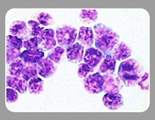
The t(15;17) is present in more than 95% of patients with acute promyelocytic leukemia (APL). This translocation generates 2 fusion proteins: promyelocytic leukemia–retinoic acid receptor alpha (PML-RARα) and RARα-PML. To understand the development of APL and to search for more effective treatments of this disease, several PML-RARα APL transgenic mouse models have been generated using myeloid specific regulatory elements, such as cathepsin G and myeloid-related protein 8 (MRP8), to direct PML-RARα expression into early myeloid cells. All of these PML-RARα mice develop a myeloproliferative syndrome early in life, and 15% to 20% of these mice develop an APL-like disease after 6 to 14 months (see Grisolano et al, Blood. 1997; 89:376-387; Brown et al, PNAS. 1997;94: 2551-2556; and He et al, PNAS. 1997;94: 5302-5307). Additional expression of the t(15;17) reciprocal fusion protein RARα-PML can increase the percentage of APL in transgenic mice by 4-fold. However, the long latency remains. These results indicate that PML-RARα is necessary but not sufficient for APL development. Additional mutations are probably required for leukemia to occur.FIG1
The current explanation of how PML-RARα is involved in leukemogenesis centers on the dominant-negative effect of PML-RARα. PML-RARα contains the DNA and retinoic acid (RA) ligand-binding domains of wild-type RARα. In the absence of a ligand, both RARα and PML-RARα repress transcription due to the interaction with nuclear receptor corepressor/silencing mediator for retinoid and thyroid receptor (NCoR/SMRT) and form complexes with histone deacetylase (HDAC). Removing acetyl groups by HDACs increases the positive charge on proteins and enhances protein interactions with negatively charged DNA to keep chromatin in a more compacted confirmation, which does not favor the initiation of transcription. Furthermore, removing acetyl groups may also enhance the binding of repressor proteins. In the presence of physiologic concentrations of RA, RA binds to RAR and changes the conformation of RARα, replacing the HDAC complex with transcription coactivators, including histone acetylase (HAT) complex. Acetylation of histones changes chromatin structure to favor gene expression. The PML portion of PML-RARα contains the oligomerization domain of PML. The oligomerized PML-RARα forms a more stable complex with NCoR/SMRT and HDAC. Dissociation of this complex requires a much higher concentration of RA. This theory explains why a high dosage of RA can be effectively used to treat APL. It also suggests that relatively higher levels of PML-RARα expression may be more effective at initiating APL development.
It has been difficult to detect PML-RARα expression in the above mentioned PML-RARα transgenic mice. Therefore, Westervelt and colleagues (page 1857) hypothesized that increasing the expression of PML-RARα may enhance the penetration of APL development in transgenic mice because the upstream fragment of the human cathepsin G used in their previously reported PML-RARα transgenic mice may lack critical regulatory elements required for high-level transgene expression. In order to fully capture cathepsin regulatory elements to express PML-RARα in early myeloid cells, Westervelt et al generated another PML-RARα mouse model by knocking the PML-RARα cDNA into the cathepsin G 5′ untranslated region. In contrast to the 15% to 20% penetration of APL in previously reported PML-RARα transgenic mouse models, more than 90% of PML-RARα knock-in mice developed APL, although the latency was similar to other transgenic models. These results suggested that the new knock-in model provides a level of PML-RARα expression that is more optimal for APL development, and most of us would have predicted that this level would be higher. However, when real-time reverse transcriptase–polymerase chain reaction (RT-PCR) analysis was used to compare the expression of PML-RARα in bone marrow cells and APL cells from this knock-in model (vs their human cathepsin G–PML-RARα transgenic mouse model) it was surprising to discover that PML-RARα was expressed at an extremely low level in the knock-in mice—less than 3% of the expression in the transgenic mice. This result goes directly against the original hypothesis (based on the dominant-negative effect of the interaction of PML-RARα with the HDAC complex) that more PML-RARα expression will enhance the development of APL. Instead, an optimal low level of PML-RARα expression appears to be favorable for APL development. Furthermore, the knock-in model may target PML-RARα expression to a more proper population of myeloid cells for APL development. It has been reported previously that PML-RARα triggers cell death in most cell lines. On one hand, PML-RARα expression may favor leukemia development; on the other hand, it may mitigate against leukemia. The disruption of such a balance with additional mutations may lead to leukemogenesis. Most importantly, this report urges the search for additional mechanisms besides the dominant-negative effect of PML-RARα via HDAC to fully explain how t(15;17) is involved in the development of APL.


This feature is available to Subscribers Only
Sign In or Create an Account Close Modal