Abstract
Adult T-cell leukemia (ATL) develops in a small proportion of individuals infected with the retrovirus human T-cell leukemia virus (HTLV-1). We evaluated the efficacy of MEDI-507 (a humanized monoclonal antibody directed against CD2) alone and in combination with humanized anti-Tac (HAT) directed toward CD25, the interleukin-2 receptor α (IL-2Rα) using a human adult T-cell leukemia xenograft model. Weekly treatments (4) with HAT significantly prolonged the survival of the ATL-bearing mice when compared with phosphate-buffered saline (PBS)–treated controls (P < .0001). Mice treated with MEDI-507 (100 μg/wk for 4 weeks) survived longer than those treated with HAT (P < .0025). Furthermore, prolonged treatment (6 months) of ATL with MEDI-507 significantly improved the outcome when compared with a short course (4 weeks) of therapy (P < .0036). Such treatment with weekly MEDI-507 for 6 months led to a prolonged survival of the ATL-bearing mice that was comparable with the survival observed in the control group of mice that did not receive a tumor or therapeutic agent. We also found that the expression of Fcγ receptors (FcRγ) on polymorphonuclear leukocytes and monocytes was required for MEDI-507–mediated tumor killing in vivo. Thus, the tumor-killing mechanism with MEDI-507 in vivo required the expression of the receptor FcRγIII on polymorphonuclear leukocytes and monocytes, suggesting that it is mediated by a form of antibody-dependent cellular cytotoxicity. These results demonstrate that MEDI-507 has therapeutic efficacy on ATL in vivo and provides support for a clinical trial involving this monoclonal antibody in the treatment of patients with CD2-expressing leukemias and lymphomas. (Blood. 2003; 102:284-288)
Introduction
Adult T-cell leukemia (ATL) develops in a small proportion of human T-cell leukemia virus (HTLV-1)–infected individuals.1 At present, there is no effective therapy for ATL, and patients progress to death with a median survival duration of 9 months for those with acute ATL and 24 months with chronic ATL.2 The conventional therapies (eg, multidrug chemotherapy regimens or zidovudine and interferon α) do not appear to prolong the life of patients with ATL.2,3 A murine model of ATL was developed by introducing leukemic cells (MET-1) from an ATL patient into nonobese diabetic (NOD)/severe combined immunodeficient (SCID) mice.4 New therapeutic agents have been tested in this model before initiating patient trials.5,6 The MET-1 ATL cells in this model are activated T cells that express CD2, CD3 dim, CD4, and CD25. In earlier studies, anti-CD25 monoclonal antibodies (eg, murine and humanized anti-Tac [HAT]) were tested in this model with promising results.4 Furthermore, clinical trials showed that HAT-based immunotherapies manifested efficacy in the therapy of ATL. In this study we target CD2 on the human xenograft MET-1 ATL cells using a humanized monoclonal antibody (mAb), MEDI-507.
CD2 is a T-cell marker that is not expressed on cells in the early stages of human T-cell development, including such cells as those with the phenotype CD44+CD25-, CD44+CD25+, and T-cell receptor T cells.7 However, it is expressed on all mature T cells and most natural killer (NK) cells.8,9 CD2 is highly expressed on activated T cells but less intensively expressed on resting T cells.10 It is also highly expressed on MET-1 ATL cells. In particular, CD2 was expressed on 96% of the abnormal cells from the MET-1 patient examined ex vivo and on 99% of the spleen cells of the MET-1–bearing NOD /SCID mice.4 The main functions of CD2 are in adhesion11,12 and activation,13,14 where the costimulatory signals for T-cell activation are provided by the interaction of CD2 with its ligand (CD58, lymphocyte functional antigen-3 [LFA-3]). The limited expression of CD2 that is restricted to mature T cells and T-leukemic cells including MET-1 leukemic cells provides the scientific rationale for its use in antibody-mediated immunotherapy of ATL. MEDI-507 is a humanized mAb directed against CD2 that was genetically engineered from the rat version of the mAb (BTI-322) by BioTransplant (Charlestown, MA) for use in the prevention of allograft rejection15-17 and in the therapy of graft-versus-host disease (GVHD) and autoimmune diseases.18 MEDI-507 (siplizumab) has been used in a series of phase 1 trials involving patients with psoriasis or with GVHD. Multiple doses of MEDI-507 were effective in psoriasis with a clinically significant disease response using the Psoriasis Area and Severity Index (PASI) observed at doses of 1.2 μg/kg or greater. Furthermore, this mAb was more effective than anti–thymocyte globulin in the treatment of GVHD with haploidentical bone marrow transplantation.19 The majority of adverse events were transient and judged to be mild. The most common adverse events included chills, headache, and decreased target lymphocyte counts.
In this study, we investigated the efficacy of MEDI-507 in a xenograft ATL model when used alone and in combination therapy with HAT (an anti-CD25 antibody). The scientific basis for the combination is that MEDI-507 and HAT target distinct epitopes, CD2 and CD25, respectively, that are expressed on the MET-1 ATL cells, an observation that suggested that they might manifest additive or synergistic efficacy. We were particularly interested in the mechanism underlying the tumor-killing action mediated by MEDI-507 on ATL in vivo. We demonstrated that the efficacy was lost in Fcγ receptor (FcRγ-/-) mice, indicating that the expression of the receptor FcRγIII that uses the Fcγ chain is required for the effective action of this mAb in the mouse leukemia model.
Materials and methods
Mouse model of ATL
Female NOD/SCID mice were purchased from Jackson Labaratories (Bar Harbor, ME). The mice were used in studies at the age of 6 to 12 weeks. Leukemia was established by intraperitoneal injection of 15 × 106 freshly isolated MET-1 cells. Mice were randomly assigned to each group when their soluble interleukin-2 receptor α (sIL-2Rα) levels reached the range of 1000 to 10 000 pg/mL serum. These levels were observed at approximately 10 to 14 days after tumor inoculation, at which time treatments were initiated. The FcRγ knock-out mice were generated in the laboratory of Jeffrey Ravetch (Rockefeller University, New York, NY). In the study directed toward defining the mechanism involved in tumor killing, very large tumor burdens were used in the FcRγ knock-out and FcRγ intact NOD/SCID mice. In these latter studies, mice with sIL-2Rα levels of 20 000 to 90 000 pg/mL serum (mean, 80 000 pg/mL) were randomly assigned to the study groups for the experiments.
Measurement of sIL-2Rα and soluble β2-microglobulin by enzyme-linked immunosorbent assay (ELISA)
Throughout the therapy experiments, the serum concentrations of soluble human IL-2Rα and human β2-microglobulin (β2μ), which were used as surrogate tumor markers, were measured using ELISA kits purchased from R&D Systems (Minneapolis, MN). The ELISAs were performed as suggested in the manufacturer's kit inserts.
Analysis of the binding of MEDI-507 to MET-1 ATL cells
The binding of MEDI-507 to CD2 was analyzed by flow cytometry before the therapeutic experiments were conducted. The phenotypic MET-1 leukemic cells were prepared in the same fashion as was used in the phenotype analysis performed in the study of Phillips et al.4 The cells were stained with the primary antibody MEDI-507 or rituximab on ice for 30 minutes, washed, and then stained with a fluorescein isothiocyanate (FITC)–labeled antibody directed against the human immunoglobulin G (IgG) Fc fragment. After washing, the cells were analyzed for the binding of MEDI-507 directed to CD2 on the MET-1 cells using a Becton Dickinson FACSort Flow Cytometer (San Jose, CA).
mAbs
The humanized mAb MEDI-507, which recognizes CD2, was a gift from BioTransplant. HAT, (daclizumab, Zenapax) a humanized mAb directed toward CD25, was obtained from Hoffmann-La Roche (Nutley, NJ). Rituximab was obtained from IDEC Pharmaceuticals (San Diego, CA).
Treatment with antibodies
For the evaluation of therapeutic efficacy, groups of 15 NOD/SCID mice each were injected with 15 million MET-1 leukemic cells intraperitoneally and were randomly assigned to groups that had comparable levels of the surrogate tumor marker, the serum soluble IL-2Rα (Tac, CD25). The animals were treated when their sIL-2Rα levels ranged from 1 to 10 000 pg/mL (10 to 14 days after introduction of MET-1 leukemic cells into the mice). The groups of mice were given PBS, HAT, MEDI-507, or the combination of MEDI-507 with HAT at a dose of 100 μg of each mAb intravenously weekly for 4 weeks. An additional group of mice was given 100 μg of MEDI-507 intravenously weekly for 6 months. Only a single dose of 100 μg per administration per mouse was used since we have noted that such a dose of a humanized mAb is sufficient to maintain saturation of the target antigens for the week between dosings. A final group of NOD/SCID mice was included that did not receive a tumor or a therapeutic agent to serve as a tumor-free and treatment-free control. In a study to define the mechanism of action of MEDI-507, the mAb was given weekly for 4 weeks by intraperitoneal injection to FcRγ knock-out mice and FcRγ intact mice. Throughout the studies, the leukemic progression was evaluated using an ELISA assay for human β2μ in the serum as well as by monitoring the survival of the mice using Kaplan-Meier analysis.
Statistics
StatView (SAS Institute, Cary, NC) was used to generate Kaplan-Meier cumulative survival plots. The unpaired t test was conducted in the analysis of β2μ levels.
Results
Demonstration of MEDI-507 binding to CD2 expressed on MET-1 ATL cells
Using fluorescence-activated cell sorter (FACS) analysis we demonstrated that MEDI-507 binds to MET-1 ATL cells (Figure 1A), in contrast with the lack of reactivity of the B-cell–specific anti-CD20 mAb, rituximab (Figure 1B).
The binding of MEDI-507 to MET-1 ATL cells was evaluated by FACS analysis. The MET-1 ATL cells were harvested as described in “Materials and methods.” The primary mAbs used were humanized anti-CD20 and MEDI-507. The secondary antibody was a rat anti–human IgG Fc fragment conjugated with FITC. In panel A, the primary antibody was MEDI-507 directed to CD2. The isotype control is represented by the solid area, whereas the line represents the humanized anti-CD2. In panel B, the solid area is the isotype control and the line represents humanized anti-CD20.
The binding of MEDI-507 to MET-1 ATL cells was evaluated by FACS analysis. The MET-1 ATL cells were harvested as described in “Materials and methods.” The primary mAbs used were humanized anti-CD20 and MEDI-507. The secondary antibody was a rat anti–human IgG Fc fragment conjugated with FITC. In panel A, the primary antibody was MEDI-507 directed to CD2. The isotype control is represented by the solid area, whereas the line represents the humanized anti-CD2. In panel B, the solid area is the isotype control and the line represents humanized anti-CD20.
Effective treatment of ATL using MEDI-507 directed toward CD2
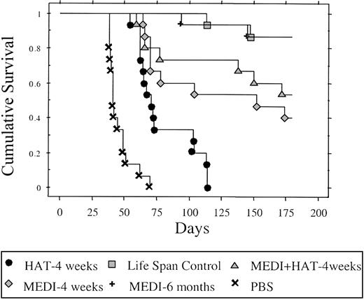
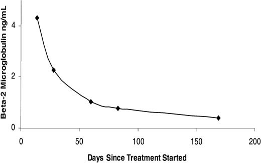
In a treatment trial in the MET-1 model of human ATL, a 4-week course of treatment with MEDI-507 (100 μg/wk intravenously), HAT, and the combination of MEDI-507 with HAT (100 μg/wk intravenously) demonstrated therapeutic efficacy by their effect on the serum levels of human β2μ, a surrogate tumor marker in the murine model (Figure 2), and on the survival of ATL-bearing mice (Figure 3). When compared with the serum concentration of human β2μ in the PBS control group of mice, on days 28 and 60, there was a significant reduction of β2μ in treated animals in the 4-week MEDI-507 (P < .0001), the 4-week HAT (P < .0001), the 4-week combination of MEDI-507 with HAT (P < .0001), and the 6-month MEDI-507 (P < .0001) groups. Human β2μ levels were undetectable in 12 of 13 surviving mice that received the 6 months of weekly treatments with MEDI-507 when measured at the end of the 6-month treatment period. The human β2μ level decreased progressively during the 6-month MEDI-507 treatment period as shown in Figure 4 for a typical mouse that initially had a large tumor burden (Figure 4). Furthermore, there was a significant (P < .0001) prolongation of the survival of the mice that were treated with the combination of HAT and MEDI-507 when compared with the PBS control (Figure 3). The median survival duration of the control group (PBS) was 45.6 days. All of the mice in the PBS group died by day 70 of the study. In contrast, 67% of the mice in the 4-week MEDI-507 group, 53% in the 4-week HAT group, 80% in the 4-week combination group receiving MEDI-507 and HAT, and 100% of the mice in the 6-month MEDI-507 treatment groups were alive at that time. The life span in the group receiving MEDI-507 for 6 months was significantly prolonged when compared with that of all other treatment groups (6-month MEDI-507 vs 4-week combination of MEDI-507 and HAT [P < .045], and 6-month MEDI-507 vs 4-week MEDI-507 alone [P < .0036]). Furthermore, the survival in the 6-month MEDI-507 group was comparable with that of the tumor-free control group of mice that did not receive either tumor or a therapeutic agent. Of the initial 15 mice, 13 were surviving in both the 6-month treatment group and in the tumor-free, treatment-free control group at day 180 following the start of treatment. This proportion of the mice surviving in the group receiving MEDI-507 weekly for 6 months was superior to that observed in any of the other treatment groups. In particular, only 6 of 15 mice in the 4-week MEDI-507 treatment alone and 8 of 15 in the group with combination treatment with 4 weeks of MEDI-507 and 4 weeks of HAT were surviving on day 180. Furthermore, all mice in the group receiving 4 weeks of HAT had died by day 114. Comparable efficacy of MEDI-507 in the therapy of ATL was observed when the study was repeated in 2 additional experiments.
The growth of MET-1 ATL cells in NOD/SCID mice bearing the MET-1 ATL leukemia was inhibited by HAT and MEDI-507. MET-1 ATL cells were transferred into mice. The groups (15 mice/group) included those receiving PBS, 4 weekly doses of 100 μg MEDI-507, 4 weekly doses of 100 μg HAT, a 4-week combination of 100 μg each of MEDI-507 with HAT, and 6 months of weekly doses of 100 μg MEDI-507. The data represent the mean concentration of human β2μ in nanograms per milliliter. The animals treated in the 4-week MEDI-507, 4-week HAT, 4-week combination of MEDI-507 with HAT, and 6-month MEDI-507 groups had significantly decreased values of β2μ when compared with those of the PBS control group (on day 28 P < .0001 and on day 60 P < .0001). Furthermore, the animals receiving MEDI-507 for 6 months had significantly decreased levels of β2μ when assessed on day 60 compared with those of the mice in the 4-week MEDI-507 treatment group (P < .0175).
The growth of MET-1 ATL cells in NOD/SCID mice bearing the MET-1 ATL leukemia was inhibited by HAT and MEDI-507. MET-1 ATL cells were transferred into mice. The groups (15 mice/group) included those receiving PBS, 4 weekly doses of 100 μg MEDI-507, 4 weekly doses of 100 μg HAT, a 4-week combination of 100 μg each of MEDI-507 with HAT, and 6 months of weekly doses of 100 μg MEDI-507. The data represent the mean concentration of human β2μ in nanograms per milliliter. The animals treated in the 4-week MEDI-507, 4-week HAT, 4-week combination of MEDI-507 with HAT, and 6-month MEDI-507 groups had significantly decreased values of β2μ when compared with those of the PBS control group (on day 28 P < .0001 and on day 60 P < .0001). Furthermore, the animals receiving MEDI-507 for 6 months had significantly decreased levels of β2μ when assessed on day 60 compared with those of the mice in the 4-week MEDI-507 treatment group (P < .0175).
Kaplan-Meier survival plot of MET-1–bearing NOD/SCID mice. The groups (15 mice/group) included those receiving intravenous PBS, 100 μg HAT, 100 μg MEDI-507 per week for 4 weeks, and a combination of 100 μg MEDI-507 with 100 μg HAT for 4 weeks. Another group received 100 μg MEDI-507 weekly for 6 months. Event-free survival was followed for 180 days. The animals treated in the 4-week HAT, 4-week MEDI-507, 4-week combination of MEDI-507 with HAT, and 6-month MEDI-507 groups had significantly prolonged survivals when compared with the PBS control group (P < .0001). Treatment of tumor-bearing mice with MEDI-507 for 6 months significantly prolonged the survival of the group when compared with the 4-week MEDI-507 treatment group (P < .0036).
Kaplan-Meier survival plot of MET-1–bearing NOD/SCID mice. The groups (15 mice/group) included those receiving intravenous PBS, 100 μg HAT, 100 μg MEDI-507 per week for 4 weeks, and a combination of 100 μg MEDI-507 with 100 μg HAT for 4 weeks. Another group received 100 μg MEDI-507 weekly for 6 months. Event-free survival was followed for 180 days. The animals treated in the 4-week HAT, 4-week MEDI-507, 4-week combination of MEDI-507 with HAT, and 6-month MEDI-507 groups had significantly prolonged survivals when compared with the PBS control group (P < .0001). Treatment of tumor-bearing mice with MEDI-507 for 6 months significantly prolonged the survival of the group when compared with the 4-week MEDI-507 treatment group (P < .0036).
Changes in β2μ levels were observed with treatment with MEDI-507 for 6 months in a MET-1 ATL–bearing NOD/SCID mouse. Human β2μ serum levels were used as a surrogate tumor marker in this human xenograft MET-1 ATL model.
Changes in β2μ levels were observed with treatment with MEDI-507 for 6 months in a MET-1 ATL–bearing NOD/SCID mouse. Human β2μ serum levels were used as a surrogate tumor marker in this human xenograft MET-1 ATL model.
FcRγ expression is required for effective MEDI-507 action
MEDI-507 clearly had a therapeutic effect in our murine model of ATL. We wished to define its mode of action. Several mechanisms could theoretically be involved. In particular, MEDI-507 could block CD2 binding to LFA-3 (CD58); it could involve complement-dependent cytotoxicity (CDC) or alternatively it could utilize antibody-dependent cellular cytotoxicity (ADCC). As considered in “Discussion,” CDC does not appear to be involved, and we focused on ADCC as a potential effector mechanism. Since NOD/SCID mice were deficient in both T and NK cells, we considered the hypothesis that monocytes and granulocytes expressing Fc receptors were the potential effector cells in vivo. An experiment involving parallel groups of mice that differed in their expression of FcRγ was conducted to define the mechanism involved in the tumor killing by MEDI-507. FcRγ knock-out ATL NOD/SCID mice were used in one study group, whereas the FcRγ was wild type in the other study group mice. In the FcRγ-/- group there was no statistical difference in the survival observed between the animals receiving 4 weekly doses of MEDI-507 and those receiving PBS (P > .702). Both PBS-treated and 4-week MEDI-507–treated animals in FcRγ knock-out MET-1 ATL–bearing NOD/SCID mice died within 22 days of initiation of treatment (Figure 5B). A contrasting pattern was observed in the FcRγ-expressing group, in which significant efficacy was manifested by the group receiving MEDI-507. In particular, all of the animals in the PBS-treated group in FcRγ intact MET-1 ATL–bearing NOD/SCID mice died within 30 days of initiation of therapy, whereas all of the animals in the 4-week MEDI-507 treatment group were still surviving at that time. Animal survival was followed for 40 days when 8 of 10 mice were still alive in the 4-week MEDI-507 treatment group (Figure 5A). Thus, MEDI-507 provides effective therapy for ATL in this model by a mechanism that requires the expression of the FcRIII receptor that involves Fcγ.
Kaplan-Meier survival plot of MET-1 FcRγ knock-out and FcRγ intact ATL-bearing NOD/SCID mice. (A) FcR intact mice. (B) FcR knock-out mice. MET-1 ATL cells were transferred into mice. Once the mice developed sIL-2Rα levels of 20 000 to 90 000 pg/mL (reflecting a very large tumor burden), therapy with MEDI-507 antibody was initiated. The groups (10 mice/group) included those receiving PBS (□) or 4 weeks of 100 μg per week of MEDI-507 (○) in the FcRγ knock-out ATL-bearing mice. In the parallel FcRγ intact ATL-bearing NOD/SCID mice, the groups (10 mice/group) included those receiving PBS (□) or 4 weekly intraperitoneal doses of 100 μg MEDI-507 (○). Event-free survival was followed for 40 days. There was no significant statistical difference in the survival between the group receiving 4 weekly doses of MEDI-507 and that receiving PBS in the FcRγ knock-out mice. In contrast, in FcRγ intact ATL-bearing NOD/SCID mice the survival of the group receiving 4 weekly doses of MEDI-507 was prolonged when compared with the control group.
Kaplan-Meier survival plot of MET-1 FcRγ knock-out and FcRγ intact ATL-bearing NOD/SCID mice. (A) FcR intact mice. (B) FcR knock-out mice. MET-1 ATL cells were transferred into mice. Once the mice developed sIL-2Rα levels of 20 000 to 90 000 pg/mL (reflecting a very large tumor burden), therapy with MEDI-507 antibody was initiated. The groups (10 mice/group) included those receiving PBS (□) or 4 weeks of 100 μg per week of MEDI-507 (○) in the FcRγ knock-out ATL-bearing mice. In the parallel FcRγ intact ATL-bearing NOD/SCID mice, the groups (10 mice/group) included those receiving PBS (□) or 4 weekly intraperitoneal doses of 100 μg MEDI-507 (○). Event-free survival was followed for 40 days. There was no significant statistical difference in the survival between the group receiving 4 weekly doses of MEDI-507 and that receiving PBS in the FcRγ knock-out mice. In contrast, in FcRγ intact ATL-bearing NOD/SCID mice the survival of the group receiving 4 weekly doses of MEDI-507 was prolonged when compared with the control group.
Discussion
The MET-1 ATL model presents many features that parallel those observed in patients with adult T-cell leukemia and thus represents a valuable model for the evaluation of the efficacy of therapeutic agents directed toward ATL.4 In earlier studies HAT showed efficacy in the MET-1 ATL model. Furthermore, in human clinical trials, therapy with the anti-CD25, anti-Tac mAb provided effective therapy for 6 of 18 patients with ATL studied.20 We found that 4 weekly treatments with MEDI-507 that is directed toward CD2 provided meaningful therapy for ATL in the MET-1 model. Furthermore, the efficacy of the 4 weekly MEDI-507 treatments was better than that of 4 weeks of HAT treatment (P < .0001). A critical finding in the present study that is of relevance to the design of a clinical trial is that the outcome in the 6-month MEDI-507 treatment group was significantly superior to that observed in the 4-week MEDI-507 treatment group, and 4-week MEDI-507 treatment group was better than that of 1-week MEDI-507 treatment group (data not shown), which implies that prolonged treatment with MEDI-507 for ATL may be of value in the efforts to achieve a sustained complete remission in the clinic. This requirement for prolonged therapy to achieve a complete remission could also be true for other mAb-based antitumor regimens. The outcome in survival of the combination of 4-week MEDI-507 and HAT was modestly better than that observed in 4-week MEDI-507 alone, but this difference did not reach a statistical significance. This modest synergy may reflect the direct agonist actions on the particular antigenic target receptor (eg, anti-CD2 agonistic action of MEDI-507). However, the fact that the synergy was modest may reflect that the underlying mechanism of actions of both MEDI-507 and HAT are the same and depend on Fcγ receptors. Both CD2 and CD25, the targets of the monoclonal antibodies, are highly expressed on the ATL MET-1 cells.
Several mechanisms could theoretically be involved in the action of MEDI-507 in the MET-1 ATL model. These include CDC and ADCC. Complement-dependent cytotoxicity appears to be excluded in the present model because the mice lack human complement, there is only a limited amount of murine complement expressed in the NOD/SCID mice used, and the complement manifests poor lysing function on MET-1 ATL cells.4 Classical ADCC mediated by NK cells also does not appear to be a likely mode of action in this model, since the NOD/SCID mice used as the recipients of the ATL cells in our study virtually lacked functional NK cells.
The analysis of the efficacy of MEDI-507 in FcRγ-/- mice was very instructive. Fcγ is required for the expression of FcRγIII, the stimulatory receptor. Efficacy of MEDI-507 was observed in FcRγ intact MET-1 ATL–bearing mice but not in FcRγ-/- knock-out mice bearing MET-1 ATL. This observation supports the view that although multiple mechanisms have been suggested for the antitumor action of antibodies in vivo, in the case of MEDI-507 there is a dominant and necessary role played by an FcRγ-dependent mechanism. The efficacy of MEDI-507 in the MET-1 ATL model parallels the previously reported requirement for FcRγ expression for an effective therapeutic response to trastuzumab and rituximab as well as to a mAb directed to a melanoma antigen in murine models of breast, B-cell, and melanoma malignancies, respectively.21,22 In the present studies comparing mAb efficacy in FcRγ knock-out with FcRγ intact animal groups, the tumor burden at the onset of therapy was more than 10 times greater than that of mice in the initial trials of this study. Thus, MEDI-507 can significantly delay the progression of human leukemic xenografts in mice with very large ATL burdens, supporting the view that the application of MEDI-507 to the therapy of ATL may be of value in the clinic. In summary, the humanized mAb MEDI-507 effectively controlled leukemia in a human leukemia xenograft model through an FcRγ-requiring process presumably mediated by effector cells, including monocytes and granulocytes that express FcRγIII. Prolonged treatment with MEDI-507 on ATL significantly improved the outcome when compared with a shorter course of therapy. These studies provide support for a clinical trial of MEDI-507 in ATL patients, and potentially in patients with other T-cell malignances that express CD2.
Prepublished online as Blood First Edition Paper, March 20, 2003; DOI 10.1182/blood-2002-11-3601.
The publication costs of this article were defrayed in part by page charge payment. Therefore, and solely to indicate this fact, this article is hereby marked “advertisement” in accordance with 18 U.S.C. section 1734.
We gratefully acknowledge BioTransplant for providing MEDI-507 for the studies.


This feature is available to Subscribers Only
Sign In or Create an Account Close Modal