We have studied adenovirus-mediated cytotoxicity after infection of malignant cells obtained from patients with chronic lymphocytic leukemia (CLL). Our studies indicate that adenoviruses can infect primary CLL cells and that infection of CLL cells with a replication-competent strain of human adenovirus 5 (Ad5dl309) results in cytotoxicity. Adenovirus-mediated cytotoxicity was also seen after infection of CLL cells with a variety of viruses attenuated by mutations in the adenovirus early region 1 (E1) or early region 2 (E2). Even viruses attenuated by deletion of the entire E1 region resulted in cytotoxicity after infection of the CLL cells obtained from some patients. Although there was variability in the degree of cytotoxicity induced by different viruses in different patients cells, a virus with a mutation in the E1B 19K gene resulted in the greatest degree of cytotoxicity in most of the CLL samples tested. These studies demonstrate that infection of CLL cells by attenuated adenoviruses with specific mutations in the E1 or E2 region results in cell death. Attenuated adenoviruses should be developed further as therapeutic agents for patients with CLL.
SEVERAL BIOLOGICAL processes may contribute to cytotoxicity after infection by wild-type or mutant adenoviruses. For example, productive infection, abortive infection, abortive infection with the induction of apoptosis, or a combination may contribute to the death of an infected cell.1-5 These possible outcomes result from complex interactions between host and virus that impact on virus replication, host cell cycle regulation, virus and host gene expression, the induction of apoptosis, and host cell survival. As the nature of some of these interactions have been elucidated, mutant adenoviruses have been developed as cytotoxic agents with specificity for malignant epithelial cells.6 7
The initial studies demonstrating specific cytotoxicity of mutant adenoviruses in selected epithelial malignancies used an adenovirus with a mutation in the E1B 55K gene product.6,7 The rationale for the specificity of the cytotoxicity induced by infection with that virus was based on the complementation of a genetic defect of the virus (E1B 55K mutation) by a genetic feature of the malignant cell (p53 mutation), which should allow the mutant virus to replicate and induce cytotoxicity selectively in malignant, but not normal (p53 wild-type) cells. The selective cytotoxicity induced by this virus was presumed to be a consequence of the requisite inactivation of p53 by the E1B 55K polypeptide during a productive infection of normal cells. In the absence of the E1B 55K polypeptide (and presence of wild-type p53), productive infection may be reduced. However, the relationship between virus replication, p53 expression, and cytotoxicity is complex, and other studies have demonstrated enhanced cytotoxicity of this virus in the presence of wild-type p53 under certain conditions.5
Although cells with p53 mutations (the targets of infection the E1B 55K mutated adenovirus) are not as prevalent in hematologic malignancies as they are in many other types of tumors, several of the lymphoid malignancies are characterized by the altered expression of other cellular proteins that might complement adenovirus mutants or interact with adenovirus proteins to result in specifically enhanced cytotoxicity. For example, malignant cells from patients with chronic lymphocytic leukemia (CLL) have a spectrum of abnormalities, including altered expression of Bcl-2, other Bcl-2 family members, cdk inhibitors, cyclins, pRB, or p53.8-11 Each of these genetic alterations may result in a cellular milieu that could enhance susceptibility to adenovirus-mediated cytotoxicity after infection with specific mutant adenoviruses. For example, the overexpression of specific cyclins, the absence of specific cdk inhibitors, and mutations in pRb might result in alterations of pRb function that would complement mutations in E1A that alter pRb binding. These changes could result in cytotoxicity after infection with viruses containing specific mutations in E1A.
Because prior studies have indicated limited virus production/cytotoxicity after adenovirus infection of normal lymphoid cells,12 13 we initiated studies to assess the degree of cytotoxicity induced by adenovirus infection of CLL cells. Despite the fact that many malignancies may have alterations that make them susceptible to adenovirus and adenovirus mutant-mediated cytotoxicity, we have chosen to direct our study to CLL because (1) the disease is incurable with standard dose chemotherapy; (2) there are documented biological/genetic changes characteristic of the disease that might facilitate selective cytotoxicity after infection with a mutant virus; (3) previous studies by others have indicated that adenovirus infection of normal lymphoid cells is generally nonproductive; and (4) there is therapeutic rationale for the induction of in vivo or ex vivo cytotoxicity. In vivo studies may demonstrate a novel therapeutic with selective cytotoxcity during systemic or local administration. Ex vivo therapy could be undertaken in conjunction with high-dose chemotherapy/autologous hematopoietic stem cell rescue (HDC/HSCR) as a means of purging autologous hematopoietic stem cell grafts of contaminating malignant cells.
We have used a panel of adenoviruses to infect CLL cells obtained from patients. These studies demonstrate adenovirus-mediated cytotoxicity after infection of primary CLL cells with viruses attenuated by mutations in E1A, E1A+E1B, E1B 19K, or E2A, raising the possibility that mutated adenoviruses may have a therapeutic role in the treatment of selected hematologic malignancies.
MATERIALS AND METHODS
Patients.
Eight B-cell CLL patients were included in this study. Diagnosis was based on clinical examination, peripheral blood count, and the morphology and immunophenotype of the leukemic cells (CD5+, CD19+, and CD23+). Samples of blood were obtained under a protocol approved by the Robert Wood Johnson Medical School Institutional Review Board. Some of the patients were treated and others were untreated. Some patient samples were available before and after the initiation of treatment. Mononuclear cells were isolated from 10 to 50 mL of fresh, heparinized peripheral blood by Histopaque (Sigma, St Louis, MO) centrifugation. Isolated cells were counted, resuspended in freezing medium (90% calf serum and 10% dimethyl sulfoxide [DMSO]), and stored in liquid nitrogen until use.
Cells and viruses.
HeLa cells and 293 cells were grown in L modified Eagle’s medium supplemented with 2 mmol/L L-glutamine, 10% fetal bovine serum (FBS), and antibiotics. CLL cells were maintained in RPMI-1640 medium supplemented with 20% FBS, L-glutamine, antibiotics, and 50 U/mL of interleukin-4 (IL-4; Boehringer Mannheim, Indianapolis, IN).
The viruses used in the experiments, Ad5dl309, E1A−, Pac3, E1B−, and Ad5dl337 were described previously.14-16 The temperature sensitive (ts) virus Ad5ts125 was generously provided by Daniel Klessig (Rutgers University, Piscataway, NJ).17 All viruses, with the exception of the Ad5ts125, were grown and titered on 293 cells at 37°C. The Ad5ts125 virus was grown at 33°C and was titered on 293 cells at 33°C and 37°C. For mock infections, Ad5dl309 was heat-inactivated at 65°C for 30 minutes.
The recombinant adenovirus containing the green fluorescent protein (GFP) gene was purchased from Quantum Biotechnologies (Montreal, Quebec, Canada). That virus (AdGFP) contains the GFP gene, under the control of a cytomegalovirus (CMV) promoter, in the E1A region of an adenovirus 5 containing a deletion in the E3 region.
Infection.
CLL cells (2 × 10⁶) were infected with the indicated viruses at a multiplicities of infection (moi) of 100 (or as otherwise indicated) for 4 to 6 hours at 37°C. Infections with Ad5ts125 were conducted at both the permissive temperature (33°C) and the nonpermissive temperature (37°C). After infection, the cells were washed 3 times with Dulbecco’s phosphate-buffered saline to remove nonadsorbed virus. The cells were then pelleted by centrifugation at 500g for 10 minutes, resuspended in 2 mL of medium, and cultured in 24-well plates. At the indicated times, samples were removed for determination of cell viability by trypan blue staining, virus yield, and apoptosis. Mock infections used heat-inactivated Ad5dl309.
Cell viability.
At the indicated times, cells in the various replicate wells were resuspended and 100 μL were removed and analyzed for cell viability by trypan blue staining and cell count. Cytotoxicity was calculated by a comparison of viable cell numbers in the infected samples as compared with uninfected controls.
Virus yield assay.
To assess virus replication, cell-free supernatants were collected at the indicated times. The virus titer was determined by plaque assay on either 293 cells or Hela cells. Briefly, 1 mL of serial 10-fold dilutions of the cell-free supernatant were added to 60-mm dishes containing a confluent layer of either 293 cells or Hela cells followed by incubation for 1 hour at 37°C. After incubation, the virus was removed and the plates were overlaid with agar as described.18 Viral plaques were counted on day 14 and the viral titer was determined.
RESULTS
Infection of primary CLL cells with adenovirus 5 (Ad5dl309).
The ability of adenoviruses to infect primary CLL cells was established by infection of CLL cells with a recombinant adenovirus expressing the GFP under the control of a CMV promoter (AdGFP; Quantum Biotechnologies). Three days after infection, green fluorescence was measured by flow cytometry. A marked shift in fluorescence was seen after infection with AdGFP (Fig 1), indicating that primary CLL cells could be infected by adenoviruses.
CLL cells can be infected with an adenovirus encoding GFP. CLL cells were infected with AdGFP, containing the GFP gene under the control of a CMV promoter (Quantum Biotechnologies). Three days later, cells were assayed for green fluorescence by flow cytometry using a cytometer equipped with an argon laser at 488 nm. The heavy line represents the fluorescence of infected cells. The light line represents the fluorescence of uninfected cells.
CLL cells can be infected with an adenovirus encoding GFP. CLL cells were infected with AdGFP, containing the GFP gene under the control of a CMV promoter (Quantum Biotechnologies). Three days later, cells were assayed for green fluorescence by flow cytometry using a cytometer equipped with an argon laser at 488 nm. The heavy line represents the fluorescence of infected cells. The light line represents the fluorescence of uninfected cells.
To determine if adenoviruses induce cytotoxicity in CLL cells, cells were obtained from the blood of a series of patients. Each patient had a lymphocytosis that was morphologically and immunophenotypically characteristic of B-cell CLL. Populations of malignant cells at 90% to 99.9% purity were infected with Ad5dl309, a replication-competent strain of human adenovirus 5 containing a deletion in E3B,14 and cell viability was monitored over the course of 20 days. The Ad5dl309 virus was chosen because it has served as the parental strain for generation of well-characterized mutant adenoviruses.
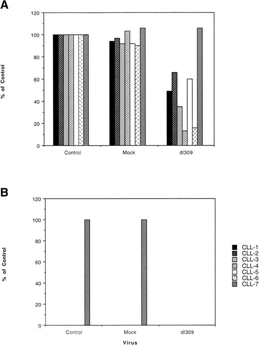
In each CLL sample, cytotoxicity after infection was detected in comparison to mock infected cells (Fig 2). At 4 days postinfection (Fig 2A), a loss of cell viability in comparison with uninfected and mock-infected was seen for samples from patients no. 1 through 6. The extent of cell death ranged from 40% to 50% for samples no. 1, 2, 3, and 5 to approximately 80% for samples from patients no. 4 and 6. Uninfected CLL samples from patients no. 1 to 6 exhibited a loss of viability by day 10, and the full extent of adenovirus-induced cytotoxicity could not be assessed under conditions allowing survival of uninfected or mock-infected cells. In contrast, CLL cells from patient 7 persisted in culture for greater than a 3-week period (>90% viability on day 20) and 100% of the cells were killed by Ad5dl309 by day 20 (Fig 2B). One hundred percent of the cells in the control and mock-infected cultures retained the immunophenotype of CLL, indicating that Ad5dl309 infection induced marked cytotoxicity for the CLL cells. The biological/genetic correlates of the ability of CLL patient no. 7 cells to persist in culture are not clear, because the patient from whom these cells were obtained has a clinically, morphologically, and immunophenotypically classic form of CLL with normal cytogenetics. The results from all 7 patients suggest that infection with adenoviruses leads to cytotoxicity of CLL cells. In some experiments, up to 10% of the cells did not have the morphology or immunophenotype of CLL. Cytotoxicity of the CLL cells in those samples was comparable to those of more homogenous patient samples.
Adenoviruses infection of CLL cells induces cytotoxicity. Primary CLL cells from 7 patients were infected with Ad5dl309 or heat-inactivated Ad5dl309 (mock-infected). At the indicated times, cell viability was determined by cell counting and trypan blue staining and compared with the viability of uninfected and mock-infected cells. (A) The degree of cytotoxicity seen 4 days after infection of primary CLL with Ad5dl309. (B) The degree of cytotoxicity seen 20 days after infection of primary CLL with Ad5dl309. In both figures, control refers to uninfected cells and mock refers to cells infected with heat-inactivated virus.
Adenoviruses infection of CLL cells induces cytotoxicity. Primary CLL cells from 7 patients were infected with Ad5dl309 or heat-inactivated Ad5dl309 (mock-infected). At the indicated times, cell viability was determined by cell counting and trypan blue staining and compared with the viability of uninfected and mock-infected cells. (A) The degree of cytotoxicity seen 4 days after infection of primary CLL with Ad5dl309. (B) The degree of cytotoxicity seen 20 days after infection of primary CLL with Ad5dl309. In both figures, control refers to uninfected cells and mock refers to cells infected with heat-inactivated virus.
Infection of CLL cells with Ad5dl309 mutants containing deletions of E1A, E1A+E1B, or E1B 19K.
The cytotoxicity detected after infection of CLL cells with Ad5dl309 raised the possibility that more attenuated viruses might also induce cytotoxicity. We thus performed a series of infections with a panel of mutated adenoviruses. In these studies, the CLL cells from individual patients were infected with Ad5dl309 mutants containing deletions of either E1A (E1A−), E1A+E1B (Pac3), or E1B 19K (Ad5dl337)15,16 19 (see Table 1). Four days after infection with the adenovirus containing a deletion of E1A (E1A−), CLL cells from patients no. 1, 3, and 5 exhibited 20% to 30% cytotoxicity, whereas CLL samples from patients no. 2, 4, and 6 demonstrated 50% to 75% cytotoxicity (Fig3A). Infection with the adenovirus deleted in both E1A and E1B (Pac3) also resulted in cytotoxicity with samples from patients no. 2, 4, 5, and 6, demonstrating a 40% to 60% loss in viability 4 days after infection (Fig 3A). Unlike the CLL cells from patients no. 1 through 6, CLL cells from patient no. 7 persisted in culture more than 20 days, and both the viruses with E1A deletion (E1A−) and E1A+E1B deletion (Pac3) resulted in complete eradication of the CLL cells under conditions that supported full viability of control (uninfected or mock-infected) cells (Fig 3B). These infections demonstrate that even highly attenuated viruses containing deletion of E1A and E1B are capable of inducing cytotoxicty after infection of CLL cells.
Attenuated adenovirus infection of CLL cells induces cytotoxicity. Primary CLL cells from 7 patients were infected with Ad5dl309, heat-inactivated Ad5dl309 (mock-infected), an E1A deleted adenovirus (E1A−), or an adenovirus with a deletion of E1A and E1B (Pac3). At the indicated times, cell viability was determined by cell counting and trypan blue staining and compared with the viability of uninfected and mock-infected cells. (A) The degree of cytotoxicity seen 4 days after infection of primary CLL with each of the adenoviruses. (B) The degree of cytotoxicity seen 20 days after infection of primary CLL cells with each of the adenoviruses. In both figures, control refers to uninfected cells and mock refers to cells infected with heat-inactivated virus.
Attenuated adenovirus infection of CLL cells induces cytotoxicity. Primary CLL cells from 7 patients were infected with Ad5dl309, heat-inactivated Ad5dl309 (mock-infected), an E1A deleted adenovirus (E1A−), or an adenovirus with a deletion of E1A and E1B (Pac3). At the indicated times, cell viability was determined by cell counting and trypan blue staining and compared with the viability of uninfected and mock-infected cells. (A) The degree of cytotoxicity seen 4 days after infection of primary CLL with each of the adenoviruses. (B) The degree of cytotoxicity seen 20 days after infection of primary CLL cells with each of the adenoviruses. In both figures, control refers to uninfected cells and mock refers to cells infected with heat-inactivated virus.
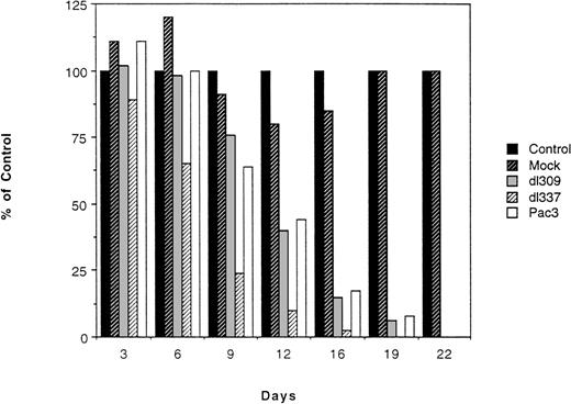
CLL cells from patient no. 7 persist in culture for more than 22 days, allowing a time course of full cytotoxicity for this patient’s cells to be undertaken (Fig 4). In this experiment, the parental virus (Ad5dl309), the highly attenuated virus containing a deletion of E1A and E1B (Pac3), and the virus containing an E1B 19K deletion (Ad5dl337) were used to infect the CLL cells from patient no. 7. Ad5dl337 was studied because it has been demonstrated to be highly cytotoxic to other cell types, resulting in cell death by apoptosis.2,3,16 20 Uninfected and mock-infected cells were viable for more than 22 days and cytotoxicity was seen after infection with each of the viruses (Fig 4). At each time point, Ad5dl337 induced the most pronounced cytotoxicity and by day 22, all of the viruses resulted in 100% cytotoxicity of patient no. 7 CLL cells.
Time course of cytotoxicity induced by adenovirus infection of CLL cells. CLL cells from patient no. 7 were infected with Ad5dl309, Pac3, or Ad5dl337 and the degree of cytotoxicity was monitored by cell counting and trypan blue staining every 3 to 4 days. Control refers to uninfected cells and mock refers to cells infected with heat-inactivated virus.
Time course of cytotoxicity induced by adenovirus infection of CLL cells. CLL cells from patient no. 7 were infected with Ad5dl309, Pac3, or Ad5dl337 and the degree of cytotoxicity was monitored by cell counting and trypan blue staining every 3 to 4 days. Control refers to uninfected cells and mock refers to cells infected with heat-inactivated virus.
To further confirm the induction of cytotoxicity in CLL cells by different mutated adenoviruses, cells from an eighth CLL patient were infected with Ad5dl309, the E1A deleted adenovirus (E1A−), Pac3, or Ad5dl337 at different moi. At an moi of 1 infectious unit per cell, the virus deleted in the E1B 19K gene (Ad5dl337) was the only virus that exhibited cell killing at day 8 (Fig 5). At higher mois, Ad5dl309, the E1A-virus, and Pac3 all exhibited cytotoxicity; however, at each moi, Ad5dl337 was the most efficacious in inducing cell death (Fig 5).
Ad5dl337 induces cytotoxicity after infection of primary CLL cells at a low moi. CLL cells from patient no. 8 were infected with Ad5dl309, Pac3, or Ad5dl337 at different moi. Cell viability was determined by cell counting and trypan blue staining 8 days after infection. Control refers to uninfected cells and mock refers to cells infected with heat-inactivated virus.
Ad5dl337 induces cytotoxicity after infection of primary CLL cells at a low moi. CLL cells from patient no. 8 were infected with Ad5dl309, Pac3, or Ad5dl337 at different moi. Cell viability was determined by cell counting and trypan blue staining 8 days after infection. Control refers to uninfected cells and mock refers to cells infected with heat-inactivated virus.
Infection of normal B cells and committed hematopoietic progenitors with Ad5dl309, E1A–, Pac3, and Ad5dl337.
To determine if normal B cells demonstrated adenovirus-mediated cytotoxicity after infection, preparations of mononuclear cells from a human tonsil were infected with Ad5dl309, Ad5dl337, E1A−, and Pac3. As demonstrated in Fig 6, the total number of viable B cells (as detected by CD19 expression) was unaffected by infection with Ad5dl309, E1A−, Pac3, or Ad5dl337 infection. In a similar experiment, blood mononuclear cells obtained from patients undergoing hematopoietic stem cell mobilization/harvesting were infected with Ad5dl309, E1A−, Ad5dl337, or Pac3 for 4 hours before plating in methylcellulose. Fourteen days later, colony formation was assessed (Fig 7). No reduction in colony formation was detected after infection with any of the viruses. Therefore, the tested viruses are not directly cytotoxic after infection of either normal B cells or committed hematopoietic progenitors.
Adenoviruses do not induce cytotoxicity after infection of normal B cells. Cells obtained from a nonmalignant human tonsil were infected with Ad5dl309, E1A−, Pac3, and Ad5dl337. At the indicated time after infection, the number of viable B cells was determined by isolating viable cells and assaying for CD19 expression by flow cytometry.
Adenoviruses do not induce cytotoxicity after infection of normal B cells. Cells obtained from a nonmalignant human tonsil were infected with Ad5dl309, E1A−, Pac3, and Ad5dl337. At the indicated time after infection, the number of viable B cells was determined by isolating viable cells and assaying for CD19 expression by flow cytometry.
Adenoviruses do not alter the ability of committed hematopoietic progenitor cells to form colonies in methylcellulose. Blood mononuclear cells obtained after treatment of patients undergoing hematopoietic stem cell mobilization with granulocyte colony-stimulating factor (G-CSF) were infected with Ad5dl309 at an moi of 100 for the indicated periods of time and plated in methylcellulose (in the presence of IL-3, granulocyte-macrophage colony-stimulating factor [GM-CSF], and erythropoietin [Epo]) using commercially available media (Stem Cell Technologies, Vancouver, British Columbia, Canada). Colonies (colony-forming unit-erythroid [CFU-e], burst-forming unit-erythroid [BFU-e], colony-forming unit–granulocyte-macrophage [CFU-GM]) were counted at 14 days. (−) Uninfected; (+) infected with Ad5dl309.
Adenoviruses do not alter the ability of committed hematopoietic progenitor cells to form colonies in methylcellulose. Blood mononuclear cells obtained after treatment of patients undergoing hematopoietic stem cell mobilization with granulocyte colony-stimulating factor (G-CSF) were infected with Ad5dl309 at an moi of 100 for the indicated periods of time and plated in methylcellulose (in the presence of IL-3, granulocyte-macrophage colony-stimulating factor [GM-CSF], and erythropoietin [Epo]) using commercially available media (Stem Cell Technologies, Vancouver, British Columbia, Canada). Colonies (colony-forming unit-erythroid [CFU-e], burst-forming unit-erythroid [BFU-e], colony-forming unit–granulocyte-macrophage [CFU-GM]) were counted at 14 days. (−) Uninfected; (+) infected with Ad5dl309.
To determine if adenoviruses were capable of infection of the B cells, a preparation of human tonsil cells (containing more than 75% B cells) was infected with AdGFP. No shift in fluorescence was seen after infection with AdGFP, suggesting that primary B cells are not infected (data not shown).
Infection of CLL cells with adenoviruses containing mutations in E2A.
These studies with primary malignant cells highlight adenovirus-mediated cytotoxicity induced by highly attenuated viruses in a cell type (B cells) that we have demonstrated to be refractory to adenovirus-mediated cytotoxicity. Others have also demonstrated that lymphoid cells are generally a nonproductive host.12,13 As a test of a further means of attenuation, a temperature-sensitive mutant containing a mutation in the E2A gene, Ad5ts12517(see Table 1), was studied to determine if the virus would induce cytotoxicity at either the permissive or restrictive temperature. Such a temperature-sensitive virus might ultimately have particular appeal for ex vivo therapeutic use, because it would be attenuated at the in vivo temperature of a patient.
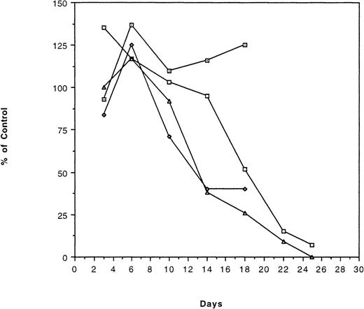
The results of the infections with the E2A temperature-sensitive mutant are demonstrated in Fig 8. As can be seen, both Ad5dl309 and Ad5ts125 induced significant cytotoxicity. Surprisingly, Ad5ts125 induced cytotoxicity at both 33°C and 37°C. Despite the cytotoxicity induced by Ad5ts125 at the restrictive temperature (37°C), it is important to note that there was a marked reduction in virus production after infection with Ad5ts125 at this temperature (see below).
Temperature-sensitive viruses induce cytotoxicity after infection of CLL cells. CLL cells from patient no. 7 were infected with a temperature-sensitive viruses containing mutations in the E2A gene (Ad5ts125) at the restrictive (37°C) and permissive (33°C) temperatures. Cytotoxicity was determined by cell counting and trypan blue staining at various times after infection. Control refers to uninfected cells and mock refers to cells infected with heat-inactivated virus. (⊡) Mock infection; (□) infection with Ad5dl309; (▵) infection with Ad5ts125 at 33°C; (◊) infection with Ad5ts125 at 37°C.
Temperature-sensitive viruses induce cytotoxicity after infection of CLL cells. CLL cells from patient no. 7 were infected with a temperature-sensitive viruses containing mutations in the E2A gene (Ad5ts125) at the restrictive (37°C) and permissive (33°C) temperatures. Cytotoxicity was determined by cell counting and trypan blue staining at various times after infection. Control refers to uninfected cells and mock refers to cells infected with heat-inactivated virus. (⊡) Mock infection; (□) infection with Ad5dl309; (▵) infection with Ad5ts125 at 33°C; (◊) infection with Ad5ts125 at 37°C.
Virus production and apoptosis after infection of CLL cells.
As a preliminary analysis of the mechanisms responsible for cytotoxcity, we have undertaken an analysis of the amount of virus present in the culture supernatant after infection of the CLL cells. In these experiments, virus was readily detected after the infection. The presence of significant amounts of virus with each of the samples reflects the presence of low-level viral production throughout the course of infection with each of the viruses. Table 2 demonstrates virus titer at days 3 and 10 after infection with Ad5dl309, Pac3, E1A−, Ad5ts125, or heat-inactivated Ad5dl309. Viral titers for Ad5dl309, the E1A− virus, and Pac3 were more than 105 pfu/mL on days 3 and 10. Pac3 is listed twice in Table2, because the supernatant from the infection on day 10 was titered on both 293 cells and HeLa cells. The absence of detectable virus when the supernatant was titered on HeLa cells reflects the inability of the Pac3 virus, which contains a deletion in E1A and E1B, to generate plaques on HeLa cells (Table 2). The Ad5ts125 is listed 4 times in Table 2, because the supernatants from infections at 33°C and 37°C were each titered at both 33°C and 37°C. For example, infection of CLL cells with Ad5ts125 at the restrictive temperature (37°C) resulted in very small amounts of virus in the supernatant at day 3 or 10 (2.5 × 10³ or 4.5 × 10³ pfu/mL), regardless of whether the supernatant was titered at 37°C or 33°C. In contrast, there was significantly more virus present in the supernatant after infection of CLL cells at the permissive temperature (33°C) when the supernatant was titered at the permissive as opposed to the restrictive temperature (1.5 × 10⁵ pfu/mL and 2.5 × 10³pfu/mL, respectively, on day 3; Table 2). Therefore, Ad5ts125 infection of CLL cells results in considerably more virus production at the permissive as opposed to the restrictive temperature and the virus produced at the permissive temperature retains the phenotype of temperature sensitivity.
Table 3 demonstrates viral yields after a series of infections with a different patient’s CLL cells. In agreement with the data presented in Table 2, the virus deleted of E1A and E1B (Pac3) and Ad5dl337 result in comparatively high virus yields in culture (more than 105 pfu/mL). In these experiments, infection of HeLa cells and 293 cells with the viruses produced during infection demonstrated the restricted host range and temperature sensitivity anticipated for infection by the temperature sensitive and Pac3 virus mutants.
Apoptosis, as measured by subdiploid DNA content, Hoechst 33258 staining, or Annexin V staining, was only detected in a small subset of the cells during infection (data not shown). Even Ad5dl337, a virus which readily induces apoptosis in other cell types,2,3,15 16 only resulted in a small increment in the number of cells with a subdiploid DNA content (as compared with mock-infected cells) and an insignificant alteration of Hoechst 33258 staining or Annexin V staining. For example, at 6 days after infection, 12% of CLL sample no. 7 cells infected with Ad5dl337 had a subdiploid DNA content compared with 7% of control or mock-infected cells. These results imply that some of the cytotoxicity induced by infection may be a consequence of apoptosis, but the degree of apoptosis, as measured by the above-described studies, was far less than anticipated if apoptosis was the sole or major mechanism of cytotoxicity. Presumably, adenovirus gene expression and virus production in CLL cells results in cytotoxicity that is only partially a consequence of apoptosis.
DISCUSSION
A panel of mutated adenoviruses was used to infect B-cell CLL cells, and even highly attenuated viruses were capable of inducing cytotoxicity. The degree of cytotoxicity induced by individual viruses varied among patients with CLL, although the adenovirus with an E1B 19K deletion, Ad5dl337, had the broadest range and extent of cytotoxicity. Although, the cells from few CLL patients persisted in culture long enough to establish the full extent of cytotoxicity under conditions supporting the continued viability of uninfected or mock-infected CLL cells, viral-induced cytotoxicty was clearly demonstrated at early time points for all of the patient samples. These studies demonstrate that adenoviruses are capable of infection of CLL cells and that infection with specific attenuated viruses results in marked cytotoxicity. These results indicate that attenuated adenoviruses could ultimately have a role in the therapy of CLL.
The variability in cytotoxicity induced by different viruses in different patient samples emphasizes the need to establish ex vivo conditions that will allow the CLL cells to persist long enough to allow a full assessment of cytotoxicity for all patients. The limited viability of CLL cells ex vivo, with 6 of the 7 initial uninfected CLL samples dying rapidly after day 10, is characteristic of CLL survival in culture. To further assess cytotoxicity under conditions reported to support primary cell viability in cell culture, we have undertaken infections during coculture of the CLL cells with irradiated CDw32 transfected L cells in the presence of IL-4 and anti-CD40 antibody.21-23 Our results confirm that this coculture system prolongs the viability of primary CLL cells in culture, however, the irradiated transfected L cells were not viable after infection with Ad5dl309, confounding an analysis of adenovirus-mediated cytotoxicity in most of the CLL samples (data not shown). Therefore, our assessment of cytotoxicity is based on early time points for the majority of the CLL samples and longer time points for those cells with greater viability in culture. The samples that persist in culture for prolonged periods of time do demonstrate 100% cytotoxicty after infection with Ad5dl309, Ad5dl337, E1A−, Pac3, and Ad5ts125 under conditions supporting the full viability of uninfected or mock-infected CLL cells. Immunophenotypic analysis has demonstrated that the viable cells in the uninfected culture were all CLL cells.
The mechanisms contributing to cytotoxcity after infection by wild-type and mutant adenoviruses are likely to be complex, involving direct cytotoxicity induced by adenovirus gene products and apoptosis induced by virus infection. Cytotoxicity after infection of malignant epithelial cells (with mutations in p53) by viruses lacking E1B 55K may be a consequence of virus replication, but the precise relationship between p53 mutation, viral replication, and cytotoxicity is likely to be complex and highly dependent on other host cell and virus features.5-7,16 In contrast to the frequent mutation of p53 in epithelial malignancies, p53 mutation is not common in CLL, with only 6% of standard CLL samples harboring mutations of p53.8-11 However, a broad spectrum of genetic aberrations have been described and many of these mutations may impact on cell cycle control or the regulation of apoptosis, and therefore, render the malignant cell an excellent target for cytotoxicity after infection with an attenuated adenovirus. For example, E1A function is essential for productive infection, thus cellular mutations that activate downstream targets of E1A (for example, mutations or deletions of pRb) may be predicted to enhance replication of an E1A-adenovirus. E1A itself has, under certain conditions, been demonstrated to mediate apoptosis3,15,16,24-28 and infection with viruses containing E1A in the absence of the antiapoptotoic functions of E1B enhances apoptosis in many cells. Both the pRb and p300 binding domains may be important for this induction of apoptosis in certain cells, whereas pRb binding may be dispensable in other cell types.16,24-27 Thus, specific E1A mutations may be predicted to facilitate apoptosis and cytotoxicity in some cell types and the proapoptotic functions of E1A may be offset by E1B. In particular, E1B 19K is of critical importance in preventing E1A-mediated apoptosis, and viruses that do not encode E1B 19K may facilitate cytotoxicity mediated by E1A and other viral genes, perhaps in conjunction with cellular changes that result in alterations of p300, pRb, Bcl-2 family members, or other cellular proteins, which impact on the development of apoptosis.2,3,15,16 24-27Therefore, viruses with mutations in E1A, E1B, or both may induce variable amounts of cytotoxicity, depending on the specific genotype of the host.
While the potent cytotoxicity induced by viruses with deletions of E1A + E1B (Pac3) or mutation of E1B 19K (Ad5dl337) in CLL cells from specific patients may at least partially be mediated by induction of apoptosis, it has not been possible to measure high levels of apoptosis during times of maximum cytotoxicity. Low-level virus production was seen after most infections, even with viruses markedly attenuated by mutations in E1A and E1B. Therefore, low-level productive infection appears to be present during most of the infections and cytotoxicity may be a consequence of a variety of processes, including both productive infection and apoptosis. Ad5dl337 consistently induced cytotoxicity in some samples of CLL to a greater extent than the parental Ad5dl309 (containing wild-type E1A and E1B). Presumably, the biological functions of E1B 19K inhibit adenovirus cytotoxicity,2,3,15,16,24 and viruses with a mutation of E1B 19K may be more cytotoxic than the parental virus in the context of low-level virus production. The cytotoxicity of the Pac3 virus is also interesting, considering the loss of both functional E1A and E1B, making Pac3 as highly attenuated as many of the adenovirus vectors commonly used for gene transfer. It is possible that other viral gene products such as the E4 open reading frame 4 (E4orf4), which has been demonstrated to be cytotoxic,29 30 may play a contributory role in the induction of cell death in the absence of E1A. Therefore, the specific cellular genotype and phenotype may complement adenoviruses with mutations necessary for productive infection, establish a milieu that fosters adenovirus-mediated apoptosis, or be cytotoxic as a consequence of other mechanisms, such as toxicity induced by adenovirus gene products. The cytotoxicity of the Pac3 virus also demonstrates the capacity of a highly attenuated virus, similar to those used as vectors for gene transfer in clinical trials of human gene therapy, to replicate and induce cytotoxicity under certain conditions.
These studies, with CLL cells from multiple patients, emphasize an advantage to screening for cytotoxicity induced by a spectrum of adenoviruses. Despite identical morphologic and immunophenotypic features, there is genetic heterogeneity among CLL samples.6-9 This heterogeneity presumably results in different patterns of cytotoxicity after infection of cells from different patients. This heterogeneity also emphasizes the importance of studying primary malignant cells as opposed to cell lines. Correlation of the cytotoxicity induced by each of the wild-type and mutant adenoviruses with the individual clinical, genetic, immunophenotypic, and cytogenetic features of the malignant cells will be important, because it may provide important insights into alterations of specific regulatory pathways in CLL cells. This may also apply to other lymphoid malignancies . For example, malignant cells from a patient with mantle cell lymphoma demonstrated cytotoxicity after infection with Ad5dl309, but no cytotoxicity was seen after infection with the Pac3 or E1A-viruses (unpublished data). Understanding the nuances of the patterns of cytotoxicity seen after infections with different mutated viruses may lead to a better understanding of the functional state of the malignant cell, particularly as it relates to the cell cycle regulatory control and the proapoptotic/antiapoptotic functions of the adenovirus E1 region.
The potential ex vivo use of these viruses for hematopoietic stem cell purging has been supported by preliminary studies from our laboratory that have demonstrated that infection of hematopoietic stem cells with Ad5dl309 and several of the adenovirus mutants used in this study does not result in cytotoxicity to committed hematopoietic progenitors, as measured by the capacity of the hematopoietic stem cells to form colonies in methylcellulose. Recombinant adenovirus vector infection of hematopoietic cells has also failed to induce toxicity, as measured by colony formation and other assays,31-37 including long-term culture initiating cell assay.38 In fact, the E1A− and Pac3 viruses used in our studies and demonstrated to be cytotoxic to CLL cells contain the same degree of attenuation as many of the adenovirus vectors used for gene transfer/therapy.31-38 Therefore, selectively cytotoxic adenoviruses might ultimately be developed for use ex vivo to purge hematopoietic stem cell grafts of CLL cells. The concept of inducing cytotoxicity with Ad5ts125 may also be relevant to this potential application of adenovirus-mediated cytotoxicity, as temperature-sensitivity may ultimately be incorporated into the viruses as an additional safety feature. Additional studies will be necessary to demonstrate that the adenoviruses used in our studies are not directly cytotoxic to primitive hematopoietic stem cells.
The studies presented in this report extend prior studies of adenovirus-mediated cytotoxicity by demonstrating that primary CLL cells can be infected with adenoviruses and are subject to cytotoxicity induced by even very highly attenuated mutant viruses. In addition, our studies demonstrate that there is no cytotoxicity after infection of normal B cells under identical conditions. These studies have implications for the biology of CLL and for the use of adenoviruses as vectors of gene-transfer and/or possible purging in lymphoid malignancies. Furthermore, the phenotypic pattern of cytotoxicity induced by different members of the panel of viruses will provide the framework to correlate cytotoxicity with specific genetic features of the malignant cells and provide additional information about the viral-host cell relationships that mediate cell death and productive infection.
Supported by the Elsa U. Pardee Foundation and National Institutes of Health Grant No. R01 CA69281.
The publication costs of this article were defrayed in part by page charge payment. This article must therefore be hereby marked “advertisement” in accordance with 18 U.S.C. section 1734 solely to indicate this fact.
REFERENCES
Author notes
Address reprint requests to Roger K. Strair, MD, PhD, The Cancer Institute of New Jersey, 195 Little Albany St, New Brunswick, NJ 08901; e-mail: strairrk@umdnj.edu.


![Fig. 7. Adenoviruses do not alter the ability of committed hematopoietic progenitor cells to form colonies in methylcellulose. Blood mononuclear cells obtained after treatment of patients undergoing hematopoietic stem cell mobilization with granulocyte colony-stimulating factor (G-CSF) were infected with Ad5dl309 at an moi of 100 for the indicated periods of time and plated in methylcellulose (in the presence of IL-3, granulocyte-macrophage colony-stimulating factor [GM-CSF], and erythropoietin [Epo]) using commercially available media (Stem Cell Technologies, Vancouver, British Columbia, Canada). Colonies (colony-forming unit-erythroid [CFU-e], burst-forming unit-erythroid [BFU-e], colony-forming unit–granulocyte-macrophage [CFU-GM]) were counted at 14 days. (−) Uninfected; (+) infected with Ad5dl309.](https://ash.silverchair-cdn.com/ash/content_public/journal/blood/94/10/10.1182_blood.v94.10.3499.422k03_3499_3508/6/m_blod42203007x.jpeg?Expires=1766087091&Signature=xzN~AhF1aI2cLkpwGcrIs96yH7bndaz2c~nfQluOGl1Jg3dd9t7Mkr5gNJIkkmF~bUIZmJD5Ffyjd-gvqwf6vX8pVNtsTu~kxMdZDnmMFQEknZaNYpXXbQwW2SEv~teZvwfrL7H3z8zUE3wvYk3nVZnXE4MnpmV0N0PjoG3u9YLi~kzBn9EUJsqFDa2AATYhtuPlqLJyBtbqxKzan7EVSlB3uYYQ1tnHGpmASPCS7uX0SqhupJqermVLX7WFY4aPyHaQhu7~5ZWMqR0iGG4Sdlju4qRdNYnR1~N0iSw0f1YzWsfStdhQu1e8w-2kyic~j6tNp1lKD5p1AHAXGO6njQ__&Key-Pair-Id=APKAIE5G5CRDK6RD3PGA)


![Fig. 7. Adenoviruses do not alter the ability of committed hematopoietic progenitor cells to form colonies in methylcellulose. Blood mononuclear cells obtained after treatment of patients undergoing hematopoietic stem cell mobilization with granulocyte colony-stimulating factor (G-CSF) were infected with Ad5dl309 at an moi of 100 for the indicated periods of time and plated in methylcellulose (in the presence of IL-3, granulocyte-macrophage colony-stimulating factor [GM-CSF], and erythropoietin [Epo]) using commercially available media (Stem Cell Technologies, Vancouver, British Columbia, Canada). Colonies (colony-forming unit-erythroid [CFU-e], burst-forming unit-erythroid [BFU-e], colony-forming unit–granulocyte-macrophage [CFU-GM]) were counted at 14 days. (−) Uninfected; (+) infected with Ad5dl309.](https://ash.silverchair-cdn.com/ash/content_public/journal/blood/94/10/10.1182_blood.v94.10.3499.422k03_3499_3508/6/m_blod42203007x.jpeg?Expires=1767423256&Signature=S0naer~w2M58xsWnTOeudxgNFzE~5DhBvEsNI0szA4Ym-mGtdXKpc1CyayhflSz3GBSOHvUxdIIMd~QzklBELhzjrrIwEfKHC8WBrdL3McduYEumvQM2lc9nDzv1L91LEb1HliQy056t4sogVZwsia-pmwkOVH5b4FJjUitxmanU1q2Pj4c3hVM2n6y4ZYiy0~FVUObtW5fjTKWK6EasgtusQ0WIDK71hsOluMtFSNn7LaUVJ-wymHP5zaPWbaBzwufCme6ASPVAefLN9nMKDusDycJtAAi1pPhxFeUucGqRG0WgFMg-4eAG3RwJCtDpn0o2nOPUe5uUijevvcp2Rg__&Key-Pair-Id=APKAIE5G5CRDK6RD3PGA)
