Abstract
Chemotherapy for adult T-cell leukemia (ATL) has been reported to fail to induce complete remission because of drug resistance in most patients. We have examined the expression of an ATL-derived factor (ADF)/thioredoxin in relation to resistance to adriamycin (ADM) in various T-cell leukemia cell lines including ATL cell lines. Immunoblot analysis demonstrated that ATL cell lines expressed ADF/thioredoxin at levels 2.8 to 12 times those of other T-cell acute lymphocytic leukemia (T-ALL) cell lines, and that ATL cell lines were 2 to 15 times more resistant to ADM than other T-ALL cell lines. Therefore, we established ADM-resistant cell lines from three different ATL cell lines, and examined the correlation between ADM resistance and expression of ADF/thioredoxin. ADM-resistant ATL cell lines were also found to be resistant to other drugs such as cisplatin and etoposide, and they expressed ADF/thioredoxin at levels 5 to 10 times those of parent ATL cell lines. Diamide and sodium selenite, which have been reported to inhibit ADF/thioredoxin, restored the sensitivity to ADM in ATL and ADM-resistant ATL cell lines. The MDR-1 gene product, a membrane P-glycoprotein (Pgp), was not expressed on ATL cell lines or ADM-resistant ATL cell lines. Topoisomerase II and glutathione peroxidase activities in T-cell leukemia cell lines were not correlated with ADM resistance. These results suggest that ADF/thioredoxin may play an important role in the drug resistance of ATL cells to ADM.
AN ADULT T-CELL leukemia (ATL)-derived factor (ADF), originally reported as a factor that induces interleukin-2 receptor-α, produced by human T-cell lymphotrophic virus 1–positive T cells and natural killer cells,1-3 has multiple functions, including the growth promotion of lymphocytes.4,5 According to cDNA cloning, ADF is considered to be a human homologue of thioredoxin,6,7 to be involved in an electron-transfer system common in a variety of organisms and tissues, and to scavenge free radicals.8-10 Recently, ADF/thioredoxin has been reported to inhibit the cell death caused by tumor necrosis factor-α (TNF-α) and anti-FAS antibody.11 On the other hand, the cellular level of ADF/thioredoxin has been reported to be involved in the drug sensitivity of bladder and prostate cancer cells to cisplatin, mitomycin C, doxorubicin, and etoposide.12 This evidence prompted us to examine whether ADF plays a key role in the resistance of ATL cells to adriamycin (ADM), which increases the amount of free radicals within the cells.
In this study, we examined the expression of ADF/thioredoxin and resistance to ADM in several T-cell leukemia, ATL, and ADM-resistant ATL cell lines. The results showed that ADF/thioredoxin expression levels correlated well with the resistance to ADM in T-cell leukemia cell lines, and that ADM-resistant ATL cell lines expressed a larger amount of ADF/thioredoxin than parent ATL cell lines. Furthermore, we found that the inhibition of ADF/thioredoxin restored the sensitivity to ADM in ATL and ADM-resistant ATL cell lines, but not in Jurkat cells.
MATERIALS AND METHODS
Reagents.Diamide (diazenedicarboxylic acid bis(N, N-dimethylamide), an electron acceptor that principally reacts with sulfhydryl groups to form disulfides, was purchased from Sigma Chemical Co (St Louis, MO). Sodium selenite was purchased from Wako Pure Chemical Industries (Osaka, Japan). Stock solutions of diamide and sodium selenite were prepared in phosphate-buffered saline (PBS) without Ca2+ and Mg2+ and stored frozen. Anti-Pgp monoclonal antibody was purchased from Oncogene Science Inc (Manhasset, NY), and anti-ADF/thioredoxin antibody was kindly provided by Dr Y. Kasahara (Fuji Rebio Co, Tokyo, Japan). Anti–glutathione peroxidase (GSH-Px) monoclonal antibody (GPX-121) was used to detect selenium-dependent cytosolic GSH-Px. The characteristics of this antibody were described previously.13 The topoisomerase II assay kit was purchased from TopoGen Inc (Columbus, OH). All of the anticancer agents used were obtained from commercial sources.
Cell lines.ATL-2 cells were provided by Dr M. Maeda (Kyoto University, Kyoto, Japan). SALT-3, ED-S, MOLT-3, and Jurkat cells were provided by Fujisaki Cell Center, Hayashibara Biochemical Laboratories Inc, (Okayama, Japan). HL-60 and HL-60/ADM cells were used as positive controls for MDR-1 gene products. These cell lines were maintained in RPMI-1640 medium supplemented with 10% fetal calf serum (FCS).
Establishment of ADM-resistant cell lines.ATL-2, SALT-3, and ED-S cells were cultured in the presence of ADM, the concentration of which was sequentially increased. Three months later, ADM-resistant cell lines started to proliferate constantly. Thereafter, these resistant cell lines were cultured for more than 12 months in the presence of ADM.
MTT assays.The resistance of cell lines to chemotherapeutic agents was estimated by colorimetric MTT assays according to the method previously described.14 Briefly, 1 × 10⁴ cells were plated in 100 μL RPMI 1640 medium supplemented with 10% FCS in 96-well flat-bottomed microplates and treated with varying doses of chemotherapeutic agents. The cultures were incubated for the first 48 hours, and then incubated with 500 μg/mL MTT-([1-(4,5-dimethylthiazol-2,5-diphenyl)tetrazolium bromide], Sigma) for the final 4 hours. After 100 μL isopropanol containing 0.04N HCl was added to each well and mixed thoroughly to dissolve the dark blue crystals, the plates were read on an MTP-100 microplate reader (Corona Electric, Tokyo, Japan) at a test wave length of 570 nm with a reference wavelength of 610 nm. Effects of sodium selenite and diamide were tested by adding sodium selenite and diamide at different concentrations (10−8 to 10−6 mol/L and 1 to 500 μg/mL, respectively) to the cultures throughout the incubation period. The IC50 values of sodium selenite and diamide for the cell lines were approximately 10−7 mol/L and 100 μg/mL, respectively. That is, sodium selenite and diamide were used at 10−7 mol/L and 100 μg/mL to overcome drug resistance.
FACS analysis.The cells (106) were incubated with 1 μg anti-Pgp monoclonal antibody for 30 minutes at 4°C, washed twice with PBS, and then incubated with fluorescein isothiocyanate–conjugated anti–mouse IgG antibody. After washing three times with PBS, the cells were analyzed by a FACScan (Becton Dickinson, Mountain View, CA). The cells stained with the second antibody alone were used as a control.
Assays for adriamycin accumulation in ATL cell lines.Cells (5 × 10⁵) were incubated with 2 μmol/L ADM for indicated intervals at 37°C. After being set on chilled ice for 2 hours, the cells were washed with ice-cold PBS twice and resuspended in fresh medium. Then, the fluorescence of ADM was sequentially analyzed by a FACScan.
Western blot analysis.Cell lysates (40 μg) were subjected to 12% sodium dodecyl sulfate–polyacrylamide gel electrophoresis (SDS-PAGE) under reducing conditions. Thereafter, the protein was transferred to polyvinylidenedifluoride membranes. After treatment with PBS containing 0.05% Tween 20 (T-PBS) and 5% skim milk in T-PBS, the membranes were incubated with 2 μg/mL anti-ADF/thioredoxin antibody and 100-fold diluted anti–GSH-Px as the first antibody and peroxidase-conjugated anti–mouse IgG antibody as the second antibody. Then, the membranes were stained using an ECL kit (Amersham Life Science, Tokyo, Japan).
GSH-Px and topoisomerase II activities.GSH-Px activities were examined by the methods described previously.15 Topoisomerase II activities were determined according to the manufacturer's instructions.
Statistical analysis.Correlations between the IC50 of ADM and the expression of ADF topoisomerase II activities or GSH-Px activities in T-cell leukemia cell lines were analyzed by Pearson and Spearman correlation coefficient analyses using StatView (Abacus Concept Inc, Berkeley, CA). Significance between the IC50 in the presence or absence of sodium selenite or diamide was analyzed by Student's t-test.
RESULTS
Sensitivity of the T-cell leukemia cell lines to ADM.The IC50 values of ADM to Jurkat and MOLT-3 (T-ALL cell lines) were 0.05 and 0.028 μmol/L, respectively (Table 1). The IC50 of ADM to ED-S, SALT-3, and ATL-2 (ATL cell lines) was 0.102, 0.399, and 0.759 μmol/L (two to four, eight to 16, and 15 to 30 times higher than T-ALL cell lines), respectively. The IC50 of ADM to ADM-resistant ATL-2 (ATL-2/ADM cl-1 and ATL-2/ADM cl-2) cell lines was 1.695 and 4.403 μmol/L (two and six times higher than parent ATL-2 cells), respectively (Table 2). The IC50 of ADM to SALT-3/ADM cl-2 and ED-S/ADM was 1.92 and 0.655 μmol/L (five and six times higher than parent SALT-3 and ED-S cells), respectively (Table 2).
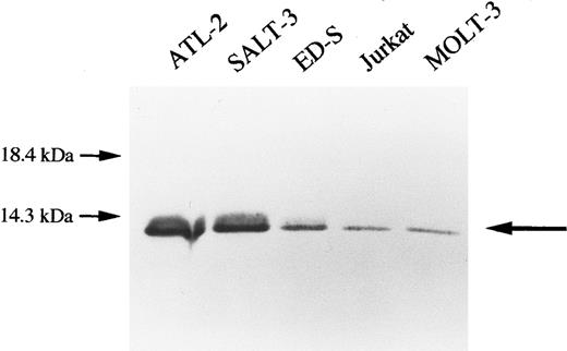
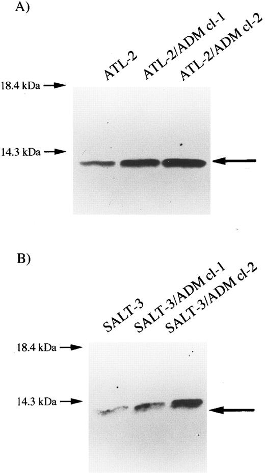
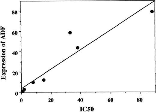
Expression of ADF/thioredoxin in T-cell leukemia cell lines.Figures 1 and 2 show the expression of ADF/thioredoxin in T-cell leukemia cell lines. ATL cell lines (ATL-2, SALT-3, and ED-S) expressed a larger amount of ADF/thioredoxin (2.8-, 9.6-, and 12.1-fold) than T-ALL cell lines (Jurkat and MOLT-3). The increase of ADF/thioredoxin expression in T-cell leukemia cell lines correlated well with the enhancement of resistance to ADM (r = .891, P < .01; Fig 3). ATL-2, which shows the highest IC50 , expressed the largest amount of ADF/thioredoxin among the cell lines. ATL-2/ADM cl-1 and ATL-2/ADM cl-2 cell lines expressed ADF/thioredoxin at levels 4.8 and 6.6 times those of parent ATL-2 cell lines, respectively. SALT-3/ADM cl-1 and SALT-3/ADM cl-2 expressed ADF/thioredoxin at levels 2.6 and 4.6 times those of parent SALT-3 cell lines, respectively. The ED-S/ADM cell line also expressed a larger amount of ADF/thioredoxin than the ED-S cell line (data not shown). The increase of ADF/thioredoxin expression in ADM-resistant ATL cell lines also correlated well with the enhancement of resistance to ADM (r = .891, P < .01; Fig 3).
Expression of ADF in T-cell leukemia cell lines. Cell lysates (40 μg) were electrophoresed by 12% SDS-PAGE under reducing conditions and transferred to polyvinylidenedifluoride membranes. Immunostaining was performed using anti-ADF antibody as the first antibody.
Expression of ADF in T-cell leukemia cell lines. Cell lysates (40 μg) were electrophoresed by 12% SDS-PAGE under reducing conditions and transferred to polyvinylidenedifluoride membranes. Immunostaining was performed using anti-ADF antibody as the first antibody.
Expression of ADF in ATL cell lines. Cell lysates (40 μg) were electrophoresed by 12% SDS-PAGE under reducing conditions and transferred to polyvinylidenedifluoride membranes. Immunostaining was performed using anti-ADF antibody as the first antibody. (A) ATL-2, ATL-2/ADM cl-1, and ATL-2/ADM cl-2; (B) SALT-3, SALT-3/ADM cl-1, and SALT-3/ADM cl-2
Expression of ADF in ATL cell lines. Cell lysates (40 μg) were electrophoresed by 12% SDS-PAGE under reducing conditions and transferred to polyvinylidenedifluoride membranes. Immunostaining was performed using anti-ADF antibody as the first antibody. (A) ATL-2, ATL-2/ADM cl-1, and ATL-2/ADM cl-2; (B) SALT-3, SALT-3/ADM cl-1, and SALT-3/ADM cl-2
Correlation between the IC50 and expression of ADF in T-cell leukemia cell lines. IC50 (relative values v that of Jurkat) and expression of ADF (relative density of the bands v that of Jurkat) were plotted. r = .891, P < .01 (Pearson's correlation coefficient analysis). Also significant by Spearman's correlation coefficient analysis (P < .01).
Correlation between the IC50 and expression of ADF in T-cell leukemia cell lines. IC50 (relative values v that of Jurkat) and expression of ADF (relative density of the bands v that of Jurkat) were plotted. r = .891, P < .01 (Pearson's correlation coefficient analysis). Also significant by Spearman's correlation coefficient analysis (P < .01).
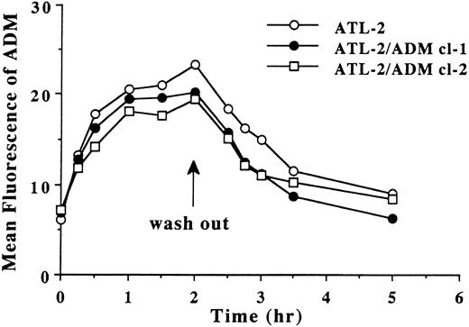
Accumulation and efflux of ADM in ATL cell lines.We examined the accumulation and efflux of ADM in ATL cell lines to determine whether transporter proteins (Pgp and MDR-associated protein) were involved in drug resistance in ATL cell lines. The accumulation and efflux curves of ADM in ATL-2 and ATL-2/ADM cells were similar (Fig 4). After the addition of ADM, the fluorescence of ADM in the cells started to increase, and after washing out ADM, it decreased gradually.
Accumulation and efflux curves of ADM in ATL-2 and ATL-2/ADM cell lines. Mean fluorescence of ADM obtained from three different experiments was plotted at indicated times.
Accumulation and efflux curves of ADM in ATL-2 and ATL-2/ADM cell lines. Mean fluorescence of ADM obtained from three different experiments was plotted at indicated times.
Resistance of ATL cell lines to other drugs.Next, we examined the resistance of ATL cell lines to other drugs, including etoposide (VP-16) as another Pgp-related agent and bleomycin (BLM) and cisplatin (CDDP) as Pgp-unrelated drugs. ADM-resistant ATL cells showed a higher IC50 to CDDP and VP-16 than parent ATL cells (Table 3). However, the levels of ADF/thioredoxin expression and the IC50 to VP-16 and CDDP of T-cell leukemia cell lines were not correlated.
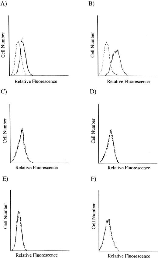
Expression of MDR-1 gene product on ATL cell lines.We examined the expression of Pgp on ATL cell lines. The positive control HL-60 and HL-60/ADM cell lines expressed Pgp, whereas none of the ATL cell lines and ADM-resistant ATL cell lines expressed Pgp (Fig 5).
Expression of Pgp on T-cell leukemia cell lines. HL-60 and HL-60/ADM cells were used as positive controls. (A) HL-60, (B) HL-60/ADM, (C) ATL-2, (D) ATL-2/ADM cl-2, (E) SALT-3, and (F) SALT-3/ADM cl-2.
Expression of Pgp on T-cell leukemia cell lines. HL-60 and HL-60/ADM cells were used as positive controls. (A) HL-60, (B) HL-60/ADM, (C) ATL-2, (D) ATL-2/ADM cl-2, (E) SALT-3, and (F) SALT-3/ADM cl-2.
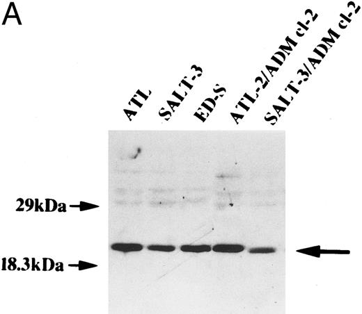
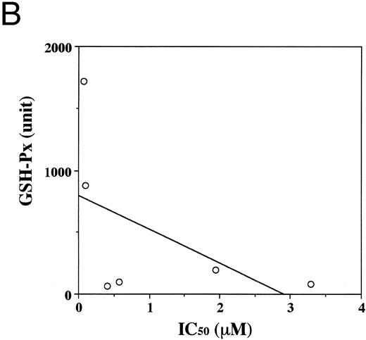
Expression of GSH-Px in T-cell leukemia cell lines.Figure 6A shows the expression of GSH-Px in ATL-2, SALT-3, ED-S, ATL-2/ADM cl-2, and SALT-3/ADM cl-2 cell lines. All cell lines expressed GSH-Px, and the expression of GSH-Px was not correlated with the resistance to ADM. GSH-Px activities also showed no correlation with the resistance to ADM (r = .22; Fig 6B).
Expression of GSH-Px in T-cell leukemia cell lines. (A) Western blot analysis. Cell lysates (4 μg) were electrophoresed by 12% SDS-PAGE under reducing conditions and transferred to polyvinylidenedifluoride membranes. Immunostaining was performed using anti–GSH-Px antibody as the first antibody. (B) Correlation between the IC50 and the activity of GSH-Px in T-cell leukemia cell lines. GSH-Px activity showed no correlation with the resistance to ADM (r = .22).
Expression of GSH-Px in T-cell leukemia cell lines. (A) Western blot analysis. Cell lysates (4 μg) were electrophoresed by 12% SDS-PAGE under reducing conditions and transferred to polyvinylidenedifluoride membranes. Immunostaining was performed using anti–GSH-Px antibody as the first antibody. (B) Correlation between the IC50 and the activity of GSH-Px in T-cell leukemia cell lines. GSH-Px activity showed no correlation with the resistance to ADM (r = .22).
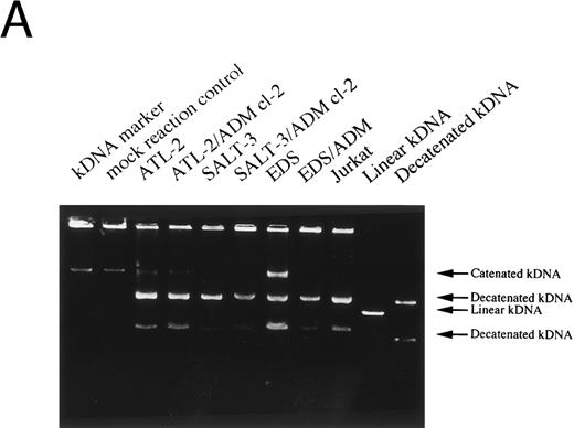
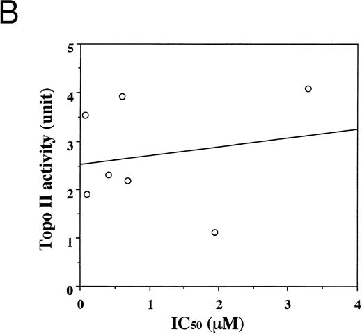
Topoisomerase II activities.Figure 7A shows the degree of decatenation of kDNA by topoisomerase II contained in the lysates of the indicated cell lines. Figure 7B shows the correlation between topoisomerase II activities and the IC50 of ADM. There was no significant correlation (r = .19).
Topoisomerase II activity. (A) Agarose gel electrophoresis; (B) correlation between topoisomerase II activity and the IC50 of ADM in T-cell leukemia cell lines. Topoisomerase II activity showed no correlation with the resistance to ADM (r = .19).
Topoisomerase II activity. (A) Agarose gel electrophoresis; (B) correlation between topoisomerase II activity and the IC50 of ADM in T-cell leukemia cell lines. Topoisomerase II activity showed no correlation with the resistance to ADM (r = .19).
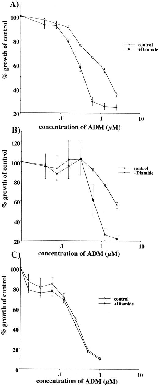
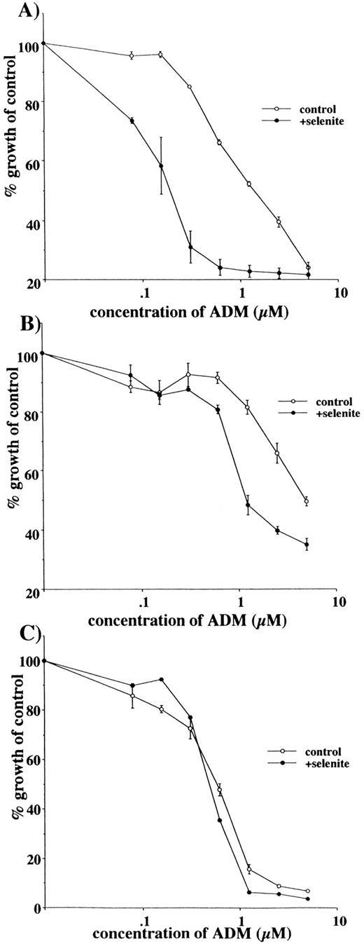
Restoration of sensitivity to ADM by sodium selenite and diamide.Both diamide and selenite have been reported to oxidize ADF/thioredoxin, which results in the inhibition of its disulfide-reductase activity.15-17 When Jurkat, ATL-2, and ATL-2/ADM cells were incubated with serially diluted concentrations of ADM in the presence of 100 μg/mL diamide, diamide restored the sensitivity to ADM of ATL-2 and ATL-2/ADM cells, but not that of Jurkat cells (Fig 8 and Table 4). Sodium selenite at 10−7 mol/L also restored the sensitivity to ADM of ATL-2 and ATL-2/ADM cells, but not that of Jurkat cells (Fig 9 and Table 4). A lower concentration of sodium selenite and diamide than the IC50 values showed some but no significant effect. However, restoration of the resistance to ADM might not be due to the direct cytotoxicity of sodium selenite and diamide, since the restoration was observed only in ATL-2 and ATL-2/ADM cell lines that expressed a large amount of ADF/thioredoxin.
Restoration of sensitivity to ADM by diamide. Viable cells were estimated by MTT assays after incubation with different doses of ADM in the presence or absence of diamide (100 μg/mL) for 3 days. Representative results were obtained from 3 different experiments that showed similar results. The mean ± SD for the IC50 obtained from 3 different experiments is shown in Table 4. (A) ATL-2, (B) ATL-2/ADM cl-2, (C) Jurkat.
Restoration of sensitivity to ADM by diamide. Viable cells were estimated by MTT assays after incubation with different doses of ADM in the presence or absence of diamide (100 μg/mL) for 3 days. Representative results were obtained from 3 different experiments that showed similar results. The mean ± SD for the IC50 obtained from 3 different experiments is shown in Table 4. (A) ATL-2, (B) ATL-2/ADM cl-2, (C) Jurkat.
Restoration of sensitivity to ADM by sodium selenite. Viable cells were estimated by MTT assays after incubation with different doses of ADM in the presence or absence of sodium selenite (10−7 mol/L) for 3 days. Representative results were obtained from 3 different experiments that showed similar results. The mean ± SD for the IC50 obtained from 3 different experiments is shown in Table 4. (A) ATL-2, (B) ATL-2/ADM cl-2, (C) Jurkat.
Restoration of sensitivity to ADM by sodium selenite. Viable cells were estimated by MTT assays after incubation with different doses of ADM in the presence or absence of sodium selenite (10−7 mol/L) for 3 days. Representative results were obtained from 3 different experiments that showed similar results. The mean ± SD for the IC50 obtained from 3 different experiments is shown in Table 4. (A) ATL-2, (B) ATL-2/ADM cl-2, (C) Jurkat.
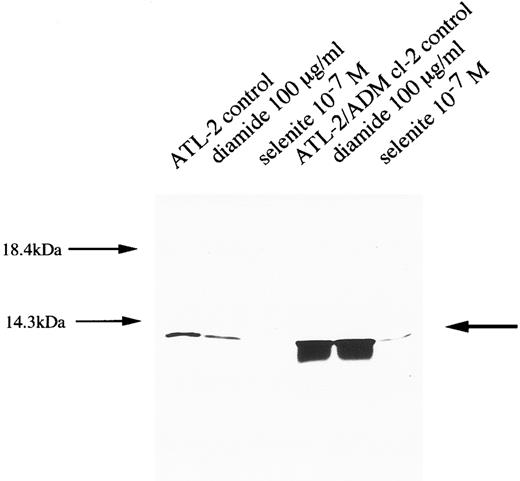
Expression of ADF in ATL cell lines after treatment with diamide and selenite. Cell lysates (40 μg) were electrophoresed by 12% SDS-PAGE under reducing conditions and transferred to polyvinylidenedifluoride membranes. Immunostaining was performed using anti-ADF antibody as the first antibody.
Expression of ADF in ATL cell lines after treatment with diamide and selenite. Cell lysates (40 μg) were electrophoresed by 12% SDS-PAGE under reducing conditions and transferred to polyvinylidenedifluoride membranes. Immunostaining was performed using anti-ADF antibody as the first antibody.
Decreased expression of ADF in ATL cell lines after incubating with diamide and sodium selenite.Treatment with diamide and sodium selenite decreased the expression of ADF/thioredoxin in both ATL-2 and ATL-2/ADM cl-2 cell lines (Fig 10).
DISCUSSION
T-cell leukemia, particularly ATL, has a poor response to chemotherapy, and the survival rate for ATL is low compared with B-cell leukemia.18-20 One of the reasons for the poor response is considered to be an emergence of drug-resistant tumor cells. Kuwazuru et al21 reported that ATL cells from patients at relapsed stages expressed Pgp, which is considered to be responsible for the drug resistance of various tumors, including B-cell leukemia.22,23 However, we hypothesized that Pgp could not be entirely responsible for the resistance of ATL to chemotherapy, since the response to chemotherapy in ATL patients is poor even at early stages. Recently, ADF/thioredoxin has been reported to have protective activity against TNF-α– and anti-FAS antibody–induced cell death.11 ADF/thioredoxin also acts in an electron-transfer system and is able to scavenge free radicals.6-10 According to this evidence, we examined whether ADF/thioredoxin contributes to the resistance of ATL cells to ADM. Our findings are as follows: (1) the resistance to ADM in T-cell leukemia cell lines, including ATL cell lines, correlated well with the level of expression of ADF/thioredoxin; (2) no ATL and ADM-resistant ATL cell lines expressed Pgp, and the chronologic accumulation of ADM in those cell lines showed no difference, which suggested that MDR-1 and MDR-related proteins (MRP) did not play important roles in the ADM resistance; (3) ADM-resistant cell lines were also resistant to VP-16 and CDDP, but there was no significant correlation between the IC50 to VP-16 or CDDP and the level of expression of ADF/thioredoxin; and (4) neither topoisomerase II activity nor GSH-Px activity correlated with ADM resistance, although they have been reported to be involved in drug resistance in some cell lines.24 25 Together, our results suggest that the enhanced expression of ADF/thioredoxin plays important roles in the intrinsic and acquired ADM resistance in T-cell leukemia cell lines, especially ATL cell lines. However, a possibility remains that MRP were also involved in ADM resistance, since we did not directly examine the expression of MRP and ADM-resistant ATL cell lines that also showed resistance to MRP-related drugs such as VP-16. And the decrease of topoisomerase II activity in SALT-3/ADM compared with SALT-3 might be partially responsible for the resistance to ADM in SALT-3/ADM cells.
Although the mechanisms by which ADF/thioredoxin plays important roles in the resistance to ADM in T-cell leukemia cells remain to be elucidated, one possible mechanism may be the decrease of DNA damage through the reactive oxygen–reducing activities of ADF/thioredoxin. ADF/thioredoxin acts in an electron-transfer system and is able to scavenge free radicals.6-10 Mitsui et al26 have recently reported that recombinant human ADF/thioredoxin reduced some reactive oxygen species such as hydrogen peroxide. Unfortunately, we have not detected the production of reactive oxygen species in ATL cell lines after incubation with ADM, because of the interference of ADM fluorescence. Furthermore, ADF/thioredoxin has been reported to have protective activity against anti-FAS antibody–induced cell death,11 in which presumably reactive oxygen species are not involved. Therefore, other mechanisms may be responsible for the acquisition of ADM resistance by ATL cells. From this point of view, our observation that ADM was distributed in the cytoplasm even after long-term incubation with ADM in ATL and ADM-resistant ATL cells is interesting (data not shown). ADF/thioredoxin also localized in the cytoplasm (data not shown), which suggests that ADF/thioredoxin may affect the proteins that are involved in transportation of ADM from the cytoplasm to the nucleus by protein-refolding activities of ADF/thioredoxin.
In this study, we showed the in vitro effects of selenite and diamide for overcoming the resistance to ADM in ATL and ADM-resistant ATL cell lines. Selenite, known to be an essential trace element for a wide range of living organisms, shows potent anticarcinogenic and antineoplastic activity,27,28 and has been used in clinical trials of liver cancer in high-risk populations.28 One of the mechanisms of selenite-mediated inhibition of carcinogenesis is growth inhibition of responsive cells.29 The effects of selenite are reported to be biphasic: stimulatory at 5 × 10⁻⁸ mol/L and inhibitory at greater than 1 × 10⁻⁶ mol/L.30 On the other hand, selenite at a relatively low concentration (< 10−7 mol/L) did not stimulate the growth of ATL cell lines including parent and ADM-resistant lines. These results suggest that selenite might be a candidate drug to overcome the drug resistance of ATL.
ACKNOWLEDGMENT
We thank Dr M. Maeda (Kyoto University), Dr J. Minowada (Fujisaki Cell Center, Hayashibara Biochemical Laboratories Inc), and Dr Y. Kasahara (Fuji Rebio Co) for providing the cell lines and anti-ADF antibody, and M. Yanome for manuscript preparation.
Supported in part by a grant from the Ministry of Education, Science, and Culture, Japan; and J.W. by Dr Y. Uehata.
Address reprint requests to Masanobu Kobayashi, MD, Laboratory of Pathology, Cancer Institute, Hokkaido University School of Medicine, Kita-15, Nishi-7, Kita-ku, Sapporo, 060 Japan.

