Abstract
Amyloid precursor protein (APP) has been reported to be highly expressed in AML1/ETO positive acute myeloid leukemia (AML1/ETO+ AML), and we found it express even higher in those with extramedullary infiltration in our previous study. But it’s still unknown what role APP plays and how it works in AML1/ETO+ AML. This study was designed to investigate the effect of APP gene on the prognosis and its molecular mechanism of extramedullary infiltration in the patients with AML1/ETO+ AML.
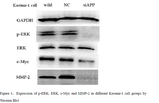
44 cases of AML1/ETO+ AML patients with median age of 29 years old, who were admitted to our hospital from February, 2006 to February, 2012 and made the diagnosis according to WHO2008 diagnosis standard, and had completed conventional induction, consolidation and intensive therapy, were investigated in this study. They were divided into high expression group (n=22) and low one (n=22) according to APP mRNA median expression level from bone marrow cells before the first chemotherapy by QRT-PCR. Some of bone marrow samples were checked by Western Blot, and 5 biopsy specimens from extramedullary infiltration were tested by APP antibody immunohistochemistry staining. Incidence of extramedullary leukemia (EML), complete response (CR), overall survival (OS), and recurrence free survival (RFS) was differentiated between the two groups. Differences of cell ultrastructure, migration, proliferation, apoptosis and expression of ERK, MMP-2, MMP-9 and CXCR4 were studied on Kasumi-1 cell line between wild, negative control (NC) and si-APP group in which the expression levels of APP gene were down regulated with application of siRNA technology.Çå
In sum, incidence of EML is significantly higher and the prognosis is poor in the patients with AML1/ETO+ AML with high expression of APP gene. We first describe that APP gene may mediate AML1/ETO+ leukemia cells in the development of extramedullary infiltration by up-regulation of the ERK/MMP-2 pathway.
No relevant conflicts of interest to declare.
Author notes
Asterisk with author names denotes non-ASH members.


