Abstract
T-cell production depends on the recruitment of hematopoietic progenitors into the thymus. T cells are among the last of the hematopoietic lineages to recover after bone marrow transplantation (BMT), but the reasons for this delay are not well understood. Under normal physiologic conditions, thymic settling is selective and either CCR7 or CCR9 is required for progenitor access into the thymus. The mechanisms of early thymic reconstitution after BMT, however, are unknown. Here we report that thymic settling is briefly CCR7/CCR9-independent after BMT but continues to rely on the selectin ligand PSGL-1. The CCR7/CCR9 independence is transient, and by 3 weeks after BMT these receptors are again strictly required. Despite the normalization of thymic settling signals, the rare bone marrow progenitors that can efficiently repopulate the thymus are poorly reconstituted for at least 4 weeks after BMT. Consistent with reduced progenitor input to the thymus, intrathymic progenitor niches remain unsaturated for at least 10 weeks after BMT. Finally, we show that thymic recovery is limited by the number of progenitors entering the thymus after BMT. Hence, T-lineage reconstitution after BMT is limited by progenitor supply to the thymus.
Introduction
T cells provide critical immune protection from a range of pathogens. The T-lineage is the slowest to recover after irradiation and bone marrow transplantation (BMT), a delay that impairs immunologic protection of the host.1 Peripheral T-cell reconstitution after BMT occurs through 2 mechanisms: one thymus-independent and one thymus-dependent. First, radioresistant host T cells and donor T cells provided in the graft homeostatically proliferate in the lymphopenic postirradiation environment.2,3 Although this population expansion can partially correct numerical T-cell defects, the resulting cells are functionally compromised.4,5 The functional recovery of the T-lineage relies on the second mechanism: the de novo generation of naive T cells in the thymus.6,7 The generation of thymus-derived naive T cells can take years and is particularly slow in adults.1,2,8 The reasons for this delay are not fully understood but have been suggested to involve impaired intrathymic development because of thymic stromal damage from conditioning regimens, age-related thymic involution, and graft-versus-host disease (GVHD).9-11
The thymus does not contain self-renewing progenitors and therefore requires the importation of circulating bone marrow (BM)-derived progenitors to sustain thymopoiesis.12-14 Thymic settling, however, is suggested to be a rare event, and the identity of thymic settling progenitors remains unclear.15-17 All progenitors descend from hematopoietic stem cells (HSCs), which are phenotypically negative for lineage markers (Lin) and are additionally Kit+Sca1+Flt3−. Directly downstream of HSCs are nonrenewing multipotent progenitors (MPPs; Lin−Kit+Sca1+Flt3low),18 which in turn give rise to lymphoid-primed multipotent progenitors (LMPPs; Lin−Kit+Sca1+Flt3high) and more downstream common lymphoid progenitors (CLPs; Lin−KitlowSca1lowFlt3highIL-7Rα+).19,20 Each of these progenitor types, plus additional progenitors, has been demonstrated to possess T-lineage potential.21,22 Prethymic hematopoiesis thus provides a multitude of progenitors with the ability to contribute to T-lymphopoiesis.
The process of thymic settling in normal hosts is selective, as only certain BM progenitors possess the capacity to enter the thymus from blood.23 The chemokine receptors CCR7 and CCR9 underlie part of this selectivity; each receptor independently supports the importation of progenitors into the thymus.23-27 Thymic settling under competitive conditions is near-absolutely restricted in the absence of both receptors.26,27 The P-selectin ligand PSGL-1 also supports thymic settling.28 Subsets of LMPPs and CLPs express functional CCR7 and CCR9, whereas HSCs and MPPs do not.23,27 Consistently, all thymic settling capacity effectively resides within the BM Lin−Kit+Flt3high pool, which includes both Kithigh LMPPs and Kitlow CLPs but excludes HSCs and MPPs.23,29,30 These Lin−Kit+Flt3high progenitors undergo Notch-dependent differentiation into early thymic progenitors (ETPs) on thymic entry.31,32 ETPs generate CD4/CD8 double-negative 2 (DN2) cells and then DN3 cells. After β-selection, DN3 cells become CD4/CD8 double-positive (DP) cells; after positive selection, DP cells give rise to CD4 and CD8 single-positive (SP) thymocytes, which emigrate from the thymus to compose the peripheral naive T-cell pool.
Our present knowledge of molecules that mediate normal thymic settling derives from unirradiated hosts, but thymic settling after irradiation is not well understood. Irradiation is often used as part of a cytoablative conditioning regimen before BMT.33 After irradiation, radioresistant intrathymic precursors proliferate and differentiate to initially reconstitute the thymus.34,35 These host thymocytes do not self-renew, and long-term thymopoiesis after BMT requires thymic settling by donor progenitors. However, the effects of irradiation on the signals and cells involved in thymic settling are not known.
We investigated the mechanisms of early thymic reconstitution after BMT. We found that the requirement for CCR7 and CCR9 in thymic settling is relaxed acutely after irradiation, although PSGL-1 retains a key role. This initial stage of CCR7/CCR9-independent thymic settling is temporary, and a strict requirement for these receptors is restored by 3 weeks after irradiation. BM progenitors that can efficiently repopulate the thymus are poorly reconstituted for at least 4 weeks after BMT. Furthermore, the intrathymic progenitor niche remains unsaturated for at least 10 weeks after BMT, suggesting that impairments before thymic entry limit T-lineage reconstitution. Indeed, we find that the delivery of more progenitors into the thymus by direct intrathymic transfer enhances thymic recovery. These data establish that the mechanisms of thymic settling after irradiation vary from those in normal mice. Furthermore, they indicate that limited entry of hematopoietic progenitors into the thymus after BMT impairs T-cell recovery.
Methods
Mice
Mice used included C57Bl/6 and B6.Ly5SJL mice (National Cancer Institute) and PSGL-1−/− mice (The Jackson Laboratory). CCR7−/−CCR9−/− mice were generated by crossing CCR7−/− and CCR9−/− strains, obtained from The Jackson Laboratory and Dr Paul Love (National Institutes of Health, Bethesda, MD), respectively. Mice used were 4-10 weeks old. Animal experiments were performed according to approved protocols of the Office of Regulatory Affairs at the University of Pennsylvania in accordance with National Institutes of Health guidelines.
Cell preparations, flow cytometry, and cell sorting
BM and thymocytes were collected as previously described.27 Antibodies in the Lin cocktail included: anti-B220 (RA3–6B2), anti-CD19 (1D3), anti-CD11b (M1/70), anti–Gr-1 (8C5), anti-CD11c (HL3), anti-NK1.1 (PK136), anti–Ter-119 (Ter-119), anti-CD3 (2C11), anti-CD8α (53.6–7), anti-CD8β (53–5.8), anti-TCRβ (H57), and anti-γδTCR (GL-3). Additional antibodies used included anti-CD45.2 (104), anti-CD45.1 (A20), anti-Kit (2B8), anti-Sca1 (D7), anti-Flt3 (A2F10), anti–IL-7Rα (A7R34), anti-CD25 (PC61.5), and anti-CD4 (RM4–5). Antibodies were purchased from eBioscience, BD Biosciences Pharmingen, and BioLegend.
BM was sorted on a FACSAria II (BD Biosciences). An LSRII (BD Biosciences) was used for fluorescence-activated cell sorter analysis. Dead cells were excluded through 4′,6-diamidino-2-phenylindole uptake. Doublets were excluded through FSC-H by FSC-W and SSC-H by SSC-W parameters. Data were analyzed using FlowJo Version 8.8.7 software (TreeStar).
Intravenous and intrathymic transfers
For intravenous transfers into unirradiated mice, 1.4 × 10⁷ T cell-depleted BM cells were injected retro-orbitally. To prevent rejection, mice were given 0.1 mg anti-CD4 (GK1.5) by intraperitoneal injection the day before BMT and every 4 days thereafter. For intrathymic transfers, 3 × 10³ sorted BM LMPPs or Lin−Kit+Flt3high cells were injected intrathymically.12 For intrathymic injections into sublethally irradiated hosts, a radiation dose of 6 Gy was used. For BM transplants, host CD45.1 wild-type (WT) mice were lethally irradiated (9.5 Gy) and inoculated with T cell-depleted donor BM 4-8 hours after irradiation. Mice were irradiated using a Cs-137 source at a rate of 43.25 cGy/min.
OP9-DL4 cultures
Sorted progenitors were placed onto OP9-DL4 stromal layers (gift of J.C. Zuniga-Pflücker, University of Toronto) as previously described.36
Vascular permeability studies
Mice were irradiated (1.5 Gy or 9.5 Gy) 24 hours before injection of Evans blue (Sigma-Aldrich) or injected intraperitoneally with 5 μg/g lipopolysaccharide (Sigma-Aldrich) 30 minutes after injection of Evans blue. Mice were intravenously injected with 20 μg Evans blue per gram of body weight. After 2.5 hours, anesthetized animals were exsanguinated and perfused with phosphate-buffered saline (PBS) to clear intravascular Evans blue. The thymus and one lobe of the lung were removed from each mouse, weighed, and homogenized in PBS. Evans blue was extracted by formamide for 24 hours at 55°C. Absorbances of extracted Evans blue at 620 nm and 740 nm were read on a spectrophotometer (BioRad SmartSpec3000). The concentrations of extracted Evans blue were calculated as previously described.37
Immunofluorescence microscopy
Thymi were cryosectioned into 30-μm sections by the Wistar Institute Histotechnology Core. Sections were stained with anti-CD31 antibody (MEC13.3) purchased from BioLegend. Images were obtained from a wide-field microscope (Zeiss Axioplan2) coupled to a camera (Zeiss AxioCam) and processed with Image J Version 1.43u software.
Statistical analysis
P values were calculated using Microsoft Excel by Student t test. All experiments are representative of 3 or more independent experiments with the exception of the experiment in Figure 3, which was performed twice.
Results
Brief CCR7/CCR9-independent thymic settling after BMT
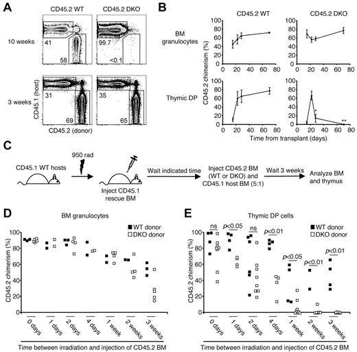
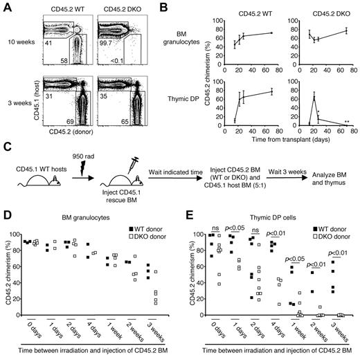
In unirradiated mice, progenitors lacking CCR9 and CCR7 are almost completely unable to settle the thymus.23,26,27 We investigated whether thymic settling relies on these same chemokine receptors after irradiation. We generated 2 cohorts of lethally irradiated mixed BM chimeras: a control group mixing WT CD45.2 BM and WT CD45.1 BM, and a second group mixing CCR7/CCR9 double knock-out (DKO) CD45.2 BM and WT CD45.1 BM. This method of generating chimeras does not allow us to distinguish donor-derived “competitor” CD45.1 thymocytes from radioresistant host-derived CD45.1 thymocytes, but we could evaluate the relative competitiveness of the 2 “test” CD45.2 cohorts. Three weeks after irradiation, CCR7/CCR9 DKO chimerism among DP cells was similar to WT chimerism (Figure 1A-B). Yet 10 weeks after irradiation, only a trace contribution to the DP pool from CCR7/CCR9 DKO cells was detected. The absence of CCR7/CCR9 DKO DP cells at this time is not the result of an intrathymic developmental defect; we previously showed that intrathymically transferred CCR7/CCR9 DKO progenitors have no defect in generating downstream progeny.27 We also examined these chimeras at 2 and 4 weeks after BMT. Donor CCR7/CCR9 DKO chimerism and donor WT chimerism within the DP pool did not become significantly different until the 4-week time point (Figure 1B). CD45.2 chimerism of the BM granulocyte (SSChighGr-1+) pool was equivalent between the 2 cohorts across all time points, ensuring similar BM engraftment. Together, these data indicate that thymic settling can occur independently of CCR7 and CCR9 acutely after irradiation.
A brief stage of CCR7/CCR9-independent thymic settling after BMT. (A) Lethally irradiated mixed BM chimeras were generated using CD45.2 WT (left column) or CD45.2 CCR7/CCR9 DKO (right column) plus CD45.1 WT competitor cells at a 2:1 ratio (106 total cells). Shown are representative fluorescence-activated cell sorter plots of thymic DP cells at the indicated time points. (B) Mean CD45.2 chimerism ± SEM of BM granulocytes (top row) and thymic DP cells (bottom row) for control WT chimeras (left column) or CCR7/CCR9 DKO chimeras (right column) at the indicated time points after transplant. *P < .05. **P < .01. N = 2 to 14 per group per time point. (C) Experimental design for evaluating the duration of CCR7/CCR9-independent thymic settling after BMT. CD45.1 WT hosts were lethally irradiated and immediately inoculated with CD45.1 BM (2.5 × 10⁵ cells) to ensure survival. After various lengths of time, mice were inoculated with CD45.2 donor BM (either WT or CCR7/CCR9 DKO) and CD45.1 BM at a 5:1 ratio (1.2 × 10⁷ total cells). After 3 additional weeks, BM and thymi were analyzed for donor chimerism. (D-E) Shown is CD45.2 donor chimerism among BM granulocytes (D) or thymic DP cells (E) in mice receiving CD45.2 BM cells at the indicated time points.
A brief stage of CCR7/CCR9-independent thymic settling after BMT. (A) Lethally irradiated mixed BM chimeras were generated using CD45.2 WT (left column) or CD45.2 CCR7/CCR9 DKO (right column) plus CD45.1 WT competitor cells at a 2:1 ratio (106 total cells). Shown are representative fluorescence-activated cell sorter plots of thymic DP cells at the indicated time points. (B) Mean CD45.2 chimerism ± SEM of BM granulocytes (top row) and thymic DP cells (bottom row) for control WT chimeras (left column) or CCR7/CCR9 DKO chimeras (right column) at the indicated time points after transplant. *P < .05. **P < .01. N = 2 to 14 per group per time point. (C) Experimental design for evaluating the duration of CCR7/CCR9-independent thymic settling after BMT. CD45.1 WT hosts were lethally irradiated and immediately inoculated with CD45.1 BM (2.5 × 10⁵ cells) to ensure survival. After various lengths of time, mice were inoculated with CD45.2 donor BM (either WT or CCR7/CCR9 DKO) and CD45.1 BM at a 5:1 ratio (1.2 × 10⁷ total cells). After 3 additional weeks, BM and thymi were analyzed for donor chimerism. (D-E) Shown is CD45.2 donor chimerism among BM granulocytes (D) or thymic DP cells (E) in mice receiving CD45.2 BM cells at the indicated time points.
The detection of CCR7/CCR9 DKO thymocytes at early times, but not 10 weeks, after BMT implies that the CCR7/CCR9-independent thymic settling induced by irradiation is temporary. To determine the length of this CCR7/CCR9-independent period, we generated irradiation chimeras into which the CCR7/CCR9 DKO BM was injected at various time points after irradiation (Figure 1C). For these experiments, CD45.1 WT hosts were lethally irradiated and initially transplanted with CD45.1 WT BM to ensure survival. After waiting various lengths of time, 107 CCR7/CCR9 DKO or WT BM cells (both CD45.2) plus 2 × 10⁶ CD45.1 WT BM cells (to provide competition) were transferred intravenously. Three weeks after transfer, the recipients were analyzed for CD45.2 donor chimerism. BM granulocytes were examined as a control for successful BM engraftment by donor cells (Figure 1D). CCR7/CCR9 DKO chimerism and WT donor chimerism were comparable at all time points except in mice receiving donor cells 3 weeks after BMT, in which CCR7/CCR9 DKO chimerism was modestly reduced. Similar levels of CD45.2 donor chimerism among thymic DP cells were observed between the groups receiving cells on the day of irradiation (Figure 1E). In mice receiving donor cells one to 4 days after irradiation, substantial CCR7/CCR9 DKO chimerism continued to be observed, although reduced, compared with the WT control. Among mice receiving CCR7/CCR9 DKO BM cells one week after BMT, only 3 of 6 recipients had donor chimerism > 1%; at the 2-week time point, this fraction fell to 1 of 6. Only trace donor chimerism was detected in all mice receiving CCR7/CCR9 DKO cells 3 weeks after BMT. These data suggest that a role for CCR7 and CCR9 in thymic settling is evident within the first 4 days, but a strict requirement for these molecules is not reinstated until between 2 and 3 weeks after irradiation. Notably, the absence of CCR7/CCR9 DKO donor chimerism in some recipients in the 1- and 2-week groups suggests that there is variability in the duration of CCR7/CCR9-independent thymic settling, and a strict requirement for these receptors can occur within one week of irradiation.
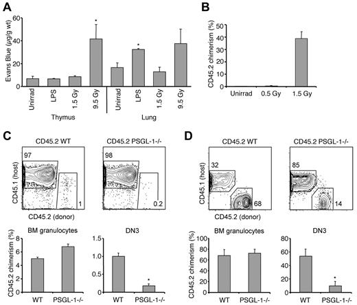
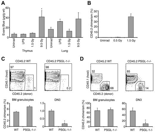
It is possible that, after irradiation, physical damage to the thymic vasculature allows for unregulated entry of circulating T-cell progenitors. Indeed, high-dose irradiation can damage lung and skin endothelium.38,39 We examined the thymic vasculature by immunofluorescence microscopy and found that thymic vascular elements appeared grossly intact 1 day after irradiation (supplemental Figure 1A, available on the Blood Web site; see the Supplemental Materials link at the top of the online article). We next examined whether irradiation affects thymic vascular permeability by intravenously injecting albumin-binding Evans blue one day after irradiation. Increased vascular permeability to Evans blue was observed in both thymus and lung after a 9.5-Gy dose of irradiation, but not after a low dose of 1.5 Gy (Figure 2A); 1.5 Gy is a sufficient dose, however, to confer CCR7/CCR9 independence on thymic settling (Figure 2B), indicating that permeability to Evans blue can be uncoupled from CCR7/CCR9-independent thymic settling. We further asked whether erythrocytes could enter an irradiated thymus, which would be expected if endothelial barrier function had been lost. After irradiation with 9.5 Gy, there was no detectable increase in the number of erythrocytes in the thymus (supplemental Figure 1B). Together, these data indicate that CCR7/CCR9-independent thymic settling after irradiation is not the result of loss of thymic endothelial barrier function.
Thymic settling remains a regulated process after BMT. (A) Evans blue was measured in thymi or lungs after no treatment, lipopolysaccharide treatment, 1.5 Gy irradiation, or 9.5 Gy irradiation. Shown is the mean quantity ± SEM of Evans blue (micrograms) per organ weight (grams). *P < .05 compared with corresponding unirradiated controls. N = 4 to 6 per group. (B) Mice exposed to 0, 0.5, or 1.5 Gy of irradiation were inoculated with CD45.2 CCR7/CCR9 DKO BM cells plus CD45.1 WT BM at a 2:1 ratio (106 total cells). Shown is the mean CD45.2 donor chimerism ± SEM of thymic DP cells after 3 weeks. *P < .05 compared with the control. N = 3 per group. (C) Top row: CD45.2 WT (left) or PSGL-1−/− (right) BM cells (1.4 × 10⁷) were adoptively transferred into unirradiated CD45.1 WT hosts. Shown are representative plots of thymic DN3 cells 3 weeks after transfer. Bottom row: Mean CD45.2 chimerism ± SEM of BM granulocytes (left) and thymic DN3 cells (right). *P < .05 compared with the WT control. N = 5 to 10 per group. (D) Top row: Lethally irradiated BM chimeras were generated using CD45.2 WT (left) or CD45.2 PSGL-1−/− BM (right) mixed with CD45.1 WT competitor cells at a 2:1 ratio (2 × 10⁶ total cells). Shown are representative plots of thymic DN3 cells 3 weeks after transplant. Bottom row: Mean CD45.2 chimerism ± SEM of BM granulocytes (left) and thymic DN3 cells (right). *P < .05 compared with the WT control. N = 5 to 10 per group.
Thymic settling remains a regulated process after BMT. (A) Evans blue was measured in thymi or lungs after no treatment, lipopolysaccharide treatment, 1.5 Gy irradiation, or 9.5 Gy irradiation. Shown is the mean quantity ± SEM of Evans blue (micrograms) per organ weight (grams). *P < .05 compared with corresponding unirradiated controls. N = 4 to 6 per group. (B) Mice exposed to 0, 0.5, or 1.5 Gy of irradiation were inoculated with CD45.2 CCR7/CCR9 DKO BM cells plus CD45.1 WT BM at a 2:1 ratio (106 total cells). Shown is the mean CD45.2 donor chimerism ± SEM of thymic DP cells after 3 weeks. *P < .05 compared with the control. N = 3 per group. (C) Top row: CD45.2 WT (left) or PSGL-1−/− (right) BM cells (1.4 × 10⁷) were adoptively transferred into unirradiated CD45.1 WT hosts. Shown are representative plots of thymic DN3 cells 3 weeks after transfer. Bottom row: Mean CD45.2 chimerism ± SEM of BM granulocytes (left) and thymic DN3 cells (right). *P < .05 compared with the WT control. N = 5 to 10 per group. (D) Top row: Lethally irradiated BM chimeras were generated using CD45.2 WT (left) or CD45.2 PSGL-1−/− BM (right) mixed with CD45.1 WT competitor cells at a 2:1 ratio (2 × 10⁶ total cells). Shown are representative plots of thymic DN3 cells 3 weeks after transplant. Bottom row: Mean CD45.2 chimerism ± SEM of BM granulocytes (left) and thymic DN3 cells (right). *P < .05 compared with the WT control. N = 5 to 10 per group.
To evaluate whether thymic entry remains regulated after irradiation, we investigated whether another signal important for normal thymic settling, namely PSGL-1,28 remains relevant. We sought to confirm a role for PSGL-1 in the unirradiated scenario through the adoptive transfer of unfractionated PSGL-1−/− BM or WT BM (both CD45.2) into unirradiated CD45.1 WT hosts. Three weeks after transfer, the donor chimerism of BM granulocytes was similar in the 2 cohorts, indicating equivalent engraftment of the BM (Figure 2C). The donor chimerism of DN3 cells in recipients of PSGL-1−/− BM was reduced compared with recipients of WT BM, consistent with a role for PSGL-1 in the unirradiated thymic settling. The role of PSGL-1 in thymic settling after BMT was tested through the generation of mixed BM chimeras using mixtures of PSGL-1−/− BM or WT BM (both CD45.2) and competitor CD45.1 WT BM. Three weeks after BMT, PSGL-1−/− donor chimerism was equal to WT donor chimerism in the BM granulocyte pool but significantly reduced among thymic DN3 cells (Figure 2D). We found similar reductions at 2 and 4 weeks after BMT as well (supplemental Figure 2). These data demonstrate that a role for PSGL-1−/− in generating thymocytes is maintained after irradiation. To distinguish between a thymic settling defect and an intrathymic developmental defect in the absence of PSGL-1, sorted PSGL-1−/− or WT LMPPs were intrathymically injected into sublethally irradiated WT hosts, bypassing the thymic settling step. Consistent with past work,28 PSGL-1−/− progenitors were not defective at generating downstream DN3 cells, thereby indicating that early intrathymic development is not reliant on PSGL-1 (supplemental Figure 3). Together, these data demonstrate that, unlike CCR7 and CCR9, PSGL-1 maintains its role in thymic settling acutely after BMT.
Collectively, these results indicate that thymic reconstitution after BMT can be divided into 3 stages: an acute stage until day 4 post-BMT during which most thymic reconstitution is CCR7/CCR9-independent; a transitional stage between day 4 and 3 weeks during which most reconstitution depends on CCR7/CCR9, although these receptors are not strictly required; and a chronic stage beginning at 3 weeks requiring CCR7/CCR9 in all cases.
Efficient thymocyte generation by BM Lin−Kit+Flt3high progenitors 2 weeks after BMT
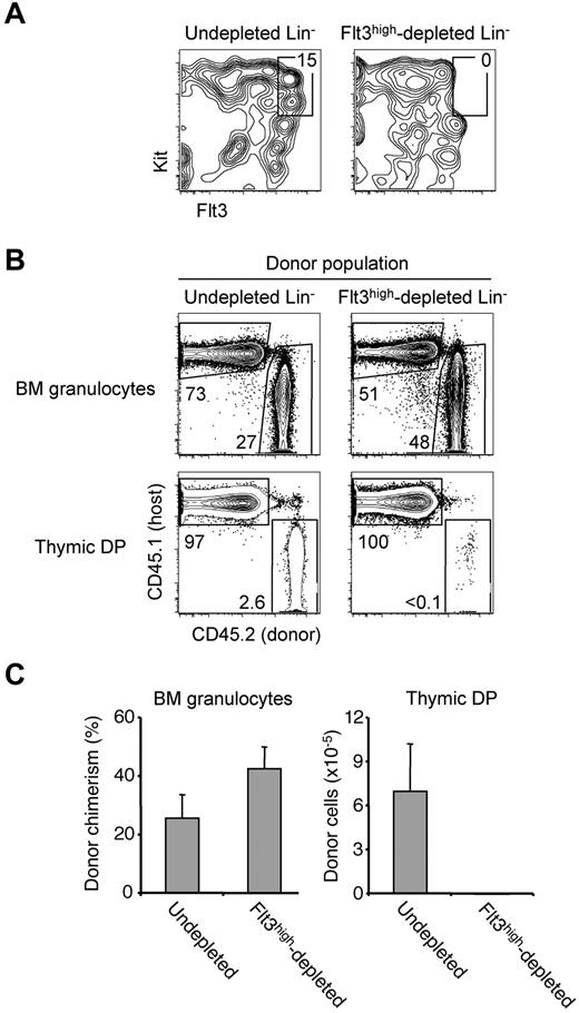
Whereas the precise identity of thymic settling progenitors used in the unirradiated scenario is unknown, these cells reside within the BM Lin−Kit+Flt3high pool.23,29,30 The observed CCR7/CCR9 independence of acute post-BMT thymic settling may allow cells outside of this population to directly settle the thymus during this time.23 Yet because thymic settling signals are normalized by 2 weeks after BMT in nearly all cases (Figure 1E), we asked whether the identity of thymic settling progenitors at this later time is similarly normalized. To test whether the ability to rapidly produce thymocytes 2 weeks after BMT is limited to the BM Lin−Kit+Flt3high pool, we sorted either total Lin− cells or Lin− cells depleted specifically of the Kit+Flt3high pool (Figure 3A). Equal numbers of these 2 populations were injected intravenously into mice that had undergone BMT 2 weeks earlier. After a further 2 weeks, BM and thymi of recipient mice were analyzed for donor contributions. Both transferred populations contributed to the BM granulocyte pool, but the Kit+Flt3high-depleted population was defective at generating thymic DP cells (Figure 3B-C). These data indicate that, just as in the unirradiated scenario, efficient thymocyte progenitor activity resides within the BM Lin−Kit+Flt3high pool 2 weeks after BMT.
Efficient T-lineage progenitor activity resides within the BM Lin−Kit+Flt3high pool 2 weeks after BMT. (A) Total Lin− cells or Lin− cells depleted of all Kit+Flt3high cells were sorted from CD45.2 WT BM. Shown are plots of sorted populations gated on live singlets. No Kit+Flt3high cells were detected in the depleted sample. (B) A total of 1.2 × 10⁵ undepleted Lin− cells (left column) or Kit+Flt3high-depleted Lin− cells (right column) were injected into CD45.1 WT recipients that had been lethally irradiated and transplanted with 2.5 × 10⁵ host-type BM cells 2 weeks earlier. After an additional 15 days, BM and thymi of recipient mice were analyzed for donor contributions. Shown are representative plots gated on BM granulocytes (top row) and thymic DP cells (bottom row). (C) Mean donor chimerism plus or minus SEM of BM granulocytes (left panel) and mean numbers ± SEM of donor thymic DP cells (right panel). N = 3 to 6 per group.
Efficient T-lineage progenitor activity resides within the BM Lin−Kit+Flt3high pool 2 weeks after BMT. (A) Total Lin− cells or Lin− cells depleted of all Kit+Flt3high cells were sorted from CD45.2 WT BM. Shown are plots of sorted populations gated on live singlets. No Kit+Flt3high cells were detected in the depleted sample. (B) A total of 1.2 × 10⁵ undepleted Lin− cells (left column) or Kit+Flt3high-depleted Lin− cells (right column) were injected into CD45.1 WT recipients that had been lethally irradiated and transplanted with 2.5 × 10⁵ host-type BM cells 2 weeks earlier. After an additional 15 days, BM and thymi of recipient mice were analyzed for donor contributions. Shown are representative plots gated on BM granulocytes (top row) and thymic DP cells (bottom row). (C) Mean donor chimerism plus or minus SEM of BM granulocytes (left panel) and mean numbers ± SEM of donor thymic DP cells (right panel). N = 3 to 6 per group.
Defective reconstitution of BM Lin−Kit+Flt3high progenitors after BMT
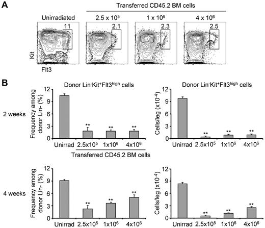
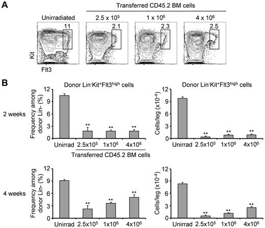
Having established that BM Lin−Kit+Flt3high cells are critical for thymic reconstitution, we evaluated whether their generation was impaired after BMT. We generated noncompetitive irradiation chimeras in which CD45.1 WT hosts were transplanted with one of 3 different doses of CD45.2 BM: 2.5 × 10⁵, 1 × 10⁶, or 4 × 10⁶. Two weeks after BMT, we analyzed the BM Lin− pool for the presence of donor Kit+Flt3high cells. Although these cells composed > 10% of the Lin− pool in unirradiated control BM, they represented < 3% of donor Lin− cells in chimeras receiving each of the 3 BM doses (Figure 4). In addition, because total BM cellularity is very low early after transplantation, these Kit+Flt3high cells had even more severe defects in absolute number (Figure 4B). Impaired reconstitution of these cells continued to be evident 4 weeks after irradiation, although the magnitudes of the defects were not as severe as at 2 weeks (Figure 4B). Furthermore, the frequency and number of Kit+Flt3high cells at this time point correlated with the BM transplant dose. Resolution of Lin−Kit+Flt3high cells into LMPPs and CLPs revealed reduced frequencies and numbers of these lymphoid progenitors (supplemental Figure 4A-B). Frequencies of MPPs and common myloid progenitors were also decreased, but the frequencies of granulocyte-macrophage progenitors and megakaryocyte-erythroid progenitors were not (supplemental Figure 4A).
Defective reconstitution of BM Kit+Flt3high progenitors after BMT. (A) Noncompetitive chimeras were generated by lethally irradiating CD45.1 WT hosts and reconstituting them with 2.5 × 10⁵, 1 × 10⁶, or 4 × 10⁶ CD45.2 WT BM cells. Two weeks after BMT, the BM was analyzed for donor contribution to progenitor populations. Shown are representative plots from chimeras transplanted with the indicated cell number; plots are gated on donor Lin− cells (or total Lin− cells for the unirradiated control). (B) Shown are the mean frequencies ± SEM of Kit+Flt3high cells among donor Lin− cells for unirradiated controls and each of the 3 chimera cohorts 2 weeks after BMT (top left panel). Multiplying these frequencies by the total leg (tibia plus femur) BM cellularity yielded the mean numbers ± SEM of donor Lin−Kit+Flt3high cells (top right panel). The same frequency (bottom left panel) and absolute number (bottom right panel) measurements were made on similar chimeras 4 weeks after BMT. **P < .01 compared with the corresponding unirradiated controls. N = 3 per group.
Defective reconstitution of BM Kit+Flt3high progenitors after BMT. (A) Noncompetitive chimeras were generated by lethally irradiating CD45.1 WT hosts and reconstituting them with 2.5 × 10⁵, 1 × 10⁶, or 4 × 10⁶ CD45.2 WT BM cells. Two weeks after BMT, the BM was analyzed for donor contribution to progenitor populations. Shown are representative plots from chimeras transplanted with the indicated cell number; plots are gated on donor Lin− cells (or total Lin− cells for the unirradiated control). (B) Shown are the mean frequencies ± SEM of Kit+Flt3high cells among donor Lin− cells for unirradiated controls and each of the 3 chimera cohorts 2 weeks after BMT (top left panel). Multiplying these frequencies by the total leg (tibia plus femur) BM cellularity yielded the mean numbers ± SEM of donor Lin−Kit+Flt3high cells (top right panel). The same frequency (bottom left panel) and absolute number (bottom right panel) measurements were made on similar chimeras 4 weeks after BMT. **P < .01 compared with the corresponding unirradiated controls. N = 3 per group.
We assessed the ability of progenitors from post-BMT mice to generate T-lineage (CD25+Thy1+) progeny in vitro using coculture with OP9 stromal cells expressing the Notch ligand DL4 (OP9-DL4).36 Unfractionated donor Lin− progenitors sorted from post-BMT mice generated fewer T-lineage progeny than progenitors from unirradiated mice (supplemental Figure 5A-B). To identify whether this deficit was solely the result of the decreased frequency of Kit+Flt3high cells, we sorted Kit+Flt3high cells and all remaining Lin− cells (ie, non-Kit+Flt3high) from unirradiated and post-BMT mice and plated them at their in vivo physiologic ratio. Kit+Flt3high cells from post-BMT mice were not defective at generating T-lineage progeny relative to their counterparts from unirradiated mice. The non-Kit+Flt3high cells produced very few T-lineage progeny (supplemental Figure 5C). These data indicate that the reduced number of T-lineage cells generated by unfractionated Lin− progenitors from post-BMT mice is attributable to the reduced frequency of Kit+Flt3high cells, and not to an intrinsic proliferative defect of these progenitors. Together, these data indicate that the reconstitution of efficient T-lineage progenitors in the BM is impaired after BMT.
An unsaturated intrathymic progenitor niche persists for at least 10 weeks after BMT
The reduced numbers of BM Lin−Kit+Flt3high progenitors competent to settle the thymus suggested that the frequency of thymic settling may be reduced after BMT. Indeed, a previous report noted that the thymus is nearly devoid of ETPs and DN2 cells for at least 4 weeks after BMT.40 However, this deficit of Kithigh intrathymic progenitors does not alone demonstrate that thymic settling is reduced; it is possible that intrathymic mechanisms impair the generation or survival of these progenitors. To distinguish between these possibilities, we investigated whether the scarcity of Kithigh thymic progenitors reflected a vacant intrathymic progenitor niche after BMT. We first confirmed the near-absence of ETPs and DN2 cells in mice 2 weeks after BMT and noted the resemblance of these thymi to those of CCR7/CCR9 DKO mice, a strain that has highly inefficient thymic settling.26,27 Both have severe defects in the frequencies of ETPs and DN2 cells, but the frequency of DN3 cells is normal (Figure 5A). We have previously shown that progenitors placed into CCR7/CCR9 DKO thymi undergo compensatory proliferation, presumably because of reduced competition for progenitor niches.27 We therefore hypothesized that, if the reduced number of Kithigh thymic progenitors in post-BMT mice reflects a vacant intrathymic progenitor niche, cells transferred into post-BMT thymi should also expand in a compensatory manner. Sorted BM Lin−Kit+Flt3high cells were intrathymically injected into hosts that had undergone BMT 2 weeks earlier or into unirradiated controls. We analyzed host mice 2 weeks after intrathymic injection, as this is a time point by which significant numbers of donor DN3 and DP cells have been generated. At this time, cells transferred into “pre-transplanted” hosts had generated 25-fold more donor DN3 cells and 190-fold more donor DP cells (Figure 5B-C). Similar experiments were performed with hosts that had been transplanted 4 or 10 weeks before intrathymic injection (Figure 5C). In both cases, cells placed in pretransplanted hosts generated significantly more downstream DN3 and DP progeny than cells injected into unirradiated hosts. The magnitudes of these differences, however, became progressively smaller as the time between BMT and intrathymic injection grew longer. These data demonstrate that progenitors placed into post-BMT thymi undergo compensatory expansion. We interpret this to signify the presence of unsaturated intrathymic progenitor niches in post-BMT thymi extending at least 10 weeks after transplantation. Hence, the reinstatement of normal thymic settling signals is decoupled from the maintenance of a saturated intrathymic progenitor niche after BMT.
Chronic vacancy of intrathymic progenitor niches after BMT. (A) Thymi from unirradiated, CCR7/CCR9 DKO, or 2 weeks post-BMT mice were analyzed for the frequency of Kithigh thymic progenitors. Shown are representative plots gated on Lin− thymocytes. (B) Sorted CD45.2 donor BM Lin−Kit+Flt3high progenitors were injected intrathymically into either unirradiated CD45.1 hosts (left column) or CD45.1 hosts that had been lethally irradiated and transplanted with 2.5 × 10⁵ CD45.1 BM cells 2 weeks earlier (right column). Recipient thymi were analyzed for donor contributions 2 weeks later. Shown are representative plots of thymic DN3 cells (top row) or DP cells (bottom row). (C) The mean numbers ± SEM of donor DN3 cells (left) and DP cells (right) in the recipient thymi described in panel B are shown (top row). The same experiment was also performed with recipients that had undergone BMT 4 weeks (middle row) or 10 weeks (bottom row) before intrathymic injection. *P < .05, compared with the corresponding unirradiated control. **P < .01 compared with the corresponding unirradiated control. N = 3 to 10 per group.
Chronic vacancy of intrathymic progenitor niches after BMT. (A) Thymi from unirradiated, CCR7/CCR9 DKO, or 2 weeks post-BMT mice were analyzed for the frequency of Kithigh thymic progenitors. Shown are representative plots gated on Lin− thymocytes. (B) Sorted CD45.2 donor BM Lin−Kit+Flt3high progenitors were injected intrathymically into either unirradiated CD45.1 hosts (left column) or CD45.1 hosts that had been lethally irradiated and transplanted with 2.5 × 10⁵ CD45.1 BM cells 2 weeks earlier (right column). Recipient thymi were analyzed for donor contributions 2 weeks later. Shown are representative plots of thymic DN3 cells (top row) or DP cells (bottom row). (C) The mean numbers ± SEM of donor DN3 cells (left) and DP cells (right) in the recipient thymi described in panel B are shown (top row). The same experiment was also performed with recipients that had undergone BMT 4 weeks (middle row) or 10 weeks (bottom row) before intrathymic injection. *P < .05, compared with the corresponding unirradiated control. **P < .01 compared with the corresponding unirradiated control. N = 3 to 10 per group.
Early thymic reconstitution is limited by the number of progenitors delivered to the thymus
The persistence of an unsaturated progenitor niche suggested that thymic reconstitution may be limited by the number of progenitors that settle the thymus after BMT.11 To address this, we generated noncompetitive irradiation chimeras in which CD45.1 WT hosts were transplanted with different doses of CD45.2 BM. A previous study used a similar approach to address this question.41 We examined recipient thymi 2 weeks after BMT and found that the numbers of donor-derived DN3 and DP cells directly correlated with the BM dose given (supplemental Figure 6A-B). Whereas total (donor plus host) DN3 cell number also correlated with BM dose, total DP cell number did not because, at 2 weeks after irradiation, the majority of DP cells are composed of the progeny of radioresistant host thymocytes.35 Mice analyzed 4 weeks after BMT also revealed dose-dependent thymic reconstitution (supplemental Figure 6C-D). These data indicate that the magnitude of early thymic reconstitution is limited by the BM dose provided at the time of transplantation.
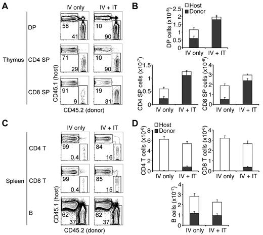
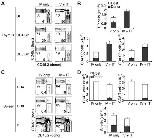
We next tested whether the enhanced thymic recovery observed after high-dose BMT was the result of an increased number of donor progenitors entering the thymus. Recipient CD45.1 WT mice were first sublethally irradiated and intravenously inoculated with 2.5 × 10⁵ CD45.2 WT donor BM cells. Half of these mice were then intrathymically injected with 3 × 10⁴ Lin− CD45.2 WT donor BM cells, whereas the other half were intrathymically injected with PBS as controls. We analyzed the thymi and spleens of these mice 4 weeks after transfer because donor peripheral T cells can be reliably quantified at this time point. Mice that had received cells intrathymically possessed significantly more donor cells among all thymic populations analyzed, which included ETPs, DN2, DP, CD4 SP, and CD8 SP cells (Figure 6A-B; supplemental Figure 7); this was true of splenic CD4 and CD8 T cells as well (Figure 6C-D). Similar donor numbers of splenic B cells were found in both cohorts as expected. These data establish that increased progenitor recruitment to the thymus after BMT improves T-cell reconstitution. Together, these findings illustrate that T-lineage reconstitution after BMT is limited by the availability of progenitors to the thymus; thus, not all delays in T-lineage reconstitution are attributable to thymus-intrinsic mechanisms.9,10
Limiting numbers of thymic progenitors delay T-lineage recovery. (A) CD45.1 WT host mice were sublethally irradiated and intravenously injected with 2.5 × 10⁵ CD45.2 WT BM cells. Immediately thereafter, one group of mice received PBS intrathymically (IV only), whereas another group received 3 × 10⁴ CD45.2 Lin− BM cells intrathymically (IV + IT). Host thymi and spleens were analyzed for donor contributions 4 weeks later. Shown are representative plots of thymic DP (top row), CD4 SP (middle row), and CD8 SP (bottom row) cells from mice in the indicated groups. (B) Mean numbers ± SEM of host and donor thymic DP (top panel), CD4 SP (bottom left panel), and CD8 SP (bottom right panel) cells from the chimeras described in panel A. P values for comparisons of donor DP cell numbers between the 2 groups, .01; of donor CD4 SP cell numbers, .002; of donor CD8 SP cell numbers, .0003. (C) Shown are representative plots of splenic CD4 T (top row), CD8 T (middle row), and B (bottom row) cells from mice in the indicated groups described in panel A. (D) Graphs show the mean number ± SEM of host and donor splenic CD4 T (top left panel), CD8 T (top right panel), and B (bottom panel) cells from the chimeras described in panel A. P values for comparisons of donor CD4 cell numbers between the 2 groups, .007; of donor CD8 cell numbers, .005; of donor B cell numbers, .41. N = 4 or 5 per group.
Limiting numbers of thymic progenitors delay T-lineage recovery. (A) CD45.1 WT host mice were sublethally irradiated and intravenously injected with 2.5 × 10⁵ CD45.2 WT BM cells. Immediately thereafter, one group of mice received PBS intrathymically (IV only), whereas another group received 3 × 10⁴ CD45.2 Lin− BM cells intrathymically (IV + IT). Host thymi and spleens were analyzed for donor contributions 4 weeks later. Shown are representative plots of thymic DP (top row), CD4 SP (middle row), and CD8 SP (bottom row) cells from mice in the indicated groups. (B) Mean numbers ± SEM of host and donor thymic DP (top panel), CD4 SP (bottom left panel), and CD8 SP (bottom right panel) cells from the chimeras described in panel A. P values for comparisons of donor DP cell numbers between the 2 groups, .01; of donor CD4 SP cell numbers, .002; of donor CD8 SP cell numbers, .0003. (C) Shown are representative plots of splenic CD4 T (top row), CD8 T (middle row), and B (bottom row) cells from mice in the indicated groups described in panel A. (D) Graphs show the mean number ± SEM of host and donor splenic CD4 T (top left panel), CD8 T (top right panel), and B (bottom panel) cells from the chimeras described in panel A. P values for comparisons of donor CD4 cell numbers between the 2 groups, .007; of donor CD8 cell numbers, .005; of donor B cell numbers, .41. N = 4 or 5 per group.
Discussion
Thymic settling into unirradiated thymi relies on contributions of CCR7, CCR9, and PSGL-1. After BMT, PSGL-1−/− progenitors retained a defect in thymic settling at all time points analyzed, demonstrating a preserved role for this molecule after irradiation. In contrast, dependence on CCR7/CCR9 can distinguish 3 distinct stages of thymic reconstitution. The acute stage extends to 4 days from BMT, during which most thymic reconstitution is independent of CCR7 and CCR9. A transitional stage, between day 4 and week 3 after BMT, relies heavily, but not absolutely, on CCR7/CCR9 for thymic reconstitution. During the chronic third stage proceeding from week 3 onwards, CCR7/CCR9 are again strictly required. We found that thymic and peripheral T-cell reconstitution directly corresponds to the number of cells that enter the thymus after BMT, indicating that T-lineage reconstitution is limited by the number of available progenitors. Together, these data illustrate that the mechanisms of early T-cell development after BMT are different from those in unirradiated mice. An important implication of this observation is that acutely irradiated hosts are not appropriate models with which to study the mechanisms of normal unirradiated thymic settling. Conversely, models of physiologic thymic settling are a poor approximation for the mechanisms at play in regeneration after BMT.
Thymic settling by circulating progenitors probably uses mechanisms analogous to those used by mature lymphocytes to enter lymph nodes, wherein chemokine signals induce the conformational change of integrins to a high-affinity state.21 The irrelevance of CCR7 and CCR9 toward thymic settling acutely after BMT presents 2 alternative mechanisms of progenitor entry into the thymus. First, cells may access the irradiated thymus independently of any chemokine receptor signals. Recent work has demonstrated that irradiation can damage BM sinusoids, making them discontinuous and consequently hemorrhagic.42,43 However, the lack of thymic vascular permeability to Evans blue after 1.5 Gy (a dose sufficient to confer CCR7/CCR9-independent thymic settling; Figure 2A-B) and the continued reliance on PSGL-1 acutely after BMT (Figure 2D; supplemental Figure 2) together indicate that a loss of endothelial barrier function does not underlie CCR7/CCR9-independent thymic settling. In addition, an absence of appropriate chemokines appears not to be the cause of CCR7/CCR9-independent settling because expression of CCL21 and CCL25, ligands for CCR7 and CCR9, respectively, is up-regulated on thymic stromal cells after irradiation.44 We speculate that a different chemokine receptor substitutes for CCR7 and CCR9 acutely after irradiation. Irradiation can cause endothelial responses similar to those seen with inflammation, such as the up-regulation of adhesion molecules.45 Such changes could lead to the expression of chemokines not normally associated with the thymic endothelium. Another possibility is that the defective reconstitution of Kit+Flt3high BM progenitors (Figure 3) limits the number of efficient CCR9+ progenitors,23 which may allow less efficient CCR9− cells to settle the thymus. Additional study is needed to identify which of these mechanisms is at work in the days after BMT.
Efficient thymocyte generation capacity 2 weeks after BMT resides in the BM Lin−Kit+Flt3high pool, which contains cells expressing CCR7 and CCR9. The available supply of these progenitors in the BM is significantly diminished after BMT. Therefore, the reimposition of a requirement for CCR7/CCR9 in thymic settling probably reduces the number of progenitors entering the thymus after BMT, effectively “starving” the thymus. We consider the chronic vacancy of the intrathymic progenitor niche to demonstrate that progenitor input into the thymus limits T-lineage reconstitution. Our observation that the intrathymic progenitor niche remains unsaturated 10 weeks after BMT further suggests the possibility that the niche may never return to a saturated state. In other words, diminished delivery of progenitors to the thymus may permanently limit T-cell recovery after BMT.
The reduced delivery of progenitors into the thymus after BMT could have multiple causes. The prethymic mechanisms in T-cell development can be considered as 3 sequential steps23 : (1) the generation of BM progenitors with thymic settling capacity, (2) the mobilization of these cells from the BM into the blood, and (3) the entry of these circulating progenitors into the thymus. We evaluated the first step and found that the rare Lin−Kit+Flt3high BM subset with efficient thymic repopulating ability is poorly reconstituted, and this impairment persists for at least a month after BMT. The reasons for the defective regeneration of the BM Lin−Kit+Flt3high pool remain unclear; they may involve the altered BM cytokine milieu after irradiation.46 Additional prethymic defects in T-lineage reconstitution remain possible, such as decreased mobilization or thymic settling of these progenitors.
Thymic reconstitution after BMT was thought to be hampered by 3 major causes: (1) thymic stromal damage from conditioning (radiation or chemotherapeutic drugs); (2) thymic involution in adults; and (3) GVHD targeting the thymus.10,11,47-49 We have investigated the ability of the thymus to generate T cells after BMT, but certain caveats exist. We used young mice as hosts; therefore, our results may not be representative of thymic reconstitution in aged humans with involuted thymi. Furthermore, we have used a syngeneic transplant model and thus have avoided any defects that GVHD could have caused. Nevertheless, we found that, despite lethal irradiation, the thymus was quite capable of supporting the extensive expansion of donor cells provided intravenously or intrathymically. This does not exclude the possibility that the thymi were impaired relative to unirradiated counterparts. Rather, we interpret this result to mean that donor thymocyte generation in the irradiated thymus benefits from the lack of competition more than it suffers from stromal damage.
The 3 proposed mechanisms of delayed T-lineage reconstitution all occur at the level of the thymus; we now demonstrate that mechanisms before thymic entry also contribute. Consistent with an earlier study,41 we found that increasing the number of progenitors delivered to the thymus, either physiologically via the blood or by direct intrathymic transfer, led to corresponding increases in thymic size and peripheral donor T-cell numbers. Hence, T-cell reconstitution is limited by what is probably an extremely diminished rate of progenitor entry into the thymus after BMT; this defect is not mutually exclusive with the intrathymic ones previously noted. The underrepresentation of Lin−Kit+Flt3high BM progenitors after BMT suggests that efforts to expand the pool of efficient T-cell progenitors in the BM would be beneficial toward T-lineage recovery after BMT. Indeed, a previous report has shown that the exogenous administration of Flt3 ligand after BMT expanded the MPP and CLP pools after BMT and also led to enhanced thymic reconstitution.11 The injected Flt3 ligand may have improved donor thymopoiesis by direct intrathymic effects,50 but our data suggest that increasing the BM supply of efficient T-lineage progenitors alone may improve thymic recovery. Elucidating new protocols to improve the reconstitution of such progenitors may be beneficial toward restoring the T-lineage after BMT.
An Inside Blood analysis of this article appears at the front of this issue.
The online version of this article contains a data supplement.
The publication costs of this article were defrayed in part by page charge payment. Therefore, and solely to indicate this fact, this article is hereby marked “advertisement” in accordance with 18 USC section 1734.
Acknowledgments
The authors thank Mark Kahn for expert advice, and Gary Koretzky, Taku Kambayashi, Tao Zou, and Amanda Schmidt for their critiques of the manuscript.
This work was supported by the National Institutes of Health (grant F30HL099271, D.A.Z.; grant T32 GM-0729, M.E.D.O.; grants R01AI059621 and RC1HL099758, A.B.) and the Leukemia & Lymphoma Society (Scholar Award, A.B.).
National Institutes of Health
Authorship
Contribution: D.A.Z. and S.L.Z. performed most of the experimental work; M.E.D.O., S.P.T., and T.D.L. contributed to the performance of the experimental work; D.A.Z. and A.B. planned the project; P.R.H. provided assistance with vascular permeability studies; D.A.Z., S.L.Z., M.E.D.O., and A.B. analyzed data; and D.A.Z., S.L.Z., and A.B. prepared the manuscript.
Conflict-of-interest disclosure: The authors declare no competing financial interests.
Correspondence: Avinash Bhandoola, 266 John Morgan Bldg, 3620 Hamilton Walk, Philadelphia, PA 19104; e-mail: bhandooa@mail.med.upenn.edu.
References
Author notes
D.A.Z. and S.L.Z. contributed equally to this study.

