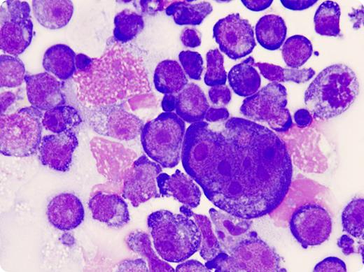
A 68-year-old man with a 10-year history of HIV, compliant with anti-retrovirals (ARV), presented with neck lymphadenopathy. A diagnosis of mixed cellularity Hodgkin lymphoma was made by biopsy. Staging investigations, including bone marrow aspirate/biopsy, cerebrospinal fluid examination, and PET/CT imaging confirmed stage IA disease. He was treated with 4 cycles of chemotherapy and involved field radiotherapy. Cumulative anthracycline doses previously administered for Kaposi sarcoma obviated the usual chemotherapy program; therefore, chlorambucil, vinblastine, procarbazine, and prednisolone were employed. A complete remission was confirmed with PET/CT.
Six months later, he developed progressive dizziness, dysarthria, and fatigue. CT brain, PET/CT body, MR brain, and spinal cord examinations identified no cause. Viral load remained undetectable and CD4 count was 65 (8%), suggesting limited immunologic recovery despite ARV compliance. An exhaustive search for an infectious cause was negative. Cerebrospinal fluid analysis demonstrated a leukocytosis (100/mm3) and an elevated protein (357 mg/dL) with Reed Sternberg cells present (shown), consistent with central nervous system relapse (CNS-HL). Despite intrathecal chemotherapy, whole brain radiotherapy, and subsequent systemic chemotherapy, he died. There was no autopsy.
CNS-HL is unusual. Leptomeningeal spread has been reported more commonly in patients with widespread relapsed disease or in immunocompromised patients.
Many Blood Work images are provided by the ASH IMAGE BANK, a reference and teaching tool that is continually updated with new atlas images and images of case studies. For more information or to contribute to the Image Bank, visit http:\\imagebank.hematology.org.


