Abstract
Abstract 3131
Although MCL is reported to be an FDG-avid NHL subtype, PET-CT is not currently recommended in the modified IWG criteria (Chesen et al, JCO 2007) to stage, survey and assess treatment response in MCL. PET-CT is however frequently used for these purposes in clinical practice in MCL. Although convincing data exists regarding the prognostic utility of PET-CT imaging in Hodgkin Lymphoma (interim & post treatment) and DLBCL (post treatment), its prognostic utility both during treatment and immediately following treatment have not systematically been evaluated in a large MCL patient cohort to support its use in clinical practice.
We conducted a two-center (JTCC & UPenn), retrospective cohort study to examine the prognostic utility of pre-treatment (PET-1), interim-treatment (PET-2 after 2–3 treatment cycles) and post-treatment PET-CT (PET-3) imaging in a uniform MCL patient cohort undergoing dose-intensive chemotherapy (R-HCVAD) or high-dose chemotherapy followed by autologous stem (ASCT) cell rescue in the 1st line setting. The primary study endpoints were PFS and OS. PET-CT images were centrally reviewed for the purposes of this study using standardized response criteria proposed by the Imaging Subcommittee of International Harmonization Project in Lymphoma (Juweid et al, JCO 2007). Radiologists were blinded to clinical outcomes at the time of PET-CT review and results were dichotomized at each time point and their concomitant SUV max at a suspected site of disease was recorded.
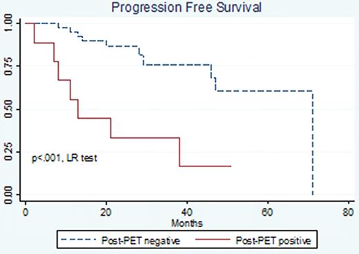
82 pts (median age 61, range 35–95) with advanced stage (98% stage IV) MCL with PET-CT data were identified for this analysis. Of these, 57 pts were treated with R-HCVAD (50) or ASCT (7) in the first-line setting. 154 PET-CT scans were reviewed. Overall treatment response rate (IWG) was 93% (76% CR + 17%PR). With median follow-up of 23 months (disease progression) and 27 months (overall survival), 3 year PFS and OS estimates were 70% (CI 53–82%) and 82% (CI 65–91%) respectively. Median baseline characteristics: WBC 7.4, ECOG PS 1, LDH/ULN .82, Ki-67 (MIB-1) 28% (range 10–90%), MIPI 3.5 (31% intermediate risk, 19% high risk). 20% and 30% of pts were blastoid variant and leukemic phase at diagnosis respectively. Pre-treatment PET-CTs were FDG-avid in 92% of MCL patients with a median SUV max of 7.8 (range 2.4–36.7) at a suspected site of disease. Pre-treatment PET-CT SUV max was positively associated with high Ki-67 (variance-weighted least-squares regression, p=.01). PET-2 and PET-3 were positive in 34% and 18% of patients respectively. Kaplan Meier and Cox regression survival analyses were used to test the association between PET-CT and survival. Interim PET-CT status was not associated with PFS (HR 1.1, CI .4-3.6, p=.8) or OS (HR .8, CI .15-4.6, p=.8). Post treatment PET-CT status was statistically significantly associated with PFS (HR 5.4, CI 2.0–14.5, p=.001, Harrell's C =.70, Figure) and trended towards significant for OS (HR 3.2, CI .8-11.9, p=.1). Post treatment PET-CT status remained an independent predictor of PFS in multivariate analysis which included MIPI score, blastoid variant and Ki-67.
Our cohort is the first large series of MCL patients treated essentially with R-HCVAD examining the correlation between PET-CT and outcome in the front-line setting. A positive PET-CT following the completion of therapy identifies patients with an inferior PFS and a trend towards inferior OS. These data do not support the prognostic utility of PET-CT in pre-treatment (no upstaging) and interim treatment settings. This information should be incorporated into the design of future prospective clinical trials. We are currently examining the relationship of novel computer-assisted quantitative FDG-PET/CT measures of total tumor volume and total tumor metabolic burden with clinical outcome in patients with mantle cell lymphoma.
No relevant conflicts of interest to declare.
Author notes
Asterisk with author names denotes non-ASH members.


