To the editor:
A recent paper in Blood reported that lineage (CD3/14/19)–negative CD56+ mononuclear cells in human peripheral blood contain 2 populations: bona fide CD7+ natural killer (NK) cells and CD7− monocyte/dendritic cells (DCs).1 Whereas this study delivers a valuable improvement in the definition of peripheral blood NK cells, it leaves unresolved the question of which monocyte/dendritic cells were found to express CD56.
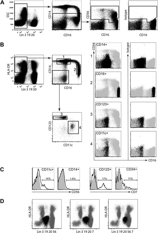
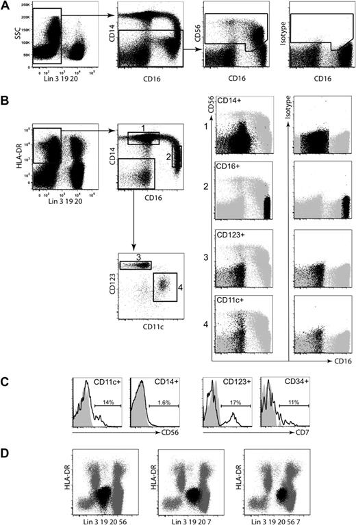
Approaching this problem from the other direction, we characterized the CD56 expression of monocytes and blood DC subsets from healthy volunteers. Figure 1 revisits the analysis of Milush et al with additional intermediate plots to show the location of monocytes and DCs.
Location of monocytes and DCs. (A) The distribution of CD56 among lineage (CD3/14/19/20)–negative cells; (B) gating strategy used to identify CD14+ monocytes, CD16+ monocytes/DC, CD123+ plasmacytoid DCs (PDCs), and CD11c myeloid DCs, and comparison of CD56 expression above isotype; (C) expression of CD56 and CD7 by defined populations; (D) comparison of HLA-DR versus lineage plots with antibodies to CD56, CD7 or both, in lineage.
Location of monocytes and DCs. (A) The distribution of CD56 among lineage (CD3/14/19/20)–negative cells; (B) gating strategy used to identify CD14+ monocytes, CD16+ monocytes/DC, CD123+ plasmacytoid DCs (PDCs), and CD11c myeloid DCs, and comparison of CD56 expression above isotype; (C) expression of CD56 and CD7 by defined populations; (D) comparison of HLA-DR versus lineage plots with antibodies to CD56, CD7 or both, in lineage.
Figure 1A shows the distribution of CD56 among lineage (CD3/14/19/20)–negative cells, as previously illustrated by Milush and colleagues, in comparison with isotype control. Anti-CD56 clone MY31 (IgG1) from Becton Dickinson was used.
Figure 1B then maps on top of these dot plots (now shown in gray as background) the major populations of CD14+ monocytes, CD16+ monocytes/DC, CD123+ plasmacytoid DCs (PDCs), and CD11c myeloid DCs,2 to reveal where CD56 expression appears above isotype.
Figure 1C shows the key results as histograms. A defined population, 12.8% to 15.4% (range; n = 3), of CD11c myeloid DCs express CD56. A small population of CD14+ monocytes also express CD56 as previously described.3,4 CD16+ monocytes and CD123+ PDCs do not express significant CD56. Because Milush and colleagues used CD14+ in their lineage cocktail, CD14+ monocytes are excluded. We therefore identify the CD56+ cells described in their article as a subset of CD11c+ myeloid DCs.
A further contribution of these authors was to show that, although certain DCs fall into lymphoid side scatter gates and express CD56, these cells do not express CD7. CD7 antibodies are therefore a potentially useful addition to the lineage cocktail for DC biologists eager to exclude NK cells from their analysis. The utility of this strategy critically requires that no other DCs express CD7. We confirmed that CD11c+ myeloid DCs are negative for CD7 but found a population, 15.2% to 22.8% (range; n = 3), of CD123+ PDCs expressing CD7 (Figure 1C). Within the lineage-negative DR+ gate, approximately 10% of CD34+ peripheral blood stem cells also expressed CD7, although these are not typically included as a subset of DCs.
Finally, we compared the addition of CD7 and CD56 singly or in combination to a lineage cocktail of CD3/14/19/20 (Figure 1D) in a plot of HLA-DR versus lineage. It is evident that the addition of both antibodies improves the separation of NK cells (depicted in black) from the DR+ lineage− region containing monocytes and DCs (background, gray). This is particularly useful for excluding activated NK cells that express HLA-DR. However, as we have now shown, the caveat is that CD7 and CD56 each removes a population of CD123+ PDCs or CD11c+ myeloid DCs, respectively. Whether these subpopulations of DCs have distinct functional properties remains to be determined.
Authorship
Approval for these studies was obtained from the Newcastle and North Tyneside Research Ethics Committee. Informed consent was provided according to the Declaration of Helsinki.
Contribution: V.B. designed and conducted experiments, performed analysis, and created the figure; L.E.S. provided manuscript input; and M.C. conceived the experiment and wrote the manuscript.
Conflict-of-interest disclosure: The authors declare no competing financial interests.
Correspondence: Matthew Collin, Institute of Cellular Medicine, Newcastle University, Framlington Place, Newcastle upon Tyne NE2 4HH, United Kingdom; e-mail matthew.collin@ncl.ac.uk.


