Abstract
The development of a new vascular network is essential for the onset and progression of many pathophysiologic processes. Cyclooxygenase-2 displays a proangiogenic activity in in vitro and in vivo models, mediated principally through its metabolite prostaglandin E2 (PGE2). Here, we provide evidence for a novel signaling route through which PGE2 activates the Alk5-Smad3 pathway in endothelial cells. PGE2 induces Alk5-dependent Smad3 nuclear translocation and DNA binding, and the activation of this pathway involves the release of active TGFβ from its latent form through a process mediated by the metalloproteinase MT1-MMP, whose membrane clustering is promoted by PGE2. MT1-MMP–dependent transforming growth factor β (TGFβ) signaling through Alk5 is also required for PGE2-induced endothelial cord formation in vitro, and Alk5 kinase activity is required for PGE2-induced neovascularization in vivo. These findings identify a novel signaling pathway linking PGE2 and TGFβ, 2 effectors involved in tumor growth and angiogenesis, and reveal potential targets for the treatment of angiogenesis-related disorders.
Introduction
Growth of new vessels to ensure the supply of oxygen and nutrients to tissues is required for the establishment and progression of a variety of pathophysiologic situations, such as tumor development and metastasis, chronic inflammatory diseases, and vascular retinopathies. In the adult, this is achieved mainly by angiogenesis, in which new vessels grow out from a preformed vascular system. The formation of new capillaries by endothelial cells (ECs) is regulated by an exquisite balance between pro- and antiangiogenic factors.1
A number of proangiogenic factors have been shown to increase the expression of cyclooxygenase-2 (COX-2).2 COX-2 is an oxygenase that catalyzes the conversion of arachidonic acid to prostaglandin H2, which is in turn metabolized by diverse specific terminal synthases to form distinct prostanoids (prostaglandins and thromboxanes).3 Evidence supporting the involvement of COX-2 in angiogenesis includes the impairment of neovessel formation in COX-2 knockout (KO) mice and the fact that specific COX-2 inhibitors block angiogenesis-like processes in in vitro and in vivo models.3 A major product of COX-2 activity is prostaglandin E2 (PGE2). PGE2 signals via 4 G-protein–coupled receptors (GPCR), named EP1 to EP4. These receptors each signal through distinct G-protein subunits, thereby initiating diverse downstream signaling pathways. EP1 usually couples to Gq, which activates PLCγ to produce an increase in intracellular calcium concentration. EP2 and EP4 couple to Gs, with subsequent activation of PKA and increased cAMP synthesis. In contrast, signaling via EP3 often results in decreased cAMP concentrations, owing to the receptor coupling to Gi subunits.4 In addition to these receptors, PGE2 transactivates tyrosine kinase receptors, either through intracellular mechanisms or, in the case of epidermal growth factor receptor, through the metalloproteinase-mediated release of one of its ligands, transforming growth factor α (TGFα). PGE2-mediated activation of the ERK and Akt pathways has been reported to occur via this route.5
PGE2 is considered to be the main mediator of COX-2 proangiogenic effects. The phenotype obtained by deleting the EP2 gene in ApcΔ716 mice, a model of familiar polyposis, clearly resembles that of COX-2–deficient mice, with diminished vascularization and reduced expression of proangiogenic factors in the polyps.6 Similarly, EP3 KO mice show decreased tumor-associated angiogenesis.7 Explanations offered for the proangiogenic properties of PGE2 in vitro include increased expression of endothelial genes such as VEGF or CXCR4,8 activation of the Rac and NO/cGMP pathways,9 and EP2-mediated EC migration and survival.10
TGFβ belongs to a family of cytokines which includes TGFβ1, 2, and 3, the bone morphogenetic proteins, the activins, the nodals, and other analogous factors. These cytokines regulate a wide array of biological responses, both during embryonic development and in the adult.11 Secreted TGFβ forms part of a multiprotein complex, from which it must be released to bind its receptors and display its biological activity. Consequently, the modulation of TGFβ bioavailability is a major regulatory step in TGFβ-mediated processes. A variety of activators of latent-TGFβ have been described, including thrombospondin, integrins (αvβ6, αvβ8), physicochemical treatments, and proteolysis by serine proteases (plasmin, cathepsin D) or matrix metalloproteinases (MMP-2, MMP-9, MT1-MMP).12 The membrane type metalloproteinase MT1-MMP, which is expressed at the cell membrane in a mature form,13 activates TGFβ in an αvβ8-dependent manner in epithelial cells.14
Cellular responses to TGFβ are elicited through 2 types of serine-threonine kinase receptor. Active TGFβ binds to its constitutively active type II receptor, which then binds and phosphorylates TGFβ type I receptor, forming a heteromeric complex. Upon activation, type I receptor phosphorylates receptor-activated Smads (R-Smads; Smad1, 2, 3, 5, and 8) at serine residues within a specific C-terminal sequence. The phosphorylated R-Smads dissociate from their anchor proteins, associate with the co-Smad Smad4, and translocate to the nucleus, where they activate transcription of their target genes, usually through the recruitment of coactivators and cooperation with other transcription factors.15 Smad signaling is curtailed by the activity of phosphatases such as PPM1A and SCPs, which promote the export of R-Smads back to the cytoplasm.16 Inhibitory Smads (Smads 6 and 7) contribute to the terminatation of TGFβ signaling, either by marking R-Smads for proteasomal degradation or by competing with them for Smad4 binding.11
Two type I receptors, Alk1 and Alk5, transmit TGFβ signals in ECs. These receptors are believed to activate distinct pathways that lead to opposite outcomes in this cell type. Alk1 phosphorylates the R-Smads Smad1 and Smad5, and seems to induce endothelial proliferation and migration. In contrast, Alk5, acting through Smad2 and Smad3, appears to impair proliferation and migration, thereby promoting the resolutive phase of angiogenesis.17
Diverse signaling pathways act as extrinsic regulators of TGFβ signals, interfering at different steps with TGFβ-mediated Smad activation. Among negative regulators, the PI3K pathway inhibits Smad activation either through direct interaction of Akt with Smad3 or through the activity of a downstream effector of the kinase mTOR.18 Similarly, the activation of Smad3 is repressed by increases in intracellular cAMP.19 In contrast, Smad pathways are activated by molecules such as IGFBP-3, NO donors, and advanced glycation end products, probably through the transactivation of TGFβ type I receptor.20 Finally, there are conflicting data on the possible regulatory action of ERK-1,2; these kinases are known to phosphorylate certain R-Smads at their linker regions, and positive and negative regulatory effects on the Smad pathway have been reported.21
In the present study, we analyze the signal transduction pathways activated by PGE2 in ECs. We show that PGE2 is able to signal through the Smad pathway, and that PGE2-induced Smad3 activation requires Alk5 transactivation, probably mediated by the MT1-MMP–induced release of bioactive TGFβ. Furthermore, we show that Alk5 and MT1-MMP activities are both required for angiogenesis mediated by PGE2, underscoring the functional importance of this novel pathway.
Methods
Cell culture and reagents
Human umbilical endothelial cells (HUVECs) were isolated and cultured as described.2 Cells were used within the first 4 passages. Mouse lung endothelial cells (MLECs) were purified from wild-type or MT1-MMP null mice as described.22 Mice (C57BL/6 strain) deficient in MT1-MMP (mmp-14) have been previously characterized.23
PGE2 was purchased from Cayman (Tallinn, Estonia), and VEGF and TGFβ1 was purchased from Peprotech (London, United Kingdom). Primary antibodies used were as follows: antiphospho–Erk-1,2 (Promega, Madison, WI), PSF (Sigma, Poole, United Kingdom), TGFβ (R&D Systems, Minneapolis, MN), Smad3 (I-20), and Smad4 (H-552) from Santa Cruz (Santa Cruz, CA), phospho-Smad3 (Biosource, Camarillo, CA), and monoclonal anti–MT1-MMP LEM-2/15.24 Alk5 inhibitor SB431542 was from Sigma, and metalloproteinase inhibitor GM6001 was from Chemicon (Temecula, CA). DETA-NONOate was from Alexis Biochemicals (Carlsbad, CA). Matrigel basement membrane matrix was from BD Biosciences (Erembodegem, Belgium), and Transignal Protein/DNA arrays was from Panomics (Redwood City, CA).
Cell lysates and Western blot
After treatments, nuclear extracts were prepared as described.25 For whole-cell extracts, HUVECs were lysed at 4°C in lysis buffer (50 mM Tris-HCl [pH 7.5], 400 mM NaCl, 1 mM EDTA, 2.5 mM EGTA [etyleneglycoltetraacetic acid], 1% Triton X-100, 1 mM DTT, 10 mM β-glycerophosphate, 1 mM sodium vanadate, 1 mM NaF, 1 mM PMSF, and 1 μg/mL each of aprotinin, leupeptin, and pepstatin). For Western blot, proteins were resolved by SDS-PAGE and transferred to nitrocellulose membranes. Membranes were probed with the required antibody, and immunoreactive bands were detected with an enhanced chemiluminiscent substrate (ECL; GE Healthcare Life Sciences, Little Chalfont, United Kingdom).
Electrophoretic mobility shift assays
Nuclear extracts (5 μg per lane) were incubated with 1.5 μg poly(dI-dC), and 2.4 μL 5 × DNA binding buffer (50 mM Tris buffer [pH 7.5], 130 mM KCl, 5 mM MgCl2, 2.5 mM ZnCl2, 25 mM DTT, 5 mM EDTA, and 25% [vol/vol] glycerol) for 10 minutes at room temperature. For supershift assays, anti-Smad3 or anti-Smad4 were added to the binding reaction and incubated for 10 minutes. Next, 0.5 to 1 ng of 32P-labeled double-stranded oligonucleotide were added. After 15 minutes at room temperature, DNA-protein complexes were resolved by electrophoresis in a 4% nondenaturing polyacrylamide gel in 0.5 × Tris-borate-EDTA buffer. The sequences of the oligonucleotide probe used in these assays were as follows: 5′-TCGAGAGCCAGACAAGGAGCCAGACAAGGAGCCAGACAC-3′, and its complementary strand 5′-TCGAGTGTCTGGCTCCTTGTCTGGCTCCTTGTCTGGCTC-3′. These sequences correspond to a Smad3/Smad4 binding site within the PAI-1 promoter.26
Determination of secreted TGFβ
HUVECs were grown to confluence and serum-starved for 16 hours (0.5% fetal calf serum [FCS]). Cells were treated with PGE2 for the times indicated, and the amounts of total and active TGFβ in culture supernatants were determined using a commercial TGF β1 enzyme-linked immunosorbent assay (ELISA) kit (Biosource).
Cell transfection and immunofluorescence
HUVECs were transiently transfected by the calcium phosphate method with the MT1-MMP-GFP construct,27 and seeded onto coverslips coated with 1% gelatin. After 24 hours, cells were either left untreated or incubated with 1 μM PGE2 for different times, and fixed with 4% paraformaldehyde. For immunofluorescence, HUVECs seeded on gelatin-coated coverslips were treated, fixed, and stained with anti–MT1-MMP antibody; secondary antibody was goat anti–mouse IgG Alexa 488 (Molecular Probes, Eugene, OR). Samples were mounted in Prolong Gold Antifade mounting medium (Invitrogen, Carlsbad, CA) and examined with an Axiovert 200M fluorescence microscope (Zeiss, Jena, Germany). Pictures were taken with a 40×/1.00 objective linked to an Axiocam HRm camera. Two blinded observers counted the numbers of clusters and lamellae per cell for each treatment. A total of 3 independent experiments were performed, and a minimum of 12 cells per condition were analyzed.
In vitro angiogenesis assays
In vitro angiogenesis assays were performed as described.2 Briefly, 10 to 20 × 103 ECs were seeded onto growth factor reduced Matrigel (BD Matrigel Basement Membrane Matrix) and exposed to treatments as required, and cords were counted after 5 to 6 hours. In the case of SB431542, treatment was begun 15 minutes before seeding. All experiments were run in triplicate. Pictures were taken with a Nikon Coolpix 4500 digital camera mounted on a Nikon Eclipse TS100 microscope using a 10×/0.40 objective.
In vivo angiogenesis assays
The corneal neovascularization assay was performed as previously described.28 Briefly, sucralfate pellets containing the required treatment agents were placed into a pocket previously cut in the corneas of anesthetized C57BL/6 mice. After 6 to 8 days, the presence and extent of vessel outgrowth from the sclerocorneal limbus toward the pellet were assessed. The angiogenic response was scored as follows: + indicates numerous vessels emerging from the limbus; +/−, weak response, with only a slight swelling of the vessels within the limbus; and −, no response. Experimental studies were approved by the local Animal Care and Use Committee and conformed to Spanish and European regulations for the use and treatment of experimental animals. Pictures were taken with a Nikon Coolpix 4500 digital camera mounted on a Leica MZ9.5 stereomicroscope at 6× magnification.
Statistical analysis
Experimental data were analyzed by the Student t test when indicated in the figure legends, or by the analysis of variance test followed by the Newman-Keuls or Dunnet tests. Differences were considered statistically significant at P levels less than .05, and individual P values are indicated in the text and/or figure legends.
Results
PGE2 induces Smad3/4 DNA binding and nuclear translocation in ECs
PGE2 binds to and activates 4 GPCRs, named EP1 to EP4. These receptors trigger diverse downstream signaling pathways, depending on the type of G-protein they associate with.4 To characterize the signal transduction pathways activated by PGE2 in ECs, we first conducted reverse transcriptase–polymerase chain reaction (RT-PCR) analysis of the expression of EP1 to EP4. HUVECs express detectable amounts of EP1, EP2, and EP4 mRNA under basal conditions (A.A., D.S., and J.M.R., unpublished data, October 2004). We could detect slight changes in the relative quantities of EP1 and EP2 in different cell batches (A.A., D.S., and J.M.R., unpublished data, October 2004).
To dissect the signaling routes potentially triggered by PGE2 in ECs, we probed protein-DNA interaction arrays with nuclear extracts from PGE2-treated HUVECs. These experiments detected a modulation of protein binding to the DNA consensus sequence of Smad3/Smad4 after 1 hour of treatment (A.A. and J.M.R., unpublished data, January 2005). To verify this finding, we carried out electrophoretic mobility shift assays with nuclear extracts from HUVECs treated with PGE2. Treatment with TGFβ1 was used as a positive control for Smad signaling.15 The nuclear extracts were incubated with a 32P-labeled probe containing the PAI-1 promoter consensus sequence for Smad3/Smad4.26 A PGE2 inducible complex was detected after 1 hour of stimulation, and the signal declined to basal levels within 4 hours of treatment (Figure 1A left panel). This complex exhibited an electrophoretic mobility and time-course profile similar to that induced by TGFβ1 (Figure 1A right panel). The presence of Smad3 and Smad4 was confirmed in supershift experiments with specific antibodies (Figure 1B).
PGE2 treatment induces binding of Smad3/4 to its DNA consensus sequence and promotes phospho-Smad3 nuclear translocation. (A) Electrophoretic mobility shift assays of nuclear extracts from HUVECs treated for the indicated times with 1 μM PGE2 (left) or 0.5 ng/mL TGFβ1 (right). Extracts were incubated with a 32P-labeled probe corresponding to the Smad3/Smad4 consensus sequence from the PAI-1 promoter. Both stimuli transiently induced a protein-DNA complex (➤) over a similar timescale. The figure shows a representative experiment of 3 performed. (B) Electrophoretic mobility shift assay of nuclear extracts from HUVECs treated for 1 hour with 1 μM PGE2 and incubated with the same probe as in panel A; preincubation of the extracts with antibodies (Ab) against either Smad3 (S3) or Smad4 (S4) gave rise to a supershift (➡) of the PGE2-inducible complex (➤). The figure shows a representative experiment of 2 performed. (C) Western blot showing the nuclear content of phospho-Smad3 (pS3; top panels) in HUVEC nuclear extracts. Cells were either treated for the indicated times with 1 μM PGE2 or 0.5 ng/mL TGFβ1 (left), or with a range of PGE2 concentrations for 1 hour (right). The nuclear protein PTB-associated splicing factor (PSF) was probed as a loading control (bottom panels). Molecular weights (kDa) are shown on the right. Experiments were repeated a minimum of 3 times.
PGE2 treatment induces binding of Smad3/4 to its DNA consensus sequence and promotes phospho-Smad3 nuclear translocation. (A) Electrophoretic mobility shift assays of nuclear extracts from HUVECs treated for the indicated times with 1 μM PGE2 (left) or 0.5 ng/mL TGFβ1 (right). Extracts were incubated with a 32P-labeled probe corresponding to the Smad3/Smad4 consensus sequence from the PAI-1 promoter. Both stimuli transiently induced a protein-DNA complex (➤) over a similar timescale. The figure shows a representative experiment of 3 performed. (B) Electrophoretic mobility shift assay of nuclear extracts from HUVECs treated for 1 hour with 1 μM PGE2 and incubated with the same probe as in panel A; preincubation of the extracts with antibodies (Ab) against either Smad3 (S3) or Smad4 (S4) gave rise to a supershift (➡) of the PGE2-inducible complex (➤). The figure shows a representative experiment of 2 performed. (C) Western blot showing the nuclear content of phospho-Smad3 (pS3; top panels) in HUVEC nuclear extracts. Cells were either treated for the indicated times with 1 μM PGE2 or 0.5 ng/mL TGFβ1 (left), or with a range of PGE2 concentrations for 1 hour (right). The nuclear protein PTB-associated splicing factor (PSF) was probed as a loading control (bottom panels). Molecular weights (kDa) are shown on the right. Experiments were repeated a minimum of 3 times.
Smad3 phosphorylation is required for the release of Smad3 and the subsequent formation and nuclear translocation of the Smad3/Smad4 complex. DNA binding by these complexes depends mainly on their rate of nuclear accumulation.29 We therefore analyzed HUVEC nuclear extracts by Western blot to determine whether PGE2-induced Smad3/Smad4 DNA-binding activity was due to increased nuclear translocation of phospho-Smad3. PGE2 augmented the nuclear content of phospho-Smad3 in a time-dependent (Figure 1C left panel) and dose-dependent (Figure 1C right panel) manner. The relative increase in nuclear phospho-Smad3 was higher at lower PGE2 doses (Figure 1C right panel), similar to the situation described for PGE2-regulated genes such as CXCR4 or MMP-9,30 and also for PGE2-induced proliferation in colon carcinoma cells.31
Alk5 mediates PGE2-induced Smad3 nuclear translocation and DNA binding
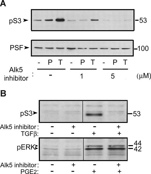
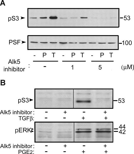
Upon receptor binding by TGFβ, the type I TGFβ receptor Alk5 phosphorylates Smad3 at specific serine residues in the SSXS motif within its C-terminal domain.11 We therefore considered whether the observed phospho-Smad3 nuclear translocation might be accounted for by PGE2-induced transactivation of Alk5. Preincubation of HUVECs with the specific Alk5 inhibitor SB431542,32 at the dose range required for TGFβ1 inhibition, totally prevented PGE2-mediated phospho-Smad3 nuclear accumulation (Figure 2A). This was a specific inhibitory effect, since other signaling pathways triggered by PGE2 in HUVECs, such as ERK-1,2 activation, were not affected (Figure 2B). These data thus indicate that Alk5 is required for PGE2-mediated Smad3 activation.
PGE2-induced phospho-Smad3 nuclear translocation and DNA binding require Alk5 kinase activity. (A) Western blot showing the nuclear content of phospho-Smad3 (pS3) in nuclear extracts of HUVECs pretreated (30 minutes) with vehicle or the specific Alk5 inhibitor SB431542 (1 or 5 μM) and then treated for 1 hour with 1 μM PGE2 or 0.5 ng/mL TGFβ1 (top panel). PSF was probed as a loading control (bottom panel). (B) Top panel: Western blot showing phospho-Smad3 content in whole-cell lysates of HUVECs pretreated as indicated with 1 μM Alk5 inhibitor and treated for 1 hour with 0.5 ng/mL TGFβ1. Bottom panel: Western blot showing the content of phospho-ERK-1,2 (pERK) in whole-cell lysates of HUVECs preincubated as indicated with 1 μM Alk5-specific inhibitor and treated for 10 minutes with 1 μM PGE2. Molecular weights (kDa) are shown on the right. The figure shows results of representative experiments of 3 performed. A dividing line shows the grouping of parts of the same gel from which irrelevant lanes have been deleted.
PGE2-induced phospho-Smad3 nuclear translocation and DNA binding require Alk5 kinase activity. (A) Western blot showing the nuclear content of phospho-Smad3 (pS3) in nuclear extracts of HUVECs pretreated (30 minutes) with vehicle or the specific Alk5 inhibitor SB431542 (1 or 5 μM) and then treated for 1 hour with 1 μM PGE2 or 0.5 ng/mL TGFβ1 (top panel). PSF was probed as a loading control (bottom panel). (B) Top panel: Western blot showing phospho-Smad3 content in whole-cell lysates of HUVECs pretreated as indicated with 1 μM Alk5 inhibitor and treated for 1 hour with 0.5 ng/mL TGFβ1. Bottom panel: Western blot showing the content of phospho-ERK-1,2 (pERK) in whole-cell lysates of HUVECs preincubated as indicated with 1 μM Alk5-specific inhibitor and treated for 10 minutes with 1 μM PGE2. Molecular weights (kDa) are shown on the right. The figure shows results of representative experiments of 3 performed. A dividing line shows the grouping of parts of the same gel from which irrelevant lanes have been deleted.
TGFβ and MT1-MMP are required for phospho-Smad3 nuclear translocation
The requirement for Alk5 in PGE2-induced phospho-Smad3 nuclear translocation suggested that the presence of active TGFβ in the medium might be necessary for this effect. We therefore analyzed, by ELISA, the amounts of TGFβ in the supernatants of HUVECs treated with 1 μM PGE2. Incubation of HUVECs with PGE2 yielded no significant increase in the relative amount of active TGFβ (Figure 3A). The possibility remains, however, that increases in active TGFβ may occur at localized areas of the cell surface, and may be undetectable by whole-supernatant ELISA. To test this, we preincubated HUVECs with a specific TGFβ1-blocking antibody before treating with PGE2. Western blot of nuclear extracts showed that PGE2-induced phospho-Smad3 nuclear translocation was completely inhibited by the anti-TGFβ1 blocking antibody (Figure 3B), suggesting that PGE2 treatment results in the activation of Alk5 receptor by active extracellular TGFβ1.
PGE2-mediated Smad3 activation requires the presence of active TGFβ and MT1-MMP. (A) The graph represents the percentage of active TGFβ versus total TGFβ, measured by ELISA in the supernatants of HUVECs treated for the indicated times with 1 μM PGE2. (B) Western blot showing the nuclear content of phospho-Smad3 (pS3) in nuclear extracts of HUVECs pretreated with a TGFβ-neutralizing antibody (αTGFβ) or a control antibody (C) before treatment for 1 hour with 1 μM PGE2 (P). A dividing line shows the grouping of parts of the same gel from which irrelevant lanes have been deleted. The figure shows a representative experiment of 3 performed. (C) Western blot showing PGE2- and TGFβ1-modulated phospho-Smad3 nuclear content as in panel B in HUVECs pretreated with the metalloproteinase inhibitor GM6001 or vehicle. The figure shows a representative experiment of 4 performed. (D) Western blot showing PGE2- and TGFβ1-modulated phospho-Smad3 nuclear content as in panel B in HUVECs preincubated with an MT1-MMP–neutralizing antibody (αMT1) or a control antibody (C). The figure shows a representative experiment of 3 performed. In all cases, blots were probed with antiphospho-Smad3 antibody (pS3; top panels). Equal loading was confirmed either by a nonspecific band (panel B; nonsignificant) or by Western blot for the nuclear protein PSF (B,C, bottom panels).
PGE2-mediated Smad3 activation requires the presence of active TGFβ and MT1-MMP. (A) The graph represents the percentage of active TGFβ versus total TGFβ, measured by ELISA in the supernatants of HUVECs treated for the indicated times with 1 μM PGE2. (B) Western blot showing the nuclear content of phospho-Smad3 (pS3) in nuclear extracts of HUVECs pretreated with a TGFβ-neutralizing antibody (αTGFβ) or a control antibody (C) before treatment for 1 hour with 1 μM PGE2 (P). A dividing line shows the grouping of parts of the same gel from which irrelevant lanes have been deleted. The figure shows a representative experiment of 3 performed. (C) Western blot showing PGE2- and TGFβ1-modulated phospho-Smad3 nuclear content as in panel B in HUVECs pretreated with the metalloproteinase inhibitor GM6001 or vehicle. The figure shows a representative experiment of 4 performed. (D) Western blot showing PGE2- and TGFβ1-modulated phospho-Smad3 nuclear content as in panel B in HUVECs preincubated with an MT1-MMP–neutralizing antibody (αMT1) or a control antibody (C). The figure shows a representative experiment of 3 performed. In all cases, blots were probed with antiphospho-Smad3 antibody (pS3; top panels). Equal loading was confirmed either by a nonspecific band (panel B; nonsignificant) or by Western blot for the nuclear protein PSF (B,C, bottom panels).
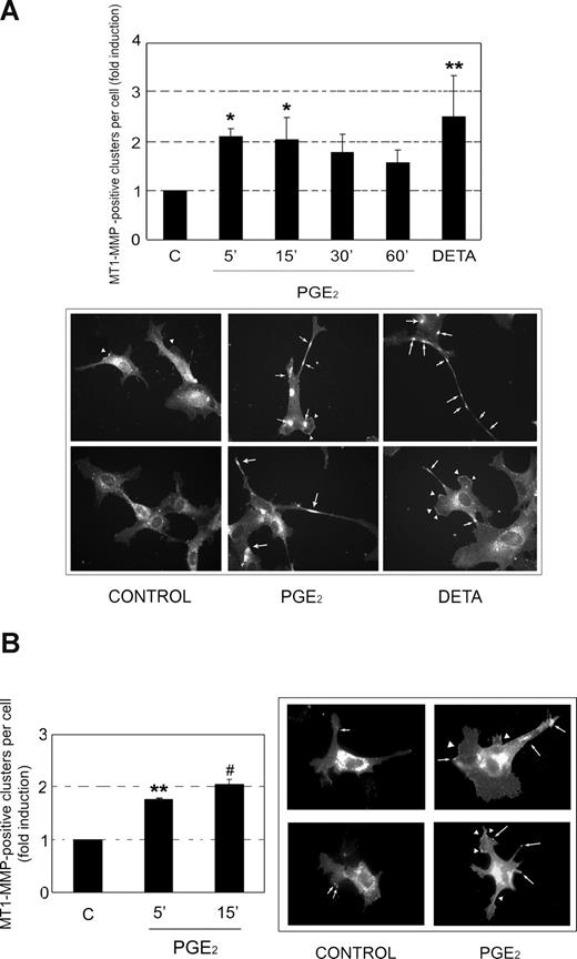
The similar time-course profiles of PGE2- and TGFβ-induced Smad3 activation (Figure 1A,C) suggest that PGE2 increases the availability of preexisting TGFβ, rather than up-regulating its expression. Several proteases have been implicated in the mobilization and subsequent activation of TGFβ, including the serine protease plasmin and certain metalloproteinases.12 To analyze the possible implication of these proteases in PGE2-mediated Smad3 activation, we initially conducted experiments with the plasmin inhibitors aprotinin and α2-antiplasmin. These compounds had no effect on PGE2-mediated phospho-Smad3 nuclear translocation (A.A. and J.M.R., unpublished data, March 2006). In contrast, the metalloproteinase inhibitor GM6001 efficiently prevented nuclear translocation of phospho-Smad3 mediated by PGE2 (Figure 3C). As expected, GM6001 did not affect Smad3 activation mediated by exogenously added active TGFβ1 (Figure 3C). Among the metalloproteinases whose catalytic activity is inhibited by GM6001,33 MT1-MMP has been shown to activate TGFβ1,14 so we analyzed the potential involvement of this enzyme in PGE2-induced TGFβ1 activation. A time-course immunoblot analysis of total HUVEC lysates revealed that PGE2 does not alter the overall amount of MT1-MMP (A.A. and J.M.R., unpublished data, November 2006). However, a specific blocking antibody against MT1-MMP fully abolished PGE2-mediated phospho-Smad3 nuclear translocation, while having no effect on the activation of Smad3 by exogenous TGFβ1 (Figure 3D). Increased activity of MT1-MMP is associated with the localization of this protease at specific cell membrane sites.34 We therefore examined whether PGE2 regulates the cellular distribution of MT1-MMP. Immunofluorescence experiments showed that PGE2 treatment induced a significant increase in the number of lamellae and clusters containing MT1-MMP at 5 and 15 minutes (Figure 4A; P < .05 vs control). This effect was similar to that obtained with the NO donor DETA-NONOate (Figure 4A; P < .01 vs control), which has been previously shown to induce MT1-MMP cluster formation and proteolytic activity in endothelial cells.34 Both down-regulation of MT1-MMP expression in HUVECs, and competition experiments with a molar excess of a specific MT1-MMP peptide, supported the specificity of MT1-MMP staining (Figures S1,S2, available on the Blood website; see the Supplemental Materials link at the top of the online article). To further confirm the PGE2-mediated increase in MT1-MMP clustering, we transfected HUVECs with a construct encoding full-length MT1-MMP fused to GFP (MT1-MMP-GFP). Treatment of transfected cells with 1 μM PGE2 induced the relocalization of MT1-MMP-GFP to membrane clusters, over a similar time scale observed for endogenous MT1-MMP (Figure 4B). These findings therefore suggest that PGE2 increases MT1-MMP activity at specific plasma membrane sites, and indicate that extracellular TGFβ1 and MT1-MMP activity are required for PGE2-induced nuclear accumulation of phospho-Smad3.
PGE2 promotes membrane clustering of MT1-MMP. (A) HUVECs treated over a time course with 1 μM PGE2, or with 100 μM DETA-NONOate (DETA) as a positive control, were immunostained with anti–MT1-MMP antibody. Top panel: the histogram shows the means plus or minus SD from 3 independent experiments of the number per cell of lamellae and membrane clusters containing MT-1-MMP. The data are presented as fold induction above control conditions (C). *P < .05 vs control; **P < .01 vs control. Bottom panel: micrographs showing representative fields of untreated control cells or cells treated with either 1 μM PGE2 for 5 minutes or 100 μM DETA-NONOate (DETA). (B) HUVECs transfected with MT1-MMP-GFP were either left untreated or incubated with 1 μM PGE2 for the times indicated. Left panel: the histogram shows the means plus or minus SD from 2 independent experiments of the number per cell of lamellae and membrane clusters containing MT-1-MMP-GFP. The data are shown as fold induction over control conditions. (C). **P < .01 vs control; #P < .001 vs control. Right panel: micrographs showing representative fields of control cells or cells treated with 1 μM PGE2 for 15 minutes. Arrows indicate MT1-MMP membrane clusters, and arrowheads denote MT1-MMP–containing lamellae.
PGE2 promotes membrane clustering of MT1-MMP. (A) HUVECs treated over a time course with 1 μM PGE2, or with 100 μM DETA-NONOate (DETA) as a positive control, were immunostained with anti–MT1-MMP antibody. Top panel: the histogram shows the means plus or minus SD from 3 independent experiments of the number per cell of lamellae and membrane clusters containing MT-1-MMP. The data are presented as fold induction above control conditions (C). *P < .05 vs control; **P < .01 vs control. Bottom panel: micrographs showing representative fields of untreated control cells or cells treated with either 1 μM PGE2 for 5 minutes or 100 μM DETA-NONOate (DETA). (B) HUVECs transfected with MT1-MMP-GFP were either left untreated or incubated with 1 μM PGE2 for the times indicated. Left panel: the histogram shows the means plus or minus SD from 2 independent experiments of the number per cell of lamellae and membrane clusters containing MT-1-MMP-GFP. The data are shown as fold induction over control conditions. (C). **P < .01 vs control; #P < .001 vs control. Right panel: micrographs showing representative fields of control cells or cells treated with 1 μM PGE2 for 15 minutes. Arrows indicate MT1-MMP membrane clusters, and arrowheads denote MT1-MMP–containing lamellae.
Alk5 kinase activity and MT1-MMP are involved in PGE2-induced angiogenesis in vitro
TGFβ is thought to play roles both in the proliferation and migration of ECs and also in the re-adoption of a quiescent EC phenotype during the resolutive phase of angiogenesis.35 To analyze whether TGFβ-mediated Smad3 activation might mediate the proangiogenic properties of PGE2, we performed in vitro Matrigel angiogenesis assays. PGE2 induced a significant increase in the number of endothelial cords on Matrigel (P < .001 vs control; Figure 5); and this was impaired by the Alk5 inhibitor SB431542 in a dose-dependent manner (P < .01 vs PGE2 alone; Figure 5). Given that Alk5 kinase activity is necessary for PGE2-mediated Smad3 activation, these results provide evidence for a role for this pathway in the regulation of angiogenesis by PGE2.
Inhibition of the kinase activity of Alk5 impairs PGE2-induced angiogenesis in vitro. Top panel: HUVECs were pretreated with vehicle or the Alk5 inhibitor SB431542 (5 μM) for 15 minutes and then seeded onto growth factor reduced Matrigel in the presence or absence (NS) of 1 μM PGE2. Formation of cords was quantified after 5 hours. Data are presented as the fold induction (FI) above cord formation by untreated cells, and are the means plus or minus SD of 3 independent experiments. The absolute value corresponding to FI = 1 was 65 cords/field. All experiments were run in triplicate. *P < .001 vs control; #P < .01 vs PGE2 alone. Bottom panel: photographs showing representative fields from experiments quantified in the top panel.
Inhibition of the kinase activity of Alk5 impairs PGE2-induced angiogenesis in vitro. Top panel: HUVECs were pretreated with vehicle or the Alk5 inhibitor SB431542 (5 μM) for 15 minutes and then seeded onto growth factor reduced Matrigel in the presence or absence (NS) of 1 μM PGE2. Formation of cords was quantified after 5 hours. Data are presented as the fold induction (FI) above cord formation by untreated cells, and are the means plus or minus SD of 3 independent experiments. The absolute value corresponding to FI = 1 was 65 cords/field. All experiments were run in triplicate. *P < .001 vs control; #P < .01 vs PGE2 alone. Bottom panel: photographs showing representative fields from experiments quantified in the top panel.
MT1-MMP is known to participate in the onset and progression of angiogenesis through a variety of mechanisms,36 and the data shown in Figure 3D indicate that MT1-MMP is required for PGE2-induced Smad3 activation. To investigate the possible participation of MT1-MMP in PGE2-mediated angiogenesis, we conducted in vitro Matrigel experiments with MLECs from MT1-MMP KO mice. PGE2 treatment induced endothelial cord formation by WT MLECs (P < .01 vs untreated wild-type [WT] control), but the same treatment failed to significantly promote cord formation by MT1-MMP–deficient MLECs (Figure 6). This effect was not due to a nonspecific impairment of the tube formation capacity of KO cells, since they were able to form cords in response to the proangiogenic factor CXCL12, whose activity does not depend on MT1-MMP and which induced a 2-fold increase in cord formation in KO cells (P < .05 vs untreated KO control).37 The reduced capacity of MT1-MMP–deficient MLECs to form cords under basal conditions can be explained by the requirement for MT1-MMP in cell migration, as described in previous reports24 (Figure 6). These results strongly suggest that MT1-MMP activity is required for PGE2-induced in vitro angiogenesis.
PGE2-induced angiogenesis in vitro requires the presence of MT1-MMP. MLECs from WT (MT1-MMP+/+) or MT1-MMP KO (MT1-MMP−/−) mice were seeded onto Matrigel in the presence or absence (NS) of PGE2 (1 μM) or the proangiogenic factor CXCL12 (10 nM). Cord formation was quantified after 6 hours. Data are presented as the fold induction above cord formation by untreated cells, and are the means plus or minus SD of 3 to 5 independent experiments. The absolute value corresponding to FI = 1 was 66 cords/field. All experiments were run in triplicate. *P < .05 vs KO control; **P < .01 vs WT control; #P < .001 vs WT control.
PGE2-induced angiogenesis in vitro requires the presence of MT1-MMP. MLECs from WT (MT1-MMP+/+) or MT1-MMP KO (MT1-MMP−/−) mice were seeded onto Matrigel in the presence or absence (NS) of PGE2 (1 μM) or the proangiogenic factor CXCL12 (10 nM). Cord formation was quantified after 6 hours. Data are presented as the fold induction above cord formation by untreated cells, and are the means plus or minus SD of 3 to 5 independent experiments. The absolute value corresponding to FI = 1 was 66 cords/field. All experiments were run in triplicate. *P < .05 vs KO control; **P < .01 vs WT control; #P < .001 vs WT control.
Alk5 kinase activity is required for PGE2-induced angiogenesis in vivo
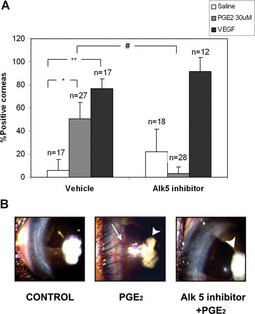
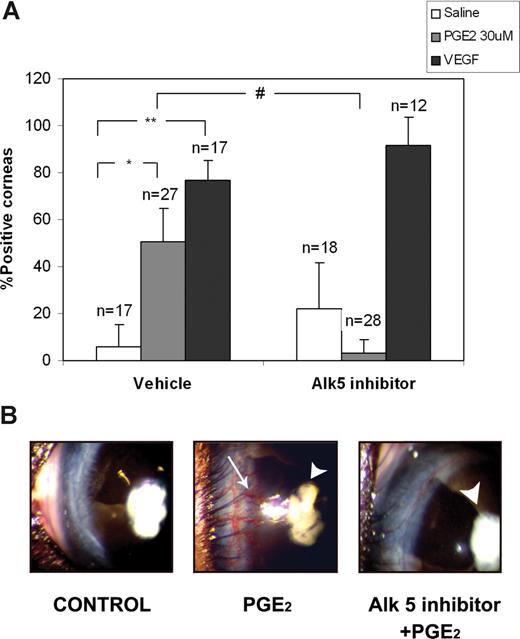
The evidence for proangiogenic activity of PGE2 in vivo mainly comes from observations in EP KO mice.7 To test directly whether PGE2 promotes neovascularization, we used the established mouse corneal in vivo angiogenesis model. In these experiments, PGE2 elicited a robust proangiogenic response observed 6 to 8 days after pellet implantation (50.70 ± 14.08% positive corneas; P < .01 vs saline), comparable to the effect of VEGF (76.66 ± 8.81% positive corneas; P < .001 vs saline; Figure 7A,B). The presence of the Alk5 inhibitor SB431542 completely prevented vascular in-growth toward pellets containing PGE2 (3.33+5.77% positive corneas; P < .01 vs PGE2), but had no significant effect on corneal neovascularization induced by VEGF (91.66+11.78% positive corneas; P > .05 vs VEGF; Figure 7A,B). The Alk5 inhibitor alone had a weak vascularizing effect in some cases, but this was not statistically significant (22.22+19.24% positive corneas; P > .05 vs saline; Figure 7A). Together, these results show that Alk5 kinase activity is required for the in vivo proangiogenic response to PGE2.
The Alk5 kinase inhibitor SB431542 impairs PGE2-induced mouse corneal angiogenesis. In vivo angiogenesis assays were performed by implanting a pellet containing saline, PGE2 (30 μM), or VEGF (125 ng) together with vehicle or 10 μM SB431542 into a previously created corneal pocket. The angiogenic response was assessed after 6 to 8 days. (A) The chart shows the mean percentage plus or minus SD of positive corneas per condition from 3 independent experiments. n indicates the total number of eyes per condition. *P < .01 vs saline; **P < .001 vs saline; #P < .01 vs PGE2 alone. (B) Representative photographs of corneas from the experiments summarized in panel A; arrowheads, pellets containing PGE2; arrow, positive angiogenic response (note the vessels emerging from the sclerocorneal limbus and reaching the pellet).
The Alk5 kinase inhibitor SB431542 impairs PGE2-induced mouse corneal angiogenesis. In vivo angiogenesis assays were performed by implanting a pellet containing saline, PGE2 (30 μM), or VEGF (125 ng) together with vehicle or 10 μM SB431542 into a previously created corneal pocket. The angiogenic response was assessed after 6 to 8 days. (A) The chart shows the mean percentage plus or minus SD of positive corneas per condition from 3 independent experiments. n indicates the total number of eyes per condition. *P < .01 vs saline; **P < .001 vs saline; #P < .01 vs PGE2 alone. (B) Representative photographs of corneas from the experiments summarized in panel A; arrowheads, pellets containing PGE2; arrow, positive angiogenic response (note the vessels emerging from the sclerocorneal limbus and reaching the pellet).
Discussion
In this study, we have identified the activation of the Alk5-Smad3 pathway in ECs as an important mediator of PGE2-promoted neovascularization in vitro and in vivo. Activation of Smad3 nuclear translocation and DNA binding by PGE2 requires the kinase activity of the type I TGFβ receptor Alk5, and is prevented by neutralizing antibodies to TGFβ and MT1-MMP. TGFβ, MT1-MMP, and Alk5 kinase activity are all required for PGE2-mediated cord formation in vitro, and inhibition of Alk5 also prevents neovascularization in an in vivo angiogenesis model. Our findings thus identify a novel transactivation signaling pathway via which PGE2 triggers the activation of MT1-MMP, which releases TGFβ from its latent complex so that it can bind its receptor Alk5, activating the Smad3 pathway and promoting angiogenesis.
Endothelial cells are known to express 2 type I TGFβ receptors, Alk1 and Alk5. Alk1 phosphorylates Smad1 and Smad5, whereas Alk5 phosphorylates Smad2 and Smad3.11 We have focused on the Alk5-Smad3 pathway, but it is possible that PGE2 sequentially activates both receptors. It should be noted, however, that although SB431542 is a known inhibitor of Alk4 and Alk7, which are not expressed in ECs, it does not affect Alk1 kinase activity.38 We can therefore clearly implicate Alk5 in PGE2-mediated Smad3 activation and angiogenesis. Even so, the cross-talk between the 2 signaling routes makes it reasonable to consider that the Alk1-activated pathway might also be involved in these processes.
Our finding that a TGFβ1-neutralizing antibody impairs PGE2-induced Smad3 nuclear accumulation (Figure 3A) discards a role for intracellular activation of Alk5, as has also been described for other PGE2-transactivated receptors.39 It is possible that PGE2 might increase the total amount of secreted TGFβ; but, given the timing of Smad3 activation (Figure 1), it is more feasible that it triggers the activation of latent TGFβ previously released to the medium. However, ELISA assays with supernatants of PGE2-treated HUVECs revealed only slight variations in the concentrations of total and active TGFβ (Figure 3A). We hypothesize that activation of latent TGFβ is a local event taking place in specialized areas of the cell surface, rendering it undetectable in whole-culture supernatants. The implication of MT1-MMP in PGE2-mediated TGFβ activation supports this view. PGE2 has been shown to regulate the expression and activity of diverse metalloproteinases in several models,40 in some cases leading to the release of trapped ligands and transactivation of their receptors. PGE2 is also known to mediate LPS-induced MT1-MMP expression in monocytes.41 However, we did not detect any changes in total MT1-MMP expression by ECs upon PGE2 treatment (A.A. and J.M.R., unpublished data, 2006); instead, our experiments indicated that PGE2 promotes the early localization of MT1-MMP to cellular lamellae and membrane clusters (Figure 4). In colon cancer cells, PGE2 transactivates the epidermal growth factor receptor (EGFR) receptor via activation of c-Src, which induces a matrix metalloproteinase activity that releases membrane-bound TGF-α.39 Furthermore, sphingosine-1P induces the Src-mediated phosphorylation of MT1-MMP, which promotes MT1-MMP relocalization and EC migration.42 Viewed in this context, it is tempting to speculate that the action of PGE2 in our model could also involve MT1-MMP subcellular relocalization upon phosphorylation by Src. It will be interesting to perform further experiments to clarify this issue. MT1-MMP is processed within the cell, and is exported to the membrane as a mature enzyme. The localization of MT1-MMP at specific plasma-membrane domains seems to be critical for its activity, and it usually associates with other membrane molecules in multiprotein complexes.43 This characteristic would fit well with the hypothesis of localized TGFβ activation. In epithelial cells MT1-MMP activates latent TGFβ in association with integrin αvβ8.14 This integrin is not expressed in ECs, but MT1-MMP is able to associate with other integrins on the endothelial surface that could potentially contribute to this process.44
We observed a clear down-regulation of PGE2-induced Smad3 activation by the metalloproteinase inhibitor GM6001 and by anti–MT1-MMP blocking antibody (Figure 3B,C). However, we also consistently detected an increase in nuclear Smad3 accumulation when ECs were treated with these inhibitors alone (Figure 3B,C). Metalloproteinases are thought to regulate several intracellular pathways.13 Increased Smad3 nuclear translocation upon pretreatment with GM6001 or anti–MT1-MMP blocking antibody could therefore be due to the modulation of additional MT1-MMP signaling routes, which would subsequently interfere with Smad3 activation.
The in vitro Matrigel experiments with MLECs derived from MT1-MMP KO mice (Figure 6) provide a direct assessment of the putative role of MT1-MMP in PGE2-induced tubulogenesis. MLECs have been used previously to investigate the role of MT1-MMP in angiogenesis induced by various stimuli.37,48 MT1-MMP KO MLECs have an intrinsically reduced migratory capacity and show impaired tube formation on Matrigel under basal conditions.23 This can be overcome by treatment with factors whose proangiogenic activity does not depend on MT1-MMP, such as LPS or CXCL-12. The 2-fold increase in cord formation induced by CXCL-12 observed in KO cells is consistent with previous reports, and is similar to the effect seen in WT cells. These results thus discard a nonspecific, general impairment of tube formation in MT1-MMP KO MLECs. The failure of PGE2 to promote tube formation in these cells, despite inducing these structures significantly in WT cells, therefore indicates that MT1-MMP activity is required for PGE2-mediated in vitro angiogenesis; however, as is also true for the Alk5 data, this finding does not imply that PGE2 proangiogenic activity can be exclusively attributed to this metalloproteinase. Angiogenesis is a tightly regulated multistep process, where each sequential phase is necessary for formation of a complete neovessel.46 Different PGE2-activated signaling pathways or genes might mediate specific phases of angiogenesis, and inhibition of any one of these steps would affect the whole process. In addition, the diverse PGE2-activated pathways may be interrelated in some cases, giving rise to a complex regulatory network. In this regard, both PGE2 and TGFβ are known to induce expression of proangiogenic factors in ECs such as VEGF.47,48 The impairment of cord formation in in vitro Matrigel experiments by the Alk5-specific inhibitor SB431542 (Figure 5) clearly implicates this TGFβ receptor in PGE2-mediated tubulogenesis. Cord formation does not depend on EC proliferation, but rather on migration events and the EC-mediated remodeling of the extracellular matrix.49 These phenomena can be mediated, at least in part, by Alk5. One possibility is that all the steps required for EC tube formation on Matrigel might be accounted for by Alk5/Smad3-induced secretion of VEGF, which also induces a potent proangiogenic response in vivo1 (Figure 7). MT1-MMP is able either to enhance VEGF expression or to release it from the extracellular matrix,36 indicating that the induction of VEGF by PGE2 and TGFβ might be linked. Similarly, eNOS has been reported to mediate PGE2-induced angiogenesis in an ex vivo model,9 which could be related to the MT1-MMP redistribution and activation promoted by NO donors34 ; however, both activation and impairment of TGFβ signaling by NO have been reported.20,50 Finally, long-term exposure of ECs to PGE2 induces expression of CXCR4, the CXCL12 receptor. CXCR4 mediates PGE2-induced angiogenesis in the presence of CXCL12,8 and TGFβ is known to increase CXCR4 expression in other cell types.51,52 Nonetheless, angiogenesis mediated by CXCL12 is not dependent on the activity of MT1-MMP, which in fact cleaves and inactivates this chemokine.53 This suggests a negative regulatory role for MT1-MMP in CXCL12-mediated angiogenesis. These lines of evidence argue against the participation of just one pathway in PGE2-induced angiogenesis.
TGFβ is a pleiotropic cytokine, which apparently elicits opposite responses in ECs depending on the type I receptor activated. Alk1 activation promotes endothelial proliferation and migration, thus favoring the early phases of angiogenesis. Conversely, Alk5 inhibits these responses and mediates the expression of genes related to matrix remodeling and differentiation of perivascular cells, supporting the resolutive phase.35 However, data from KO mice appear to contradict these studies, attributing roles to both receptors opposite to those observed in vitro.54 This contradiction can be partly explained by the interplay between Alk1- and Alk5-activated pathways. Alk1 negatively regulates Alk5-mediated responses and, conversely, some reports indicate that Alk5 signaling is required for full activation of pathways downstream of Alk1.55 Another factor that might contribute to the explanation of this discrepancy is the expression level of the accessory TGFβ receptor endoglin, since this receptor differentially modulates the routes activated by Alk1 and Alk5.56
A detailed understanding of the mechanisms that regulate neovascularization is necessary for improved strategies for the resolution of angiogenesis-controlled diseases. COX-2 is a major mediator of neovessel formation, and specific inhibitors of this enzyme have proved to be highly effective in the prevention and treatment of some of these disorders.57 PGE2 is the main mediator of COX-2 proangiogenic activity, but little is known about the signals triggered by this prostanoid in ECs. In this article, we have provided the first description in any cell type of PGE2 signaling through the Alk5-Smad3 axis. Activation of this pathway in ECs involves the activity of MT1-MMP and the release of active TGFβ. We have also uncovered a possible novel mechanism for PGE2-mediated MT1-MMP regulation, through the modification of subcellular MT1-MMP distribution. Our results support a functional role for this pathway, since TGFβ, MT1-MMP, and Alk5-Smad3 activation are required for PGE2-mediated angiogenic processes. These findings thus identify a novel signaling pathway that links PGE2 and TGFβ, 2 effectors involved in tumor growth and angiogenesis, and reveal potential targets for the development of new therapeutic approaches for the treatment of angiogenesis-related disorders.
The online version of this article contains a data supplement.
The publication costs of this article were defrayed in part by page charge payment. Therefore, and solely to indicate this fact, this article is hereby marked “advertisement” in accordance with 18 USC section 1734.
Acknowledgments
We thank Drs S. Apte and K. Tryggvason for providing MT1-MMP KO mice; Drs C. Bernabéu, E. Cano, P. Gómez del Arco, S. Lamas, S. Martínez-Martínez, and A. Rodríguez for critical reading of the manuscript; and Drs F. Rodríguez-Pascual, A. Sobrado, and P. Gonzalo for reagents. Dr S. Bartlett provided excellent editorial assistance.
The Centro Nacional de Investigaciones Cardiovasculares (CNIC; Spain) is supported by the Spanish Ministry of Health and Consumer Affairs and the Pro-CNIC Foundation. This work was also supported by grants from the Ministerio de Educación y Ciencia (SAF2006-08348), the Ministerio de Sanidad y Consumo (RD06/0014/005), the Comunidad Autónoma de Madrid (CAM-S2006/BIO-0194: CARDIOVREP), and the European Union (LSHM-CT-2004-005033: EICOSANOX), all to J.M.R.; and by grant SAF2005-02228 from the Ministerio de Educación y Ciencia to A.G.A.
Authorship
Contribution: A.A. designed and performed research, analyzed data, and wrote the paper; J.M.L.-O., L.G., D.L.-M., I.M., D.S., and A.J.Q. performed research. A.G.A. designed research and analyzed data. J.M.R. designed and supervised research and analyzed data.
Conflict-of-interest disclosure: The authors declare no competing financial interests.
Corresponding author: Juan Miguel Redondo, Department of Vascular Biology and Inflammation, Centro Nacional de Investigaciones Cardiovasculares, Melchor Fernández Almagro 3, 28029 Madrid, Spain; e-mail: jmredondo@cnic.es.


