Abstract
Human mast cells are found in skin and mucosal surfaces and next to blood vessels. They play a sentinel cell role in immunity, recognizing invading pathogens and producing proinflammatory mediators. Mast cells can recruit granulocytes, and monocytes in allergic disease and bacterial infection, but their ability to recruit antiviral effector cells such as natural killer (NK) cells and T cells has not been fully elucidated. To investigate the role of human mast cells in response to virus-associated stimuli, human cord blood–derived mast cells (CBMCs) were stimulated with polyinosinic·polycytidylic acid, a double-stranded RNA analog, or infected with the double-stranded RNA virus, reovirus serotype 3 Dearing for 24 hours. CBMCs responded to stimulation with polyinosinic·polycytidylic acid by producing a distinct chemokine profile, including CCL4, CXCL8, and CXCL10. CBMCs produced significant amounts of CXCL8 in response to low levels of reovirus infection, while both skin- and lung-derived fibroblasts were unresponsive unless higher doses of reovirus were used. Supernatants from CBMCs infected with reovirus induced substantial NK cell chemotaxis that was highly dependent on CXCL8 and CXCR1. These results suggest a novel role for mast cells in the recruitment of human NK cells to sites of early viral infection via CXCL8.
Introduction
Mast cells are long-lived resident tissue cells found close to blood vessels, and are numerous at sites in close proximity to the external environment such as the skin and airways (reviewed in Galli et al1 and Metz and Maurer2 ). From these strategic locations they can quickly recognize and respond to invading pathogens. They are also relatively resistant to ultraviolet (UV) and gamma irradiation.3-5 Upon activation, mast cells produce a wide array of mediators, including granule-associated products, such as histamine, and preformed and de novo synthesized cytokines, chemokines, and lipid mediators. They can activate and recruit effector cells, including eosinophils,6 neutrophils,7 and monocytes.8 Their role in innate immune responses to bacterial infections has been clearly delineated, however their involvement in viral infections is not well understood. Mast cells express Toll-like receptor 3 (TLR3), which recognizes viral double-stranded RNA (dsRNA),9 and they can produce type I interferons when activated through this receptor.10 Studies examining the permissiveness of mast cells to viruses show that they can be infected by, and respond to, dengue virus,11,12 HIV,13,14 and respiratory syncytial virus.15 Human mast cells produce the chemokines CCL3, CCL4, and CCL5 when infected with dengue virus,16 and mouse mast cells produce CCL4 and CCL5 when infected with Newcastle disease virus, all of which are known natural killer (NK) cell and T-cell chemoattractants.17
NK cells are large granular lymphocytes that can kill virally infected cells, and are crucial for the clearance of viruses during infections (reviewed in Lodoen and Lanier18 ). The chemokines and chemokine receptors necessary for the infiltration of NK cells into virally infected tissues have recently begun to be uncovered. NK cells have been reported to express multiple chemokine receptors including CCR1, CCR2, CCR5, CXCR1, CXCR3, and CXCR4.17,19-22 Studies examining murine cytomegalovirus infection in mice show that CCL2 and CCL3 are necessary for NK cell infiltration into the liver and subsequent viral clearance.23,24 Experiments using a central nervous system infection model of mouse hepatitis virus have also demonstrated the importance of CXCL10 in this process.25 The chemokine receptors CCR2, CCR5, and CXCR3 are generally considered the most important in NK cell recruitment in the context of viral infection.
The major cellular sources of chemokines inducing NK cell recruitment during viral infection are just beginning to be understood. Infection of myeloid and plasmacytoid dendritic cells (DCs) with influenza virus can lead to DC-induced recruitment of NK cells and CD4+ memory T cells.26,27 In addition, plasmacytoid DCs infected with herpes simplex virus can recruit NK cells and activated T cells via CCL4 and CXCL10.28 However, these cells would not be found in large numbers at many initial sites of infection. The ability of tissue-resident human mast cells to recruit NK cells or T cells to sites of viral infection has not been previously examined.
In this study, we used 2 different virus-associated stimuli to evaluate the response of human mast cells to viral infection: polyinosinic·polycytidylic acid (poly(I:C)) and mammalian reovirus that has a dsRNA genome.29 Our results demonstrate that mast cells can induce the selective chemotaxis of NK cells in response to both of these stimuli by a mechanism highly dependent on the chemokine CXCL8.
Methods
Mast cell cultures
HMC-1 cells were cultured in Iscove modified Dulbecco medium (IMDM; Invitrogen, Burlington, ON) containing 10% fetal bovine serum (FBS; Medicorp, Montreal, QC, and Sigma-Aldrich, Oakville, ON), 100 U/mL penicillin G, and 100 μg/mL streptomycin (Invitrogen). Human cord blood–derived mast cells (CBMCs) were generated according to an adaptation of the method described by Saito et al.30 Briefly, mononuclear cells were obtained from umbilical cord blood following appropriate ethical approval from the Isaac Walton Killam (IWK) Health Center (Halifax, NS), and passaged twice a week for 5 to 6 weeks in RPMI 1640 (Invitrogen) containing 20% FBS, penicillin G, streptomycin, 10 mM N-2-hydroxyethylpiperazine-N′-2-ethanesulfonic acid (HEPES; Invitrogen), 20% human skin fibroblast culture supernatant, primarily as a source of interleukin-6 (IL-6; American Type Culture Collection [ATCC], Manassas, VA), 10−7 M prostaglandin E2 (Sigma-Aldrich), and 75 ng/mL stem cell factor (SCF; Peprotech, Rocky Hill, NJ). Only cultures that were 95% or more pure mast cells by metachromatic staining using toluidine blue (pH 1.0) were used.
Mast cell activations
HMC-1 cells or CBMCs were washed and activated (106/mL) with 1 or 10 μg/mL poly(I:C) or control diluent (Calbiochem, San Diego, CA) for 24 hours in 5% CO2 at 37°C. Prior to activation, CBMCs were cultured overnight in the absence of prostaglandin E2, and subsequent steps were performed with activation medium consisting of RPMI 1640 containing 1% FBS, HEPES, penicillin G, streptomycin, and 10 ng/mL SCF. Cell-free supernatants were harvested by centrifugation at 300g at 6 and 24 hours.
Reovirus infection of human mast cells
Mammalian reovirus serotype 3 Dearing was purified as previously described.31 Reovirus adsorption was performed by incubating CBMCs with reovirus at various multiplicities of infection (MOI) in RPMI 1640 for 1 hour at 37°C, followed by 2 washes before cell resuspension in activation medium containing SCF for 24 hours. For UV inactivation, reovirus was treated with 6 × 999 900 μjoules/cm2 UV light using the UV Stratalinker 1800 (Stratagene, La Jolla, CA). UV inactivation was confirmed by standard plaque assay. Cell-free supernatants were harvested as stated in “Mast cell activations.” In some experiments, CBMCs were stained with 7-AAD (Biotium, Hayward, CA) and examined by flow cytometry to determine cell viability. For the detection of reovirus by flow cytometry, CBMCs were fixed and permeabilized, Fc receptors were blocked using purified human immunoglobulin G (IgG), and staining was done with antireovirus polyclonal rabbit antibody (Ab) followed by anti–rabbit IgG conjugated to Cy5 (Jackson Immunoresearch, West Grove, PA). The cells were analyzed using a BD FACSCalibur (BD Biosciences, San Diego, CA) and Winlist Software (Verity Software House, Topsham, ME).
Stimulation of human keratinocytes and fibroblasts
Primary adult keratinocytes were cultured in fully supplemented serum-free KGM-2 medium (Lonza, Rockland, ME). Keratinocytes were activated in basal medium containing EGF, 1% FBS, penicillin, and streptomycin. CCL-204 cells were obtained from ATCC and cultured in RPMI 1640 containing 10% FBS, 1% nonessential amino acids (Invitrogen), penicillin G, and streptomycin. Human dermal fibroblasts (HDFs; a gift from Dr Andrew Issekutz, IWK Hospital) were cultured in MEM-α (Invitrogen) containing 10% FBS, penicillin G, and streptomycin. The A549 human lung epithelial cell line (ATCC) was cultured in DMEM/F12 (Invitrogen) supplemented with 10% FBS, penicillin G, and streptomycin. All cells were grown to confluence in 6-well plates and activated with poly(I:C) at 1 and 10 μg/mL or with reovirus at 2 and 20 MOI. All reovirus binding incubations were done in the presence of appropriate medium without FBS for 1 hour, followed by 2 washes. All final incubations were done with appropriate culture medium containing 1% FBS, penicillin G, and streptomycin. Supernatants were frozen at 6 and 24 hours.
NK cell, T-cell, and CD56+ T-cell isolation
To prepare a mixed population of NK cells and T cells for use in 24-well chemotaxis assays, peripheral blood mononuclear cells (PBMCs) from healthy donors were isolated from heparinized human peripheral blood by Ficoll-Paque separation (GE Healthcare, Piscataway, NJ). Monocytes and B cells were depleted by passage of cells through a nylon wool (Polysciences, Warrington, PA) column.
CD56+ cells were isolated using magnetic-activated cell sorting (MACS). Briefly, PBMCs were incubated with purified human IgG to block Fc receptors, labeled with CD56 microbeads, and MACS was performed according to the manufacturer's protocol (Miltenyi Biotec, Auburn, CA). These cells were 90% or more pure.
In some experiments, CD56+ CD3− NK cells and CD56+ CD3+ T cells were obtained by fluorescence-activated cell sorting (FACS). After blocking Fc receptors, PBMCs were labeled with biotin-anti-CD56 monoclonal antibody (mAb) (clone MEM-188; Cedarlane, Burlington, ON), streptavidin-phycoerythrin (BD Biosciences), and anti–CD3-Alexa647 mAb. Cells were sorted once by FACSAria (BD Biosciences) and were 95% or more pure. All cells were cultured overnight in RPMI 1640 containing 10% autologous human platelet-poor plasma, HEPES, penicillin G, and streptomycin before use in chemotaxis assays.
Chemotaxis assays
Nylon wool–purified lymphocytes were resuspended in RPMI 1640 containing 0.5% human serum albumin. Transwells with a 5-μm pore size were used (Corning, Corning, NY). Test supernatant or recombinant chemokine (600 μL) was placed in the bottom chamber and 0.5 × 10⁶ donor blood lymphocytes in a volume of 100 μL were added to the top. Poly(I:C) (10 μg/mL) was added to medium alone to control for potential chemotactic/chemokinetic effects of the poly(I:C). All chemotaxis assays were carried out for 2 hours at 37°C. To enumerate total migrating cells, each well received 15 μm polystyrene beads (Polysciences) as an internal standard, and migrated cells were blocked with purified human IgG, and labeled with anti-CD56 (clone B159; BD Biosciences) and anti–CD8-Alexa647 (clone OKT8) mAbs by immunofluorescence, and analyzed on a FACSCalibur (BD Biosciences). Total migrated subsets are expressed as a percentage of the starting cell subset population, and were calculated based on absolute numbers of each cell subset.
In some experiments, a 96-well plate format (Neuroprobe, Gaithersburg, MD) was used for measuring chemotaxis with conditions similar to the 24-well transwells. Each well received 30 μL test supernatant or recombinant chemokine in the bottom chamber and 0.1 × 10651Cr-labeled MACS-isolated CD56+ lymphocytes, or FACS-isolated CD56+ CD3− NK or CD56+ CD3+ T cells in a volume of 60 μL in the top. All reovirus-CBMC supernatants were UV inactivated prior to use in chemotaxis assays. Total migrated cells in the bottom chamber were enumerated by a gamma counter, and data are presented as percentage migrated cells of the starting population in the upper chamber. Fold increase of chemotaxis is calculated as the percentage of cells migrating to a test stimulus divided by the percentage chemotaxis to medium alone.
Chemokine blockade conditions
In specific experiments as indicated, NK cells were pretreated with 100 ng/mL pertussis toxin (Sigma-Aldrich), 20 μg/mL anti-CXCR3 (clone 1C6; BD Biosciences), 13 μg/mL anti-CXCR4 (clone 12G5; eBioscience, San Diego, CA), or 5 μg/mL anti-CXCR1 (clone 42705) and/or anti-CXCR2 (clone 48311; R&D Systems, Minneapolis, MN) mAbs or isotype-matched control Abs for 30 minutes at room temperature prior to chemotaxis assays. To block CXCL8, CBMC activation supernatants were pretreated with anti-CXCL8 (clone 6217; R&D Systems) or isotype-matched mAbs for 30 minutes at 37°C prior to chemotaxis. Percentage inhibition was calculated after subtracting the percentage of background migration to medium from the percentage migration to test stimuli, before determining the inhibition of NK cell migration with chemokine/chemokine receptor blockade relative to no treatment.
Enzyme-linked immunosorbent assay
Enzyme-linked immunosorbent assay (ELISA) kits were used according to manufacturer's protocol to detect interferon alpha (IFN-α; PBL Biomedical Laboratories, Piscataway, NJ), CXCL8 and CCL4 (R&D Systems), and CXCL2 (Peprotech). Sandwich ELISAs using commercial matched Ab pairs were obtained to further detect human CXCL8 (R&D Systems), CXCL9 and CXCL10 (BD Biosciences), CXCL11 (Peprotech), and IL-6 (clones 5IL6 and 7IL6; Endogen, Woburn, MA). Granulocyte macrophage colony-stimulating factor (GM-CSF) was measured using mAb clones 3209.1 (R&D Systems) and 1089 (Endogen). CCL5 was detected by sandwich ELISA using a mAb from Endogen with a polyclonal Ab from R&D Systems. Leukotriene C4 (LTC4) was measured using mAb clone 6E7 (Neomarkers, Freemont, CA) as previously described.32 CCL2 was measured using a mAb as a gift from Dr Teizo Yoshimura (National Cancer Institute, Bethesda, MA) in combination with rabbit anti–human CCL2 (Endogen) and horseradish peroxidase–conjugated antirabbit Abs detected with o-phenylenediamine substrate.
Statistical analysis
All data are represented as the mean plus or minus the standard error of the mean (SEM), unless otherwise stated. All analyses were performed using a paired Student t test. Where data are presented as percentage chemotaxis, statistical analysis was done on arcsine transformed data. P values of less than .05 were considered significant.
Results
Poly(I:C)-activated mast cell chemotaxis of CD56+ lymphocytes
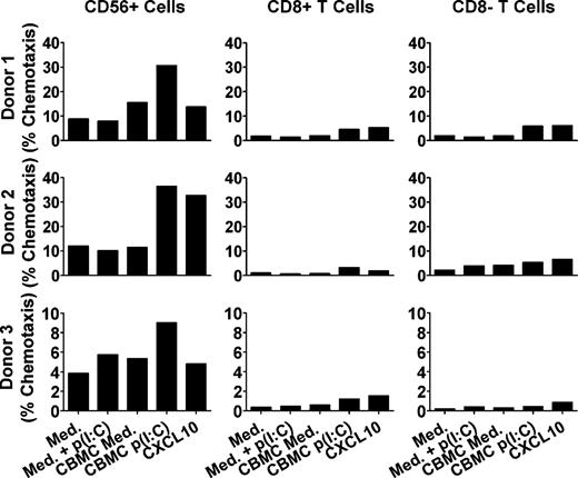
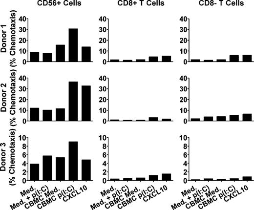
The effect of activation of mast cells with poly(I:C), a viral dsRNA analog, on the chemotaxis of various lymphocyte subsets was determined. CBMCs were incubated for 24 hours with or without poly(I:C), and the resultant supernatants were placed in the bottom chamber of chemotaxis wells with relevant controls in parallel wells. Freshly isolated, nylon wool–purified lymphocytes were placed in the top chamber and allowed to migrate for 2 hours, followed by immunofluorescence staining and flow cytometric analysis to examine the migrated cell subsets. CD8−, CD8+, and CD56+ lymphocytes migrated in response to poly(I:C)-activated CBMC supernatants, although the level of migration observed varied among lymphocyte populations and between donors (Figure 1). The CD56+ lymphocytes migrated much more to poly(I:C)-stimulated CBMC supernatants (mean: 25% ± 8% of the starting population; 3-fold over medium alone; P < .05; n = 3) compared with CD8+ T cells (3% ± 1%; P < .01; n = 4) and CD8− T cells (4% ± 1%; P < .05; n = 4). Due to the variable level of chemotaxis between different donors, future data sets are represented as mean fold increase in percentage chemotaxis over medium alone.
Supernatants from poly(I:C)-stimulated CBMCs induce CD56+ cell and T-cell migration. Supernatants from CBMCs incubated with poly(I:C) (p(I:C)) or medium (med) for 24 hours were placed in the bottom chambers of 24-well chemotaxis assays. CXCL10 alone (50 ng/mL) was used as a positive control, and medium containing poly(I:C) was used as a control for potential chemotactic/chemokinetic effects of the poly(I:C). Human nylon wool–purified peripheral blood lymphocytes were placed in the top chamber. After 2 hours, flow cytometry was performed to analyze the cell subsets migrating into the bottom chamber. Graphs show the mean percentage chemotaxis of CD56+ cells and T cells from each of 3 separate blood donors performed in duplicate, where each blood donor population migrated toward supernatants from different CBMC cultures (n = 3). Mean migration to poly(I:C)-CBMC supernatants: CD56+ cells, 25.4%; CD8+ T cells, 3.3%; and CD8− T cells, 4.3%.
Supernatants from poly(I:C)-stimulated CBMCs induce CD56+ cell and T-cell migration. Supernatants from CBMCs incubated with poly(I:C) (p(I:C)) or medium (med) for 24 hours were placed in the bottom chambers of 24-well chemotaxis assays. CXCL10 alone (50 ng/mL) was used as a positive control, and medium containing poly(I:C) was used as a control for potential chemotactic/chemokinetic effects of the poly(I:C). Human nylon wool–purified peripheral blood lymphocytes were placed in the top chamber. After 2 hours, flow cytometry was performed to analyze the cell subsets migrating into the bottom chamber. Graphs show the mean percentage chemotaxis of CD56+ cells and T cells from each of 3 separate blood donors performed in duplicate, where each blood donor population migrated toward supernatants from different CBMC cultures (n = 3). Mean migration to poly(I:C)-CBMC supernatants: CD56+ cells, 25.4%; CD8+ T cells, 3.3%; and CD8− T cells, 4.3%.
Chemokines produced by poly(I:C)-activated CBMCs
To examine the possible CD56+ lymphocyte chemoattractants produced by poly(I:C)-stimulated CBMCs, supernatants were analyzed for chemokine content by ELISA. The levels of CCL2 (16-fold), CCL4 (13.7-fold), CXCL2 (53.5-fold), CXCL8 (77.8-fold), and CXCL10 (2.5-fold) were all significantly increased at 24 hours compared with control (medium incubated) CBMCs (Table 1). This response was highly selective, with no evidence of CCL5, CXCL9, CXCL11, IFN-α, IL-6, or LTC4 up-regulation. The mast cell line HMC-1 was also examined for chemokine production in response to poly(I:C), which confirmed production of CXCL10 by human mast cells (medium, 22 ± 11 pg/mL; poly(I:C), 1788 ± 608 pg/mL; P < .05). Protein array analysis of 2 different poly(I:C)-treated CBMC cultures compared with control also revealed that CBMCs produced greater than 2-fold increases in CCL17, CCL23, and CXCL13 production, while a number of other cytokines and chemokines were unchanged (data not shown).
CD56+ lymphocyte migration occurs by a mechanism independent of CXCR3
To investigate the mechanism of CD56+ lymphocyte migration, the involvement of G protein–coupled receptors was first tested. MACS-isolated CD56+ human peripheral blood lymphocytes were treated with pertussis toxin, an inhibitor of Gα proteins, before being added to chemotaxis assays. Pertussis toxin completely inhibited migration to poly(I:C)-CBMC supernatants, indicating that the CD56+ lymphocyte migration was dependent on G protein–coupled receptors (Figure 2 top). Although the overall percentage migration of MACS-isolated CD56+ lymphocytes was lower than that seen with nylon wool–isolated lymphocytes, the fundamental findings were consistent and significant (figure legends).
CD56+ cell migration occurs independently of CXCR3. Radiolabeled MACS-isolated CD56+ cells were pretreated with either pertussis toxin (PTX; top) or an anti-CXCR3 (20 μg/mL) or isotype control mAb (bottom) preceding chemotaxis in 96-well chemotaxis assays to supernatants from poly(I:C) [p(I:C)]– or medium (med)–stimulated CBMCs for 2 hours. Poly(I:C) was added to medium alone to control for potential chemotactic/chemokinetic effects of the poly(I:C). The chemokine CXCL10 (200 ng/mL) was used as a positive control for anti-CXCR3 mAb inhibition of chemotaxis. Mean migration to poly(I:C)-CBMC supernatants: top, 12.9%; bottom, 8.2%. Graphs represent the mean (± SEM) of the fold increase in chemotaxis over medium of 3 to 8 separate experiments. Dashed line indicates baseline chemotaxis to medium only. *P < .05, **P < .01, compared with CBMC medium; †P < .05, ††P < .01, compared with no treatment.
CD56+ cell migration occurs independently of CXCR3. Radiolabeled MACS-isolated CD56+ cells were pretreated with either pertussis toxin (PTX; top) or an anti-CXCR3 (20 μg/mL) or isotype control mAb (bottom) preceding chemotaxis in 96-well chemotaxis assays to supernatants from poly(I:C) [p(I:C)]– or medium (med)–stimulated CBMCs for 2 hours. Poly(I:C) was added to medium alone to control for potential chemotactic/chemokinetic effects of the poly(I:C). The chemokine CXCL10 (200 ng/mL) was used as a positive control for anti-CXCR3 mAb inhibition of chemotaxis. Mean migration to poly(I:C)-CBMC supernatants: top, 12.9%; bottom, 8.2%. Graphs represent the mean (± SEM) of the fold increase in chemotaxis over medium of 3 to 8 separate experiments. Dashed line indicates baseline chemotaxis to medium only. *P < .05, **P < .01, compared with CBMC medium; †P < .05, ††P < .01, compared with no treatment.
NK cells express the G protein–coupled receptor CXCR3, and the CXCR3 ligand CXCL10 was up-regulated by poly(I:C)-stimulated CBMCs. However, incubation of CD56+ cells with a neutralizing anti-CXCR3 mAb did not significantly decrease CD56+ lymphocyte migration to poly(I:C)-activated CBMC supernatants, compared with isotype-matched control mAb treatment (Figure 2 bottom). In contrast, the anti-CXCR3 mAb inhibited 79% (± 8%) of CD56+ cell migration to CXCL10.
CD56+ lymphocytes also migrated strongly to the CXCR4 ligand CXCL12. However, pretreatment with a known blocking anti-CXCR4 mAb did not inhibit CD56+ lymphocyte migration to poly(I:C)-CBMC supernatants beyond the level of inhibition caused by a matched control mAb, while it decreased chemotaxis to the positive control CXCL12 by 84% (± 25%; data not shown).
CXCL8 is crucial for CD56+ lymphocyte migration to poly(I:C)-CBMC supernatants
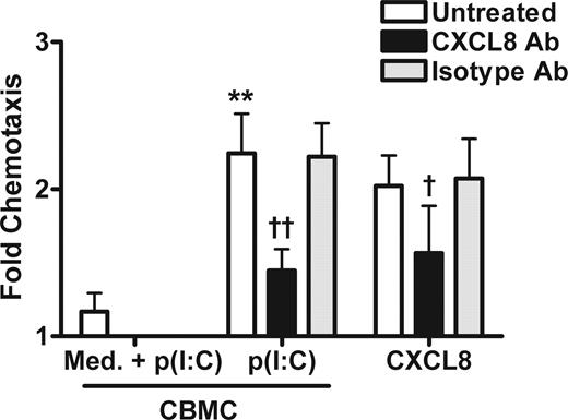
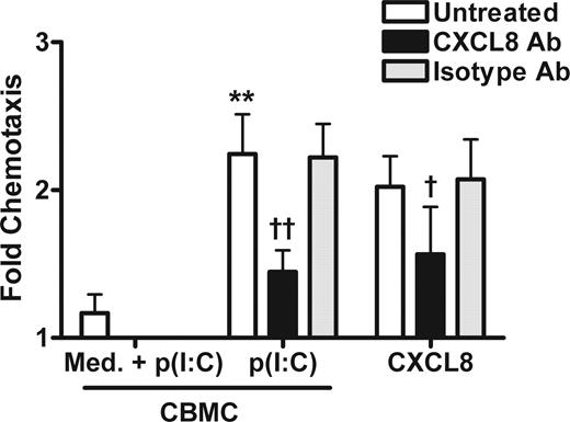
The chemokine CXCL8 has been described as a potential chemoattractant for NK cells,19,33 and there was a high level of this chemokine produced by CBMCs in response to poly(I:C). To test the involvement of CXCL8, poly(I:C)-CBMC supernatants were pretreated with a known blocking CXCL8 mAb at a concentration predicted to be effective based on the manufacturer's recommendations. Ab neutralization of CXCL8 in poly(I:C)-CBMC supernatants significantly inhibited the migration of MACS-isolated CD56+ lymphocytes by a mean of 65% (± 13%), compared with 0% (± 15%) decrease in chemotaxis in the presence of an isotype-matched control mAb (Figure 3). Pretreatment with a 10-fold higher concentration of the CXCL8 mAb caused a similar decrease (70% ± 4%; data not shown), suggesting that maximal inhibition was achieved. Anti-CXCL8 mAb treatment also inhibited CD56+ lymphocyte migration to human recombinant CXCL8 by 60% (± 24%).
CBMC-derived CXCL8 is crucial for CD56+ cell migration. Supernatants from poly(I:C) (p(I:C))– or medium (med)–stimulated CBMCs were incubated with anti-CXCL8 (0.4 μg/mL) or isotype control mAbs before being placed in the bottom chamber of 96-well chemotaxis assays. Poly(I:C) was added to medium alone to control for potential chemotactic/chemokinetic effects of the poly(I:C). Radiolabeled MACS-isolated CD56+ cells were placed in the top chamber, and chemotaxis was done for 2 hours. CXCL8 (30 ng/mL) was used as a positive control for mAb inhibition of chemotaxis. Mean migration to poly(I:C)-CBMC supernatants was 10.7%. Graphs represent the mean (± SEM) of the fold increase in chemotaxis over medium of 4 to 6 separate experiments. **P < .01 compared with CBMC medium; †P < .05, ††P < .01, compared with no treatment.
CBMC-derived CXCL8 is crucial for CD56+ cell migration. Supernatants from poly(I:C) (p(I:C))– or medium (med)–stimulated CBMCs were incubated with anti-CXCL8 (0.4 μg/mL) or isotype control mAbs before being placed in the bottom chamber of 96-well chemotaxis assays. Poly(I:C) was added to medium alone to control for potential chemotactic/chemokinetic effects of the poly(I:C). Radiolabeled MACS-isolated CD56+ cells were placed in the top chamber, and chemotaxis was done for 2 hours. CXCL8 (30 ng/mL) was used as a positive control for mAb inhibition of chemotaxis. Mean migration to poly(I:C)-CBMC supernatants was 10.7%. Graphs represent the mean (± SEM) of the fold increase in chemotaxis over medium of 4 to 6 separate experiments. **P < .01 compared with CBMC medium; †P < .05, ††P < .01, compared with no treatment.
NK cells but not CD56+ T cells migrate in a CXCL8-dependent manner
CD56 is expressed on both human NK cells and a small subset of T cells commonly referred to as CD56+ T cells. To determine which of these populations were participating in the observed CD56+ lymphocyte migration, human PBMCs were sorted by FACS into CD56+ CD3− NK cells and CD56+ CD3+ T cells. Chemotaxis assays revealed that the purified CD56+ CD3− NK cell population migrated strongly to poly(I:C)-activated CBMC supernatants (15% ± 1% chemotaxis; n = 6; Figure 4 top), and to 15 ng/mL CXCL8 alone (12% ± 1% chemotaxis; n = 2). NK cell migration to CBMC-poly(I:C) supernatants was decreased by 65% (± 6%) upon treatment of supernatants with an anti-CXCL8 mAb, compared with 4% (± 10%) inhibition with isotype-matched control mAb treatment. NK cell migration to recombinant CCL2 (2 ng/mL, 9.3% ± 3.2%) or CCL4 (0.4 ng/mL, 9.9% ± 2.0%) in the concentration range detected in CBMC-poly(I:C) supernatants was variable compared with medium control (5.3% ± 2.5%), with little dependence on dose (data not shown). Chemokinesis experiments were also performed with CBMC supernatants in both the top and bottom chambers, where NK cells migrated when CBMC-poly(I:C) supernatants were present in the bottom chamber only, indicating this was a chemotactic response (data not shown).
NK cells but not CD56+ T cells migrate in a CXCL8-dependent manner. Supernatants from CBMCs stimulated with poly(I:C) [p(I:C)] or medium control (med) were treated with an anti-CXCL8 or isotype control mAb previous to placement in the bottom chamber of 96-well chemotaxis assays. Poly(I:C) was added to medium alone to control for potential chemotactic/chemokinetic effects of the poly(I:C). FACS-isolated CD56+ CD3− NK cells (top) or CD56+ CD3+ T cells (bottom) were placed in the top chamber. CXCL8 (30 ng/mL) was used as a positive control for anti-CXCL8 mAb inhibition of chemotaxis during the 2-hour migration. Mean migration to poly(I:C)-CBMC supernatants: NK cells, 14.8%; CD56+ T cells, 16%. Graphs represent the mean (± SEM) of the fold increase in chemotaxis over medium of 2 to 6 separate experiments, except for CD56+ CD3+ T-cell migration to CXCL8 (n = 1). ***P < .001 compared with CBMC medium; †††P < .001 compared with no treatment.
NK cells but not CD56+ T cells migrate in a CXCL8-dependent manner. Supernatants from CBMCs stimulated with poly(I:C) [p(I:C)] or medium control (med) were treated with an anti-CXCL8 or isotype control mAb previous to placement in the bottom chamber of 96-well chemotaxis assays. Poly(I:C) was added to medium alone to control for potential chemotactic/chemokinetic effects of the poly(I:C). FACS-isolated CD56+ CD3− NK cells (top) or CD56+ CD3+ T cells (bottom) were placed in the top chamber. CXCL8 (30 ng/mL) was used as a positive control for anti-CXCL8 mAb inhibition of chemotaxis during the 2-hour migration. Mean migration to poly(I:C)-CBMC supernatants: NK cells, 14.8%; CD56+ T cells, 16%. Graphs represent the mean (± SEM) of the fold increase in chemotaxis over medium of 2 to 6 separate experiments, except for CD56+ CD3+ T-cell migration to CXCL8 (n = 1). ***P < .001 compared with CBMC medium; †††P < .001 compared with no treatment.
Despite an overall high percentage chemotaxis observed with CD56+ CD3+ T cells, this was not significantly increased with poly(I:C) activation of CBMCs compared with CBMC-medium control (P = .050), and was not inhibited by anti-CXCL8 treatment (Figure 4 bottom). These results suggest that NK cells were responsible for the majority of the selective CXCL8-driven chemotaxis observed in Figure 3.
Mammalian reovirus infects CBMCs and induces CXCL8 production
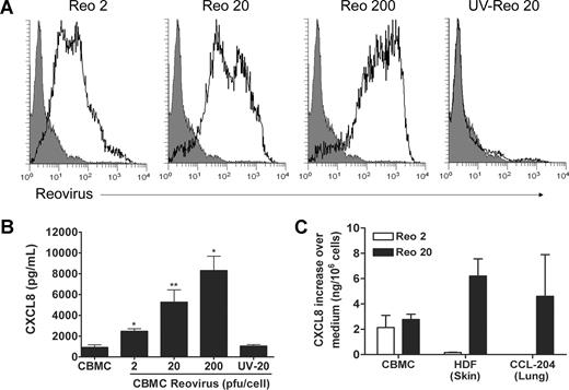
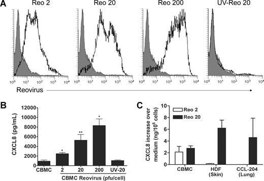
To evaluate the consequences of mast cell exposure to a virus that induces a potent and effective immune response, CBMCs were infected with mammalian reovirus serotype 3 Dearing for 24 hours at 2, 20, and 200 plaque forming units (pfu)/cell MOI. Intracellular immunofluorescence staining of the infected cells revealed a viral dose-dependent increase in staining for viral proteins (Figure 5). UV-inactivated reovirus used at an MOI of 20 did not induce detectable viral protein expression when analyzed under similar conditions. Plaque assay analysis confirmed UV inactivation of the virus (data not shown). Infected CBMCs produced substantial amounts of CXCL8 in a virus dose-dependent manner in response to all MOIs (Figure 5). UV-inactivated virus treatment of CBMCs resulted in CXCL8 production in 5 of 6 cultures tested, however the mean level of CXCL8 was lower than that seen with replication competent virus, and was not statistically significant. Viability analysis using 7-AAD revealed there was no effect on mast cell viability at 24 hours (data not shown). Based on the level of CXCL8 production and evidence of viral infection, an MOI of 20 pfu/mast cell was chosen for most future experiments.
Mammalian reovirus infects CBMCs and induces CXCL8 production. CBMCs were infected with mammalian reovirus type 3 Dearing at an MOI of 2, 20, or 200 pfu/cell, or with UV-inactivated reovirus at an MOI of 20 pfu/cell for 24 hours. (A) CBMCs were stained with an antireovirus Ab by immunofluorescence and detected by flow cytometry after exposure to reovirus (thick black line) or medium control (filled gray). Histograms are representative of 2 to 3 experiments using different CBMC cultures. (B) Supernatants from these infections were also analyzed for CXCL8 content by ELISA. Bar graph shows mean (± SEM) of 3 to 9 experiments. *P < .05, **P < .05, compared with CBMC medium. (C) Human primary HDFs, and CCL-204 lung fibroblasts were grown to confluence in 6-well plates. These, along with CBMCs, were treated with medium control or with reovirus at an MOI of 2 or 20 pfu/cell for 24 hours under similar FBS conditions. CXCL8 content was measured in the supernatants by ELISA. Graphs show the mean (± SEM; ng/106 cells) of the increase in CXCL8 content over medium control of 3 to 6 experiments. CCL-204 did not produce CXCL8 above medium control after treatment with 2 MOI of reovirus.
Mammalian reovirus infects CBMCs and induces CXCL8 production. CBMCs were infected with mammalian reovirus type 3 Dearing at an MOI of 2, 20, or 200 pfu/cell, or with UV-inactivated reovirus at an MOI of 20 pfu/cell for 24 hours. (A) CBMCs were stained with an antireovirus Ab by immunofluorescence and detected by flow cytometry after exposure to reovirus (thick black line) or medium control (filled gray). Histograms are representative of 2 to 3 experiments using different CBMC cultures. (B) Supernatants from these infections were also analyzed for CXCL8 content by ELISA. Bar graph shows mean (± SEM) of 3 to 9 experiments. *P < .05, **P < .05, compared with CBMC medium. (C) Human primary HDFs, and CCL-204 lung fibroblasts were grown to confluence in 6-well plates. These, along with CBMCs, were treated with medium control or with reovirus at an MOI of 2 or 20 pfu/cell for 24 hours under similar FBS conditions. CXCL8 content was measured in the supernatants by ELISA. Graphs show the mean (± SEM; ng/106 cells) of the increase in CXCL8 content over medium control of 3 to 6 experiments. CCL-204 did not produce CXCL8 above medium control after treatment with 2 MOI of reovirus.
Comparison of CXCL8 production by structural cells and CBMCs in response to poly(I:C) and reovirus
Primary cultures of human keratinocytes, dermal fibroblasts (HDFs), A549 airways epithelial cells, and CCL-204 lung fibroblasts were treated with poly(I:C) under the same conditions used for CBMCs. Poly(I:C) stimulation of CBMCs induced a sustained CXCL8 response at 6 and 24 hours (Table 2). Keratinocytes and HDFs produced similar large amounts after 24 hours of poly(I:C) stimulation. However, neither CCL-204 nor A549 cells produced substantial CXCL8 in response to poly(I:C) at any time point, and HDFs had little response at 6 hours.
Since reovirus induces a highly lytic infection in epithelial cells,34 fibroblasts were examined for reovirus-induced CXCL8 production. Stimulation with reovirus for 24 hours revealed that at higher MOI of 20, all cells produced similar levels of CXCL8 (Figure 5). Notably, only CBMCs but not lung or airways fibroblasts produced CXCL8 in response to 2 MOI of reovirus. Little to no production of CXCL8 occurred at 6 hours after reovirus treatment with any cells tested (data not shown).
Supernatants from reovirus-infected CBMCs induce the migration of CD56+ NK cells
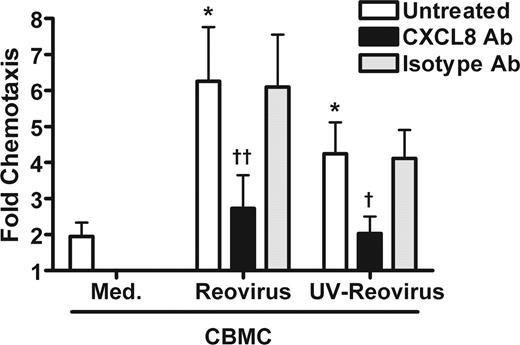
Chemotaxis assays were performed using FACS-isolated CD56+ CD3− NK cells and supernatants from reovirus-infected CBMCs. Significant NK cell chemotaxis was induced by supernatants from CBMCs exposed to both replication competent and UV-inactivated reovirus, with lower migration occurring with the latter (Figure 6). Chemotaxis of NK cells to supernatants from both replication competent and UV-inactivated reovirus-treated mast cells was inhibited by pretreatment of the supernatants with a neutralizing anti-CXCL8 mAb (CBMC-reovirus, 71% ± 7% inhibition; CBMC-UV-reovirus, 55% ± 23% inhibition), while little inhibition occurred upon treatment with an isotype-matched control mAb (CBMC-reovirus, 4% ± 1% inhibition; CBMC-UV-reovirus, 0% ± 11% inhibition; Figure 6).
Supernatants from reovirus-infected CBMCs induce CXCL8-dependent NK cell migration. CBMCs were infected with reovirus or UV-inactivated reovirus, or with medium (med) as a negative control for 24 hours. The resultant supernatants were left untreated, or treated with an anti-CXCL8 (4 μg/mL) or isotype control mAb before being placed in the bottom chamber of 96-well chemotaxis assays. FACS-isolated CD56+ CD3− NK cells were placed in the top chamber, and chemotaxis was assessed after 2 hours. Mean migration to supernatants: reovirus, 17.2%; UV-reovirus, 12.3%. Graph shows the mean (± SEM) of the fold increase in chemotaxis over medium of 4 experiments using different CBMC cultures. *P < .05 compared with CBMC medium; †P < .05, ††P < .01, compared with no treatment.
Supernatants from reovirus-infected CBMCs induce CXCL8-dependent NK cell migration. CBMCs were infected with reovirus or UV-inactivated reovirus, or with medium (med) as a negative control for 24 hours. The resultant supernatants were left untreated, or treated with an anti-CXCL8 (4 μg/mL) or isotype control mAb before being placed in the bottom chamber of 96-well chemotaxis assays. FACS-isolated CD56+ CD3− NK cells were placed in the top chamber, and chemotaxis was assessed after 2 hours. Mean migration to supernatants: reovirus, 17.2%; UV-reovirus, 12.3%. Graph shows the mean (± SEM) of the fold increase in chemotaxis over medium of 4 experiments using different CBMC cultures. *P < .05 compared with CBMC medium; †P < .05, ††P < .01, compared with no treatment.
NK cell migration to reovirus-CBMC supernatants is highly dependent on CXCR1
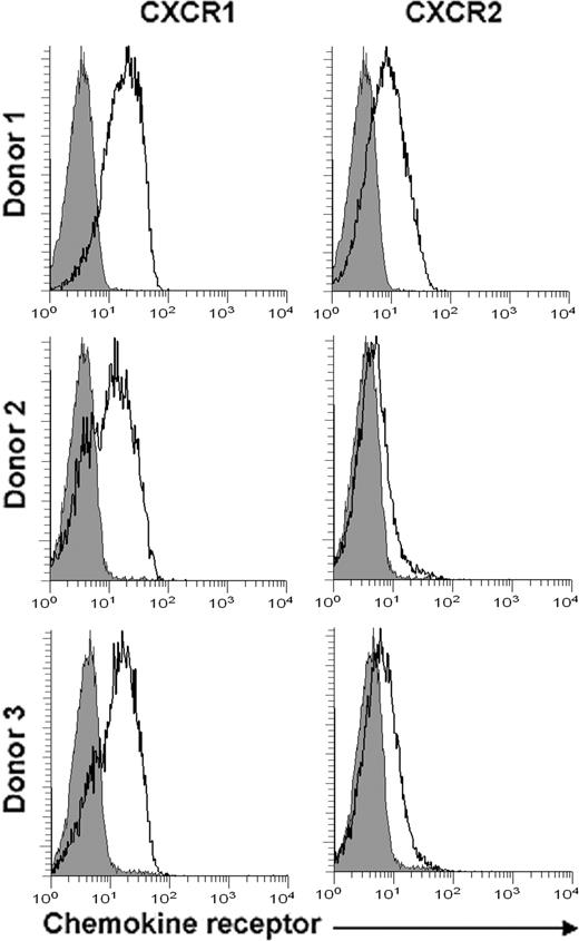
To examine which of the CXCL8 receptors was important primarily for the NK cell migration to reovirus-CBMC supernatants, we examined the expression of CXCR1 and CXCR2 on freshly isolated PBMCs. CXCR1 was expressed on a mean of 64% (± 7%) of NK cells, with all 3 blood donors showing strong expression (Figure 7 left panels). In contrast, only 24% (± 9%) of all NK cells expressed CXCR2, with considerable variability among donors (Figure 7 right panels). To test their role in migration, CXCR1 and/or CXCR2 on NK cells were inhibited by neutralizing mAb cell treatments prior to use in chemotaxis assays. Anti-CXCR1 mAb treatment of NK cells decreased total chemotaxis to reovirus-CBMC supernatants by 61% (± 3%) and to UV-reovirus-CBMC supernatants by 49% (± 7%), compared with 12% (± 5%) and 10% (± 3%) inhibition by isotype control mAb, respectively (Table 3). Reovirus-increased chemotaxis was inhibited by 105% (± 11%) and UV-reovirus–increased chemotaxis was inhibited by 96% (± 3%) with anti-CXCR1 mAb treatment of NK cells. Treatment of NK cells with a combination of both anti-CXCR1 and anti-CXCR2 Abs (reovirus-CBMCs, 52% ± 5%; UV-reovirus-CBMCs, 46% ± 13%) did not inhibit NK cell migration beyond that of CXCR1 blockade alone. Blockade of CXCR2 alone resulted in only 28% (± 7%) and 22% (± 1%) of migration to reovirus and UV-reovirus supernatants, respectively (data not shown). These data indicate that CXCR1 is the most critical receptor for NK cell chemotaxis in response to viral infection of mast cells.
NK cells express more CXCR1 than CXCR2. CXCR1 and CXCR2 expression was determined on untreated freshly isolated human PBMCs from 3 separate blood donors by immunofluorescence with gating on CD56+ CD3− cells. Histograms show isotype control (filled gray) or CXCR1 or CXCR2 staining (thick black line), as indicated.
NK cells express more CXCR1 than CXCR2. CXCR1 and CXCR2 expression was determined on untreated freshly isolated human PBMCs from 3 separate blood donors by immunofluorescence with gating on CD56+ CD3− cells. Histograms show isotype control (filled gray) or CXCR1 or CXCR2 staining (thick black line), as indicated.
Discussion
We have demonstrated that human mast cells respond to stimulation with the dsRNA analog poly(I:C) by selectively producing several chemokines, including high levels of CXCL8. This study also demonstrated that mammalian reovirus, an RNA virus that is normally effectively controlled by the immune response, can infect human mast cells and induce the production of large amounts of CXCL8. Both of these CXCL8 responses are sufficient to induce the chemotaxis of CD56+ NK cells, and NK cell–expressed CXCR1 plays a major role in this response.
We used several methods to isolate NK cells in this study and in each case substantial and consistent NK cell migration was observed to poly(I:C)-activated mast cell supernatants, although the overall percentage of cells migrating varied. This is likely due to the increased manipulation during isolation, as the CD56+ cells partially purified by nylon wool column and identified after assay by flow cytometry migrated most strongly (Figure 1), compared with the CD56+ lymphocytes isolated by MACS and the sorted NK cells isolated by FACS.
This is the first study to demonstrate reovirus infection of human mast cells. Mammalian reovirus can infect many cell types, including macrophages,35 fibroblasts,34 and epithelial cells.36 Reovirus can also induce CXCL8 production in monocytes, although in our experiments human mast cells produced 5-fold higher CXCL8 levels when infected with reovirus than reported for monocytes.37 Our experiments revealed that fibroblasts can produce CXCL8 in response to reovirus. All cell types examined produced CXCL8 in response to higher doses of reovirus (20 MOI). However, treatment with reovirus at 2 MOI induced CXCL8 selectively in CBMCs compared with skin or airways fibroblast populations, suggesting a role for CBMCs early in infection before multiple rounds of viral replication have taken place. Considering their close proximity to blood vessels in the mucosa, mast cells are well equipped to recruit NK cells when low levels of virus are present early in infection. Other local cell populations may assist in such effector cell recruitment at later stages of infection.
CXCL8 production by CBMCs stimulated with poly(I:C) was at similar levels per cell to that of skin-derived fibroblasts or keratinocytes, suggesting that within the skin, mast cells are not the only major source of this chemokine in response to exogenous dsRNA. However, airway-derived epithelial cells and fibroblasts had only minimal CXCL8 responses compared with CBMCs. Mast cells may potentially be a much more critical source of this chemokine and inducer of NK cell migration within the context of viral infection in the airways than the skin.
Our study shows that the profile of mediators produced by mast cells in response to poly(I:C) activation is distinct from that induced by stimuli such as Escherichia coli seen in other studies.38 For example, E coli exposure of human mast cells induces the production of CCL1, CCL2, CCL18, and CCL23,38 compared with the production of CCL2, CCL23, CXCL8, and CXCL10 in response to poly(I:C) as demonstrated in this study. CXCL8 production by human mast cells stimulated via IgE receptor cross-linking has been demonstrated by some,39,40 yet refuted by others.38 In this context, our data support the concept of pathogen-type specific chemokine responses by human mast cells, perhaps allowing an early selective recruitment of effector cells appropriate for eradicating the particular pathogen. It should be noted that mast cells can secrete PGD2, which can inhibit NK cell chemotaxis, cytokine production, and cytotoxicity, and could be produced during in vivo viral infection.41 However, to date mast cells have not been reported to produce this mediator as a consequence of exposure to viral products or active viral infection.
We chose reovirus for this study because it contains a dsRNA genome, which provided an appropriate model to determine the effects of dsRNA stimulation. Human mast cells have previously been shown to express TLR3, a receptor for poly(I:C) and dsRNA.10 UV inactivation is known to cross-link the nucleic acid component of the virus. Although production of CXCL8 was higher in mast cells infected with replication competent reovirus, it was also produced by 5 of 6 mast cell cultures exposed to UV-inactivated reovirus, indicating that while dsRNA may contribute to the response, reovirus can also induce CXCL8 production by a mechanism independent of de novo synthesized nucleic acids. Such replication-independent CXCL8 production has also been observed in human fibroblasts.42 The literature suggests that the reovirus genome is protected by core viral proteins, implying the genomic nucleic acids may not be capable of inducing a host cellular response.43 Therefore, the mechanism by which reovirus induces CXCL8 production in human mast cells may be more complex than with poly(I:C). Possible mechanisms of CXCL8 induction include the recognition of virus proteins, such as the cell attachment protein sigma 1.
Reovirus also has oncolytic properties. Because mast cells have been found at higher densities surrounding solid tumors, it may be possible that administration of reovirus at the tumor site can induce mast cell chemokine production and NK cell recruitment, leading to tumor cytolysis. In addition to the direct oncolytic properties of reovirus, this outlines a second possible mechanism by which reovirus can eliminate solid tumors.
The results in this study suggest a mechanism by which mast cells could provide the chemotactic stimulus necessary to induce NK cell recruitment to sites of viral infection, and this is the first study to do so. It is important to mobilize innate effector cells such as NK cells early during viral infection, as they limit replication while acquired immune effectors develop. While transendothelial migration studies would be useful to confirm our chemotaxis experiments, such studies would be compromised by histocompatibility differences between donor NK cells and human umbilical vein endothelial cells.
Virally stimulated plasmacytoid DCs have previously been shown to recruit NK cells, however in contrast to our current findings this recruitment was dependent on CCL4 and CXCL10.28 Although chemokines such as CXCL10 are classic NK cell chemoattractants, there is also evidence to support the ability of CXCL8 to recruit this cell type.19,33 NK cells have been suggested to express the CXCL8 receptors CXCR1 and CXCR2,17,19,21 and our studies confirmed these data, although there is some controversy in this area.20 Since CXCL8 is also a powerful neutrophil chemoattractant, our data suggest a possible role for mast cells in the concurrent recruitment of neutrophils that may contribute to the immune response to viral infection.44,45 Previous studies have demonstrated that reovirus serotype 3 Dearing can induce neutrophilia in the lungs of infected mice.46
Previously, Orinska et al have demonstrated the ability of mast cells to participate in the recruitment of CD8+ T cells in an in vivo mouse model of poly(I:C) stimulation in the peritoneum.47 Such T-cell recruitment required TLR3 activation. Our experiments showed that only a low percentage of CD8+ T cells underwent chemotaxis in response to poly(I:C)-activated human mast cell supernatants, in contrast to selective chemotaxis of a high percentage of NK cells. This underlines the potential differences between mice and humans, and perhaps between in vivo and in vitro systems, in mast cell–mediated cell recruitment.
Mast cells have also been shown to recruit multiple other effector cell types to inflamed tissues, including blood monocytes and granulocytes during infection or in models of allergic disease.48,49 Notably, supernatants obtained from mast cells activated with poly(I:C) induced substantial chemotaxis of NK cells, compared with CD56+ CD3+ T cells. While this response failed to reach statistical significance, the trend observed may suggest a role for mast cells in the recruitment of these cells in vivo.
Mast cells are located in a prime location to respond quickly to invading pathogens. Mast cells remain relatively unaffected by radiation and are able to maintain cell numbers3 and chemokine production, as demonstrated by UVB-induced CXCL8 production by human CBMCs,4 and poly(I:C) induced IFN-α after gamma-irradiation of mouse mast cells.44 Therefore, after irradiation, mast cells may be one of few cell types that are functionally intact. This radioresistance enables them to remain capable of putting forth inflammatory signals necessary for innate effector cell recruitment should viral infection occur. Mast cell promotion of NK cell chemotaxis extends the recognized ability of mast cells to serve as sentinel cells for recruitment of effector cells in bacterial and nematode parasite infection to include NK cell recruitment in viral infections. This further underscores the ability of local mast cells to serve as sentinel cells for a variety of different types of pathogen challenge.
The publication costs of this article were defrayed in part by page charge payment. Therefore, and solely to indicate this fact, this article is hereby marked “advertisement” in accordance with 18 USC section 1734.
Acknowledgments
The authors thank Yi-Song Wei for her excellent technical assistance.
This work was supported by operating grants from the Canadian Institutes for Health Research (Ottawa, ON) and the National Cancer Institute of Canada (Toronto, ON). S.M.B. was supported by student research awards from the Nova Scotia Health Research Foundation (Halifax, NS) and the Cancer Research Training Program (Halifax, NS).
Authorship
Contribution: S.M.B. performed the research and drafted the paper; J.S.M. was instrumental in designing the research and drafting the paper; T.B.I. participated in designing the research, contributed vital reagents, and revised the paper; P.W.K.L. participated in designing the research and contributed vital reagents; M.S. participated in performing the reovirus experiments and revised the paper; K.M. participated in designing and performing the chemotaxis experiments and revising the paper.
Conflict-of-interest disclosure: The authors declare no competing financial interests.
Correspondence: Jean S. Marshall, Room 7C, Dept of Microbiology and Immunology, Dalhousie University, 5850 College Street, Halifax, Nova Scotia, Canada, B3H 1X5; e-mail: jean.marshall@dal.ca.

![Figure 2. CD56+ cell migration occurs independently of CXCR3. Radiolabeled MACS-isolated CD56+ cells were pretreated with either pertussis toxin (PTX; top) or an anti-CXCR3 (20 μg/mL) or isotype control mAb (bottom) preceding chemotaxis in 96-well chemotaxis assays to supernatants from poly(I:C) [p(I:C)]– or medium (med)–stimulated CBMCs for 2 hours. Poly(I:C) was added to medium alone to control for potential chemotactic/chemokinetic effects of the poly(I:C). The chemokine CXCL10 (200 ng/mL) was used as a positive control for anti-CXCR3 mAb inhibition of chemotaxis. Mean migration to poly(I:C)-CBMC supernatants: top, 12.9%; bottom, 8.2%. Graphs represent the mean (± SEM) of the fold increase in chemotaxis over medium of 3 to 8 separate experiments. Dashed line indicates baseline chemotaxis to medium only. *P < .05, **P < .01, compared with CBMC medium; †P < .05, ††P < .01, compared with no treatment.](https://ash.silverchair-cdn.com/ash/content_public/journal/blood/111/12/10.1182_blood-2007-10-118547/6/m_zh80130820730002.jpeg?Expires=1769445295&Signature=EdUFsSyqX7NVjOm7zu8T6THCA0NymXWNZkYOzMaRyJnld-j~NAf~JA3peI0zpZ9ncNcTc9imeTD~tBBaGLSkQxyzc1RNpFZm01M8Wq2mSA2ZHtx01V0v1k3duHKymSHMYagICoawOpQBeU6DIOg-ZHJoTIIAZKDYMlp0BNy1YmV3Y0nQxeRb23FAAHTa9ZFLaJ8~7LFw465nQkkeInxtRvKxJUj3LVCiEDSVsBfnybzHatx1CuxCD-HlvTCPXVWmY8LHarMuEQkWT~y~zbziB09YynOLV7A4aRhxeifRZOQdfrOMAwSRg6UfHLI1SSXko-zuZQgujfZb5PRxYAljUA__&Key-Pair-Id=APKAIE5G5CRDK6RD3PGA)
![Figure 4. NK cells but not CD56+ T cells migrate in a CXCL8-dependent manner. Supernatants from CBMCs stimulated with poly(I:C) [p(I:C)] or medium control (med) were treated with an anti-CXCL8 or isotype control mAb previous to placement in the bottom chamber of 96-well chemotaxis assays. Poly(I:C) was added to medium alone to control for potential chemotactic/chemokinetic effects of the poly(I:C). FACS-isolated CD56+ CD3− NK cells (top) or CD56+ CD3+ T cells (bottom) were placed in the top chamber. CXCL8 (30 ng/mL) was used as a positive control for anti-CXCL8 mAb inhibition of chemotaxis during the 2-hour migration. Mean migration to poly(I:C)-CBMC supernatants: NK cells, 14.8%; CD56+ T cells, 16%. Graphs represent the mean (± SEM) of the fold increase in chemotaxis over medium of 2 to 6 separate experiments, except for CD56+ CD3+ T-cell migration to CXCL8 (n = 1). ***P < .001 compared with CBMC medium; †††P < .001 compared with no treatment.](https://ash.silverchair-cdn.com/ash/content_public/journal/blood/111/12/10.1182_blood-2007-10-118547/6/m_zh80130820730004.jpeg?Expires=1769445295&Signature=uaXhuMDTAzkKHD2CX4uD9WGtS3f~6U2sXQ8HV9OIh7Ay5RxRromHT2JUH2PD4Dj9fw-w7WEIgmf8wIAVC5MP9ENT4-51xu17L9US4HudNUOqmpjBa3MadO~B29vFtK2HkQYvzVu7JORosNbGskeC6ogy1EJIrc~cFCfo1iLJaucGj4e4HIqtIo-Huv3~TnEX1Q49tAW4M5iCUotO0gW8do5ETsm5PqSw9bnat7PR6Zt8s~lOSakdsv8ex4ogetGEJkkDLOBsnsogCczRBmyfLAdvHlXFCQb2sZrlaudxchjEG0XKhHrlC191nRnsmrAXuylDbsEUcCQExgGOLYvJ0A__&Key-Pair-Id=APKAIE5G5CRDK6RD3PGA)

![Figure 2. CD56+ cell migration occurs independently of CXCR3. Radiolabeled MACS-isolated CD56+ cells were pretreated with either pertussis toxin (PTX; top) or an anti-CXCR3 (20 μg/mL) or isotype control mAb (bottom) preceding chemotaxis in 96-well chemotaxis assays to supernatants from poly(I:C) [p(I:C)]– or medium (med)–stimulated CBMCs for 2 hours. Poly(I:C) was added to medium alone to control for potential chemotactic/chemokinetic effects of the poly(I:C). The chemokine CXCL10 (200 ng/mL) was used as a positive control for anti-CXCR3 mAb inhibition of chemotaxis. Mean migration to poly(I:C)-CBMC supernatants: top, 12.9%; bottom, 8.2%. Graphs represent the mean (± SEM) of the fold increase in chemotaxis over medium of 3 to 8 separate experiments. Dashed line indicates baseline chemotaxis to medium only. *P < .05, **P < .01, compared with CBMC medium; †P < .05, ††P < .01, compared with no treatment.](https://ash.silverchair-cdn.com/ash/content_public/journal/blood/111/12/10.1182_blood-2007-10-118547/6/m_zh80130820730002.jpeg?Expires=1769445296&Signature=NG2dnds28o1sOpxgKhT7-54LquCpWv3CRxBSU8Xlb0fdwa6Pa8u2DvWQUMw7dPR2CitlDnU9qw~TA3tia78Edu05xbUxTOK1G-Gj8-zrxNGYRIh0WWheT~ndsIAy5JvGFyd50UO9l4pdwwY4pThcFkBRSovyHMUSxjTWRcw9eeuhMe2iKgPSIZzolyKVra9DOFmYgd9xbF4lkdgfGwnGlonlCslmrh-hha1l8b3MgAarSx7diKQxQ7smId5wbTgnwsJBnvMNp2yxgGpTZQWVhoNRSwvNLFFh8bZsdhi-1SCn5QIZY58vEJUS0PzJ185JgDVG~gMdkburE-jsbJHb8w__&Key-Pair-Id=APKAIE5G5CRDK6RD3PGA)
![Figure 4. NK cells but not CD56+ T cells migrate in a CXCL8-dependent manner. Supernatants from CBMCs stimulated with poly(I:C) [p(I:C)] or medium control (med) were treated with an anti-CXCL8 or isotype control mAb previous to placement in the bottom chamber of 96-well chemotaxis assays. Poly(I:C) was added to medium alone to control for potential chemotactic/chemokinetic effects of the poly(I:C). FACS-isolated CD56+ CD3− NK cells (top) or CD56+ CD3+ T cells (bottom) were placed in the top chamber. CXCL8 (30 ng/mL) was used as a positive control for anti-CXCL8 mAb inhibition of chemotaxis during the 2-hour migration. Mean migration to poly(I:C)-CBMC supernatants: NK cells, 14.8%; CD56+ T cells, 16%. Graphs represent the mean (± SEM) of the fold increase in chemotaxis over medium of 2 to 6 separate experiments, except for CD56+ CD3+ T-cell migration to CXCL8 (n = 1). ***P < .001 compared with CBMC medium; †††P < .001 compared with no treatment.](https://ash.silverchair-cdn.com/ash/content_public/journal/blood/111/12/10.1182_blood-2007-10-118547/6/m_zh80130820730004.jpeg?Expires=1769445296&Signature=NMmLDRYqd63Zv1811eoVsDFRZCbDSHxcZ-M72iouG3Tlcr24OYJaxG44M7oOETymdujXp1j7V8NdUL-277O1hQzvYlb93gtMNVCT-uylohSt3ADI1DcB2EB3SGfO64Z1WS3i70SSeyDzSx3NJqphq8NCfOw2BMteCm622-L-ZI75NaqURSXYJEdXobu7bMMypzx5allQ4CODOz~hyQ2c2RFbPs1G6lTuYYMf1MJ4CeM1jjJoDnQgqcGD1tkOGLsiYFLmq3pQ0satexGdgWhoLTtdGNefxZ-G418fa05w87GiMdgiZbm4g1hXeyIikF~GYgzanPh5D0IQ7TwhMDRHoQ__&Key-Pair-Id=APKAIE5G5CRDK6RD3PGA)
