Abstract
Patients with thalassemia major develop life threatening cardiac complications in their teens and twenties from iron overload. Cardiac MRI allows us to diagnosis preclinical cardiac iron deposition, but it is not known at what age T2* screening should be initiated. Historical data and pilot MRI studies suggest that patients must reach a critical transfusional exposure prior to cardiac iron uptake. This study was a two-institution characterization of the prevalence of cardiac iron overload in 77 pediatric patients.
Methods: Study was performed at the Ospedale Regionale Microcitemie in Cagliari and the Childrens Hospital Los Angeles (CHLA). Retrospective review of medical records was authorized by the IRB for pediatric patients undergoing cardiac MRI prior to July of 2007. Subjects at both institutions had assessment of cardiac T2* and cardiac function using validated techniques on a 1.5 T General Electric CVi scanner; patients at CHLA also underwent MRI-based liver iron measurements. Complete transfusional iron burden was measured in the Italian patients. Serial MRI data was recorded in 30 patients, but associations between cardiac T2* and age were determined using linear and logistic regression using only results from the first patient MRI scan.
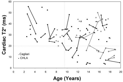
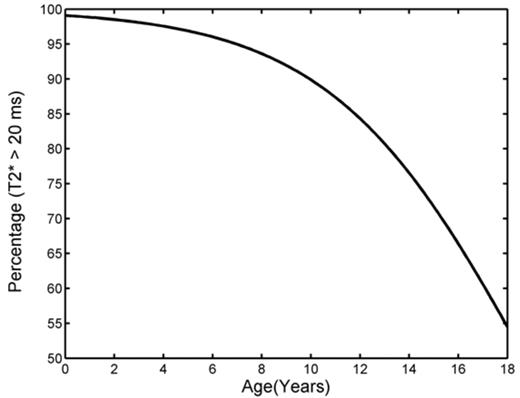
Results: Patient ages ranged from 8.0 to 18 years of age at Cagliari (n=36) and 2.5 to 17.9 years of age at CHLA (n=41), reflecting the use of MRI to monitor liver iron in patients < 8 years of age. Patients were moderately iron loaded with hepatic iron concentrations of 12.7 ± 9.8 (CHLA) and ferritin values of 2329 ± 1162 (Cagliari). Median cardiac T2* was 30.2 and ranged from 3.4 to 72.8. Cardiac T2* and its reciprocal were uncorrelated with liver iron (CHLA) and ferritin levels (Cagliari). Figure 1 demonstrates the decrease in cardiac T2* with increasing chronologic age. Serial data are connected by lines. Although linear regression was weakly positive (r2 = 0.06, p = 0.03), the relationship was fundamentally nonlinear. No patient below the age of 9.5 years of age demonstrated an abnormal cardiac T2* (< 20 ms) while 36% of patients between the ages of 15–18 years had detectable cardiac iron. Figure 2 demonstrates the logistic regression curve modeling the prevalence of detectable cardiac iron as a function of age (r2= 0.13, p< 0.002). Chronologic age was highly correlated (r2 = 0.88) with both transfusional iron burden and with duration of transfusion therapy, making it impossible to separate the relative importance of these three variables.
Conclusion: Thalassemia major patients did not accumulate cardiac iron until early in their second decade of life. Consequently, cardiac iron monitoring can be safely deferred until children are able undergo MRI examination without sedation.
Author notes
Disclosure:Consultancy: Dr. Wood and Dr. Coates have served as consultants to Novartis. Research Funding: Drs. Wood, Coates, and Galanello have received research funding from Novartis and Apotex. Honoraria Information: Drs. Wood, Coates, and Galanello have received honoraria from Novartis and Apotex. Membership Information: Dr. Wood is an advisor to Exjade Speaker’s Bureau. Dr. Coates is a member of Exjade Speakers Bureau. Financial Information: Dr. Orias receives an academic fellowship from Novartis. Dr. Agus ireceives an academic fellowship by Apotex. Off Label Use: MRI assessment of cardiac T2* is not an FDA-approved procedure.


