TO THE EDITOR:
Multiple myeloma (MM) remains an incurable plasma cell (PC) malignancy.1,2 MM cells residing in the bone marrow (BM) engage in extensive crosstalk with the BM microenvironment. These interactions are leveraged by immunotherapies, but many patients acquire resistance, suffer dose-limiting toxicities, or remain unresponsive, highlighting an unmet need.
Natural killer (NK) cells demonstrate strong cytotoxicity against stressed cells, such as tumor cells and malignant PCs.3 The efficacy of some existing MM therapies, including immunomodulatory drugs and monoclonal antibodies (mAbs), rely on antibody-dependent cellular cytotoxicity (ADCC) driven by endogenous NK cells.4 Yet, MM often escapes NK cell–mediated cytotoxicity and induces NK cell dysfunction.4,5
Dysfunctional NK cells are a hallmark of MM disease6,7 with decreased cytotoxicity, reduced activating receptors expression (NKG2D,8,9 CD16, 2B4,10 and DNAM-111), and increased inhibitory receptors expression (KIR2DL112 and PD113). To overcome this, the adoptive transfer of ex vivo–activated or engineered autologous or allogeneic NK cells has been used.14 Cytokine-induced memory-like NK cells are of particular interest because they acquire memory-like features, including elevated activity upon restimulation, greater interferon gamma (IFN-γ) responses, and antitumor cytotoxicity.15,16 Memory-like NK cells safely induce clinical remission in adult and pediatric patients with acute myeloid leukemia17-19 but have not been studied in MM.
The successful adoptive transfer of memory-like NK cells in MM depends on the ability of the cells to recognize malignant PCs. NKG2D, a major NK cell–activating receptor, is upregulated in memory-like NK cells compared with that in conventional NK cells (cNK cells).17 NKG2D recognizes malignant PCs via its ligands major histocompatibility complex class I chain–related protein A (MICA) and major histocompatibility complex class I chain–related protein B (MICB).11,20,21 MM cells evade recognition by proteolytically shedding surface MICA/B, which leads to NKG2D internalization.9
The novel mAb 7C6 binds to the proteolytic MICA α3 domain, stabilizing surface MICA/B and enhancing NK cell–mediated ADCC.22,23 We hypothesized that 7C6 would simultaneously blockade of MICA/B shedding and increase ADCC by memory-like NK cells, resulting in enhanced MM cytotoxicity.
Previous studies have shown that surface MICA/B decreases and soluble MICA/B increases stepwise from monoclonal gammopathy of undetermined significance to overt MM due to shedding induced by the coordinated action of disulfide isomerase ERp5 and proteases, including a disintegrin and metalloproteinase domain-containing protein 10 (ADAM10) and ADAM17.9,24 We investigated the extent of surface MICA/B shedding in MM cells. MICA/B shedding involves ERp5 disulfide isomerase, which removes a disulfide bond from the α3 domain of MICA/B,25 allowing subsequent cleavage by proteases from the ADAM family.26 Therefore, MICA, MICB, ADAM10, and ERp5 transcript levels in human MM cell lines (HMCLs) were analyzed in the Cancer Cell Line Encyclopedia (CCLE) (21Q4).27MICB and PDIA6 (encoding ERp5) transcription were the highest in HMCLs among the 1389 tumor cell lines (Figure 1A); this was not the case for MICA (Figure 1A). In public single-cell sequencing data from tumors of patients with newly diagnosed MM,28PDIA6 transcription appeared higher in primary PCs from MM BM than in healthy control PCs (supplemental Figure 1A). Although MICA/B surface protein levels appeared to be significantly higher in primary PCs from patients with newly diagnosed multiple myeloma than in patients with monoclonal gammopathy of undetermined significance, it should be noted that it is difficult to maintain good viability of primary PCs ex vivo, which likely affects MICA/B shedding and thus surface MICA/B levels. Furthermore, the MICA and MICB transcript and protein levels in primary PCs were low, limiting our ability to interpret changes with disease progression (supplemental Figure 1B-C). Overall, prerequisites for proteolytic MICA/B cleavage were present in MM.
Inhibition of MICA/B shedding enhances memory-like NK cell–mediated cytotoxicity against MM. (A) MICA, MICB, PDIA6 (ERp5), and ADAM10 gene expression levels in 1389 tumor cell lines from the Cancer Cell Line Encyclopedia database. Gene expression levels are presented as log2(transcripts per million [TPM] + 1). (B) MICA/B surface expression in 10 different HMCLs, as measured by flow cytometry. (C) MICA/B gene expression levels in 9 different HMCLs from the publicly available Broad CCLE data set. (D) Dose-dependent increase in MICA/B surface expression in HMCL MM.1S in response to 7C6 treatment. Significance was calculated using the t test. (E) Shed MICB was determined using sandwich enzyme-linked immunosorbent assay. A decrease in soluble MICB was seen in response to the 7C6 mAb treatment. For functional assays, effector cells (conventional or memory-like NK cells) were cocultured with MM.1S at a 5:1 effector-to-target ratio for 6 hours. (F) Increased CD107a degranulation (n = 7), IFN-γ production (n = 10), and tumor necrosis factor-α (TNF-α) production (n = 7) measured by flow cytometry in response to 7C6 treatment of cNK cells. (G) Summary of cytotoxicity (n = 9) of cNK and memory-like NK cells upon 7C6 mAb treatment of MM.1S cells. Data are depicted as the mean ± standard deviation. Significance was calculated using the Wilcoxon test for comparison of IC and 7C6 treatment conditions, and the Mann-Whitney U test was used for the remainder of the comparisons; ns, P > .05; ∗P ≤ .05; ∗∗P ≤ .01; ∗∗∗P ≤ .001; ∗∗∗∗P ≤ .0001. E:T, effector:target; IC, isotype control; ns, not significant.
Inhibition of MICA/B shedding enhances memory-like NK cell–mediated cytotoxicity against MM. (A) MICA, MICB, PDIA6 (ERp5), and ADAM10 gene expression levels in 1389 tumor cell lines from the Cancer Cell Line Encyclopedia database. Gene expression levels are presented as log2(transcripts per million [TPM] + 1). (B) MICA/B surface expression in 10 different HMCLs, as measured by flow cytometry. (C) MICA/B gene expression levels in 9 different HMCLs from the publicly available Broad CCLE data set. (D) Dose-dependent increase in MICA/B surface expression in HMCL MM.1S in response to 7C6 treatment. Significance was calculated using the t test. (E) Shed MICB was determined using sandwich enzyme-linked immunosorbent assay. A decrease in soluble MICB was seen in response to the 7C6 mAb treatment. For functional assays, effector cells (conventional or memory-like NK cells) were cocultured with MM.1S at a 5:1 effector-to-target ratio for 6 hours. (F) Increased CD107a degranulation (n = 7), IFN-γ production (n = 10), and tumor necrosis factor-α (TNF-α) production (n = 7) measured by flow cytometry in response to 7C6 treatment of cNK cells. (G) Summary of cytotoxicity (n = 9) of cNK and memory-like NK cells upon 7C6 mAb treatment of MM.1S cells. Data are depicted as the mean ± standard deviation. Significance was calculated using the Wilcoxon test for comparison of IC and 7C6 treatment conditions, and the Mann-Whitney U test was used for the remainder of the comparisons; ns, P > .05; ∗P ≤ .05; ∗∗P ≤ .01; ∗∗∗P ≤ .001; ∗∗∗∗P ≤ .0001. E:T, effector:target; IC, isotype control; ns, not significant.
If proteolytic shedding indeed occurs in MM, it would result in a discordance between MICA/B transcription and surface protein expression. We measured surface MICA/B levels by flow cytometry in 10 HMCLs and observed heterogeneous expression (Figure 1C). We next collected MICA/B transcript levels for these HMCLs from CCLE (Figure 1D). There was no correlation between transcript levels and protein expression (supplemental Figure 1D), suggesting that posttranslational processes determine MM MICA/B surface expression. This agrees with studies showing NKG2D ligand expression is finely regulated by transcriptional, posttranscriptional, and posttranslational mechanisms.29 Finally, to determine whether MICA and MICB transcript levels could be of prognostic value in MM, we analyzed the Multiple Myeloma Research Foundation CoMMpass data. High MICB (but not MICA) expression was significantly associated with inferior progression-free survival and overall survival (supplemental Figure 2A-B).
Antibodies targeting the MICA/B proteolytic cleavage site, the α3 domain,22 prevent shedding in solid malignancies. We treated HMCL with the novel anti-α3 domain mAb, 7C6. Flow cytometry of HMCL MM.1S revealed that 7C6 induced a dose-dependent increase in surface MICA/B (Figure 1D) and a corresponding decrease in shed MICA/B (Figure 1F). These results suggest that proteolytic cleavage of MICA/B occurs in MM and is effectively inhibited by 7C6 mAb. To assess the functional consequences of MICA/B stabilization, we examined NK cell activation in coculture assays of NK cells with KMS-12-BM, MM.1S, and RPMI-8226 pretreated with 7C6 or isotype control. We hypothesized that MICA/B stabilization would increase NK cell activation against HMCLs with sufficient MICA/B surface levels (MM.1S and RPMI-8226). Indeed, MICA/B stabilization led to increased NK cell activation, with increased cytotoxicity, degranulation (CD107a), and IFN-γ production against MM.1S (Figure 1G) and RPMI-8226 (supplemental Figure 3). This demonstrates that a MICA/B-stabilizing antibody can enhance human NK cell activation by MM cells.
The therapeutic potential of MICA/B stabilization in MM depends on the intrinsic functionality of NK cells in patients with MM. However, dysfunctional NK cells with reduced NKG2D expression have been described in MM8,9 and could hamper the efficacy of MICA/B stabilization, whereas adoptive transfer of memory-like NK cells could mitigate this limitation. We measured the reactivity of memory-like NK cells to MM target cells directly by coculturing memory-like NK cells generated from healthy human peripheral blood mononuclear cells with MM.1S. Memory-like NK cells exhibited enhanced cytotoxicity against MM target cells compared with cNK cells from the same donors (Figure 1I; supplemental Figure 4A). We repeated the coculture experiments by pretreating MM.1S with 7C6 or isotype control. Memory-like NK cells demonstrated significantly higher cytotoxicity against 7C6-pretreated MM.1S (Figure 1H; supplemental Figure 4B), likely due to increased MICA/B and NKG2D expression (supplemental Figure 5). Increased IFN-γ and tumor necrosis factor-α secretion was observed upon combining memory-like NK cells with 7C6 treatment, whereas CD107a degranulation was only significantly increased when compared with the control and 7C6 treatment (supplemental Figure 4C-F). These data demonstrated that human memory-like NK cells are superior to cNK cells in response to MM cells. MICA/B shedding further increased anti-MM memory-like NK cell activity.
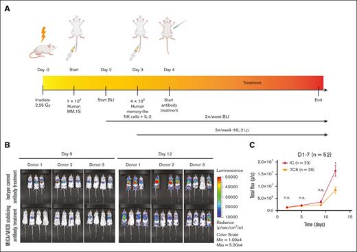
To test the combined effect of MICA/B stabilization and memory-like NK cell adoptive transfer, we used a xenograft MM model17,22,30 (supplemental Methods; Figure 2A). Animal experiments were in accordance with and approved by the Institutional Animal Care and Use Committee. Luciferase-expressing MM.1S cells were engrafted into irradiated NOD scid gamma (NSG) mice, and memory-like NK cells were adoptively transferred on day 3. NK cells were isolated from 7 independent donors and then differentiated into memory-like NK cells by activating briefly (12-16 hours) with interleukin-12 (IL-12), IL-15, and IL-18. Memory-like NK cells were adoptively transferred into 7 independent cohorts of tumor-bearing NSG mice. IL-2 was injected intraperitoneal every other day for the duration of the study to support NK cell survival. Starting 1 day after NK cell transfer, 7C6-hIgG1 or isotype control antibodies were administered once per week, and tumor progression was tracked via bioluminescence imaging. Memory-like NK cells more effectively controlled early MM tumor cell outgrowth in mice treated with 7C6-hIgG1 mAb than in those treated with control antibodies (Figure 2B-C). However, early MM control did not result in improved survival of these mice (supplemental Figure 6A; P = .89). This was partly explained by donor-dependent responses to 7C6 therapy (supplemental Figure 6B-C). Of note, when the survival of “responding” donors was analyzed separately, a statistical difference in survival was detected (data not shown) but was limited by the small number of tested mice. Whether the donor-dependent responses result from MICA/B surface variation, in vivo persistence, exhaustion of adoptively transferred NK cells, or technical variance remains unknown.
Combined MICA/B stabilization and memory-like NK cell transfer leads to early tumor control in vivo. (A) Experimental design. (B) Representative bioluminescence imaging (BLI) of recipient mice 9 and 12 days after engraftment with MM.1S-luc. Note the scale bar with lower intensities for the BLI signal in the 7C6-treated mice. (C) Enumeration of serial BLI measurements that show reduced tumor burden in mice receiving 7C6 mAb treatment compared with the isotype control. Differences were determined using the Mann-Whitney test; ns, P > .05; ∗P ≤ .05; ∗∗P ≤ .01; ∗∗∗P ≤ .001; ∗∗∗∗P ≤ .0001. IC, isotype control; ns, not significant.
Combined MICA/B stabilization and memory-like NK cell transfer leads to early tumor control in vivo. (A) Experimental design. (B) Representative bioluminescence imaging (BLI) of recipient mice 9 and 12 days after engraftment with MM.1S-luc. Note the scale bar with lower intensities for the BLI signal in the 7C6-treated mice. (C) Enumeration of serial BLI measurements that show reduced tumor burden in mice receiving 7C6 mAb treatment compared with the isotype control. Differences were determined using the Mann-Whitney test; ns, P > .05; ∗P ≤ .05; ∗∗P ≤ .01; ∗∗∗P ≤ .001; ∗∗∗∗P ≤ .0001. IC, isotype control; ns, not significant.
Here, we demonstrated that MICA/B shedding occurs in MM, and inhibition by the novel 7C6 mAb enhances memory-like NK cell–mediated responses against MM cells. Our data support the hypothesis that this combination of targeting MICA/B on MM cells and introducing memory-like NK cells results in improved NK cell–mediated immune surveillance in MM, making it a strong case for evaluation as a potential immunotherapeutic approach in MM.
The review boards of all participating centers approved the study, and the study was performed in accordance with the Declaration of Helsinki.
Acknowledgments: Anna V. Justis, a medical writer employed by the Dana-Farber Cancer Institute, provided writing and editorial support to the authors.
This work was supported by National Institutes of Health grant R01CA238039 (K.W.W.). R.R. is a member of the Parker Institute for Cancer Immunotherapy at the Dana-Farber Cancer Institute, and his work is supported, in part, by the Parker Institute for Cancer Immunotherapy. R.R. is a recipient of the Career Development Award from the Leukemia and Lymphoma Society. I.M.G. was supported by funding from the Dr. Miriam and Sheldon G. Adelson Medical Research Foundation. L.F.d.A. was funded by Cancer Research Institute award CRI3483, Leukemia and Lymphoma Society award 6647-23, US Department of Defense project number CA210940, the Tisch Cancer Institute Development Funds Award (National Cancer Institute grant P30CA196521), and the Elsa U. Pardee Foundation.
Contribution: S.T., K.W.W., R.R., and I.M.G. conceptualized the study; S.T., L.F.d.A., K.W.W., R.R., T.C., G.G., and I.M.G. developed the methodology; S.T., N.K.S., S.P., L.L., H.D., J.V.C., N.P., M.R., Y.S., L.B.F., A.C., J.-B.A., R.B., and E.L. conducted the investigations; S.T. wrote the original draft of the manuscript; K.W.W. and I.M.G. acquired funding; T.C., R.R., and I.M.G. supervised the study; and all authors reviewed and edited the final manuscript.
Conflict-of-interest disclosure: I.M.G. has acted as a consultant for Bristol Myers Squibb, Novartis, Amgen, Takeda, Noxxon Pharmaceuticals, Celgene, Sanofi, Genentech, GlaxoSmithKline, Gene Network Sciences Healthcare, Karyopharm Therapeutics, Adaptive Biotechnologies, Janssen, Medscape, and AbbVie, and has received honoraria from Celgene, Bristol Myers Squibb, Takeda, Amgen, Janssen, Karyopharm Therapeutics, Cellectar Biosciences, Adaptive Biotechnologies, Sanofi, and Medscape. P.S. reports consulting fees and research support from Celgene, Janssen, Karyopharm Therapeutics, and SkylineDx. K.W.W. serves on the scientific advisory boards of SQZ Biotech, Nextech Invest, Bisou Bioscience, and TScan Therapeutics; receives sponsored research funding from Novartis; and is a scientific cofounder of Immunitas Therapeutics. R.R. receives sponsored research funding from CRISPR Therapeutics and Skyline Therapeutics; owns equity in InnDura Therapeutics; and serves on the scientific advisory board of Glycostem Therapeutics and XNK Therapeutics. L.F.d.A. is a coinventor on an issued patent for an α3 domain-specific antibody and has served as a consultant for Cullinan Oncology. The remaining authors declare no competing financial interests.
Correspondence: Rizwan Romee, Division of Cellular Therapy and Stem Cell Transplantation, Dana-Farber Cancer Institute, 450 Brookline Ave, Boston, MA, 02215; email: rizwan_romee@dfci.harvard.edu; and Irene M. Ghobrial, Department of Medical Oncology, Dana-Farber Cancer Institute, 450 Brookline Ave, Boston, MA, 02215; email: irene_ghobrial@dfci.harvard.edu.
References
Author notes
S.T. and S.P. are joint first authors.
I.M.G. and R.R. are joint senior authors.
Only publicly available data sets have been used in this manuscript and referenced accordingly.
The full-text version of this article contains a data supplement.

![Inhibition of MICA/B shedding enhances memory-like NK cell–mediated cytotoxicity against MM. (A) MICA, MICB, PDIA6 (ERp5), and ADAM10 gene expression levels in 1389 tumor cell lines from the Cancer Cell Line Encyclopedia database. Gene expression levels are presented as log2(transcripts per million [TPM] + 1). (B) MICA/B surface expression in 10 different HMCLs, as measured by flow cytometry. (C) MICA/B gene expression levels in 9 different HMCLs from the publicly available Broad CCLE data set. (D) Dose-dependent increase in MICA/B surface expression in HMCL MM.1S in response to 7C6 treatment. Significance was calculated using the t test. (E) Shed MICB was determined using sandwich enzyme-linked immunosorbent assay. A decrease in soluble MICB was seen in response to the 7C6 mAb treatment. For functional assays, effector cells (conventional or memory-like NK cells) were cocultured with MM.1S at a 5:1 effector-to-target ratio for 6 hours. (F) Increased CD107a degranulation (n = 7), IFN-γ production (n = 10), and tumor necrosis factor-α (TNF-α) production (n = 7) measured by flow cytometry in response to 7C6 treatment of cNK cells. (G) Summary of cytotoxicity (n = 9) of cNK and memory-like NK cells upon 7C6 mAb treatment of MM.1S cells. Data are depicted as the mean ± standard deviation. Significance was calculated using the Wilcoxon test for comparison of IC and 7C6 treatment conditions, and the Mann-Whitney U test was used for the remainder of the comparisons; ns, P > .05; ∗P ≤ .05; ∗∗P ≤ .01; ∗∗∗P ≤ .001; ∗∗∗∗P ≤ .0001. E:T, effector:target; IC, isotype control; ns, not significant.](https://ash.silverchair-cdn.com/ash/content_public/journal/bloodadvances/8/20/10.1182_bloodadvances.2023010869/2/m_blooda_adv-2023-010869-gr1.jpeg?Expires=1765896440&Signature=iKZ3jlV~mKCnv5GYcHMP1V1GWefKiZ2rG1TlmF9w0eb1t5604Pdxd-gDS5z9xaHdWV0WyE8ofUlRunOpwEa9~55iV-BsXbhoenjuNTwGPvu~xpP2wPKkvSumhb4gidRMhmR-YyyLz3xp0NWqzO4p~aJZmFsEBRDogNmrEednSOIC~K9wmfEMUir~~psMyl7dG-bytQAUDd4ZE2XtuvpA4i3dDhiVLDdOLbTgaQGuncHZVh54h3qZG~4qLx7EON4ZTXlb~pWRR00WhJLB24Ew7ZAhPIov2o~6PBzj~kTger-PkDokEaCa-Nnga1C6CHEUNLCAgwTTyV2SES-t9sDRvQ__&Key-Pair-Id=APKAIE5G5CRDK6RD3PGA)
