Key Points
Autologous CART-19 can be safely added to ibrutinib.
Addition of CART-19 to ibrutinib leads to frequent, durable, and deep remissions in CLL.
Abstract
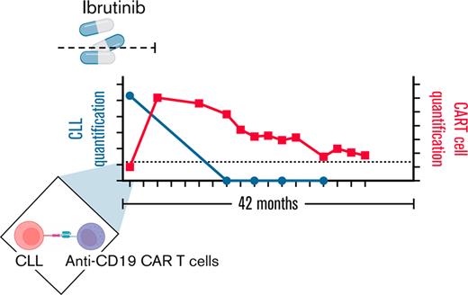
In chronic lymphocytic leukemia (CLL) patients who achieve a complete remission (CR) to anti-CD19 chimeric antigen receptor T cells (CART-19), remissions are remarkably durable. Preclinical data suggesting synergy between CART-19 and the Bruton’s tyrosine kinase (BTK) inhibitor ibrutinib prompted us to conduct a prospective single-center phase 2 trial in which we added autologous anti-CD19 humanized binding domain T cells (huCART-19) to ibrutinib in patients with CLL not in CR despite ≥6 months of ibrutinib. The primary endpoints were safety, feasibility, and achievement of a CR within 3 months. Of 20 enrolled patients, 19 received huCART-19. The median follow-up for all infused patients was 41 months (range, 0.25-58 months). Eighteen patients developed cytokine release syndrome (CRS; grade 1-2 in 15 of 18 subjects), and 5 developed neurotoxicity (grade 1-2 in 4 patients, grade 4 in 1 patient). While the 3-month CR rate among International Working Group on CLL (iwCLL)-evaluable patients was 44% (90% confidence interval [CI], 23-67%), at 12 months, 72% of patients tested had no measurable residual disease (MRD). The estimated overall and progression-free survival at 48 months were 84% and 70%, respectively. Of 15 patients with undetectable MRD at 3 or 6 months, 13 remain in ongoing CR at the last follow-up. In patients with CLL not achieving a CR despite ≥6 months of ibrutinib, adding huCART-19 mediated a high rate of deep and durable remissions. ClinicalTrials.gov number, NCT02640209.
Introduction
Chimeric antigen receptor (CAR) T cells are now approved in the United States for the treatment of adults with relapsed/refractory non-Hodgkin lymphoma (NHL), multiple myeloma (MM), and children and adults with acute lymphoblastic leukemia (ALL). Regulatory approvals in these diseases were obtained after multicenter single-arm phase 2 studies showing CR rates of approximately 40% to 60% and 80%, respectively, and event-free survivals approximating 30% to 50% at 12 months.1-4 The first patients successfully treated with CAR T cells for chronic lymphocytic leukemia (CLL) were reported more than 10 years ago and predated reports in ALL, MM, or aggressive NHL.5-7 CLL is diagnosed in 191 000 new patients and estimated to cause 61 000 deaths per year worldwide and remains largely incurable with any treatment other than allogeneic stem cell transplantation despite the advent of new therapies.8-10 Previous studies showed that CAR T cells induce CRs in a minority (21-47%) of patients with CLL, but most CLL patients who do achieve a CR do not relapse.11-13
Preclinical data from our group support potency-enhancing interactions between anti-CD19 chimeric antigen receptor T cells (CART-19) and ibrutinib. Among other biologic changes, T cells from patients with CLL treated with ibrutinib for ≥5 cycles exhibit superior proliferative capacity, survival and effector cytokine production in vitro, and an improved immunosuppression profile through Bruton’s tyrosine kinase (BTK)-dependent and -independent mechanisms.14 Coadministration of ibrutinib with CART-19 led to a greater than additive antitumor effect in murine xenograft studies.15,16
Thus, we hypothesized that treating patients with autologous CAR T cells after ≥6 months of exposure to ibrutinib would have beneficial effects on manufacturing feasibility, be well-tolerated, and lead to high rates of durable remission.16 We, therefore, initiated a single-center study in patients with advanced or high-risk CLL who had not achieved a CR to ibrutinib monotherapy. Here we report the results of a planned analysis of data from this study in 18 patients with ≥15 months of follow-up.
Methods
Study design
To be eligible for participation in this phase 2 study, patients had to have documented CD19-positive CLL or small lymphocytic lymphoma, be currently receiving ibrutinib for ≥6 months without grade 2 or higher nonhematologic toxicity, and the best response to ibrutinib must not have exceeded partial response (PR). Previous treatment with anti-CD19 therapy was allowed. Before December 2016, patients must have failed ≥1 prior regimen before ibrutinib unless they carried a chromosome 17p or TP53 aberration or were at high risk of ibrutinib failure (mutations in BTK or PLCγ2). Ibrutinib was approved in first-line for CLL in March 2016. After December 2016, the protocol was amended to allow patients receiving first-line ibrutinib without documentation of 17p/TP53 aberration or BTK/PLCγ2 mutations.
Humanized anti-CD19 binding domain T cells (huCART-19) cells were generated ex vivo from autologous T cells transduced with a lentiviral vector to express a CAR containing a CD3-ζ domain to provide a T-cell activation signal and a 4-1BB (CD137) domain to provide a costimulatory signal.17 The targeting domain was derived from a humanized anti-CD19 single-chain variable fragment (scFv). The anti-CD19 scFV of CTL019 (now produced commercially as tisagenlecleucel) was humanized by grafting the complementarity-determining regions of the heavy and light chains onto human germline acceptor framework regions.18
During manufacturing of huCART-19 cells, as well as upon and after administration, patients continued to receive ibrutinib. Discontinuation of ibrutinib after attainment of remission was not defined in the protocol but could occur in the event of treatment-related toxicity or upon discussion between the patient and investigator upon achievement of measurable residual disease (MRD)-undetectable remission after huCART-19 that was sustained for ≥6 months. Protocol treatment comprised either fludarabine/cyclophosphamide or bendamustine (investigator choice) lymphodepleting chemotherapy followed by infusion of up to 5 × 10⁸ huCART-19 cells in 3 fractionated daily doses (10%, 30%, and 60%), such that the 30% and 60% doses could be withheld in the event of cytokine release syndrome (CRS).13 Infusions occurred in the outpatient setting.
The study was designed by the investigators, sponsored by an Investigation New Drug approval (IND) held by the University of Pennsylvania, funded by Novartis Pharmaceuticals, and registered on clinicaltrials.gov as NCT02640209 with a study activation date of 29 January 2016. It was approved by the University of Pennsylvania’s institutional review board. Patients provided written informed consent. Data were analyzed and interpreted by the investigators, and all authors reviewed the manuscript and vouch for the accuracy and completeness of the data and analyses and for adherence of the study to the protocol.
Endpoints
The primary endpoint was the safety of huCART-19 administration in combination with ibrutinib. The secondary endpoints included dose feasibility (number of manufacturing failures) and efficacy measures, including the International Working Group on CLL (iwCLL) complete response (including a complete response with incomplete marrow recovery [Cri]) within 3 months in evaluable patients19; MRD negativity rate was assessed by deep sequencing of the immunoglobulin heavy chain locus, best overall response, progression-free survival (PFS), overall survival (OS), time to response, duration of response, time to alternative therapy, and huCART-19 pharmacokinetics. Additional details are provided in the supplemental Appendix.
Statistical analysis
Primary safety endpoints, including the frequency and severity of study-related adverse events that are possibly, likely, or definitely related to study treatment, were evaluated for all infused subjects who received a minimum of 1 × 10⁷ CART-19 cells specified by the protocol as evaluable. Manufacturing failures were calculated as the proportion of enrolled subjects whose product did not meet the manufacturing release criteria or the minimum protocol-specific dose of 1 × 10⁷ huCART-19 cells. The 3- and 12-month CR and CRi rates and the associated exact 90% confidence intervals (CIs) were determined among the infused iwCLL-evaluable subjects.19 MRD negativity rate at 3, 6, and 12 months was calculated as the proportion of subjects with <1 estimated diploid genome (or cell) carrying the baseline CLL clonotype cell using the Clonoseq assay (Adaptive). The best overall response rate was calculated as the proportion of patients achieving CR/CRi or PR at any timepoint. Kaplan-Meier method was used to estimate OS and PFS among all infused subjects. Comparisons of OS and PFS between subject subgroups were made as post hoc analyses using a log-rank test. This pilot study was initially targeted to enroll 15 evaluable subjects but was amended to 25. The trial was stopped after 20 subjects were infused due to funding constraints. With a sample size of 20, we have a 90% probability to exclude a true safety event rate that is >14% if no event is observed. If 1 or 2 events are observed in 20 subjects, the upper 90% exact CI for the true event rate would be 0.21 and 0.28, respectively. The unacceptable rate for safety is ≥30%. Exploratory analysis for the correlative data was primarily descriptive. Due to the pilot nature of the study, a 90% confidence level was used, and no adjustment for multiple comparisons was performed. Missing data were minimum. The data cutoff date was 28 April 2021.
All statistical tests were performed with GraphPad Prism version 9.1.0 software or Stata version 16. All reported CI are exact CI.
Results
Patient characteristics
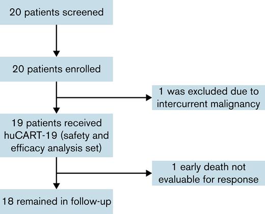
Twenty patients were enrolled (Figure 1). One patient did not receive huCART-19 due to the development of intercurrent Richter transformation and lung adenocarcinoma. There was no bridging therapy. A total of 19 patients received an infusion of huCART-19, with a median time from enrollment to infusion of 60 days (range, 28-252 days). The median age was 62 years (range, 42-76 years), with a median of 2 prior therapies (range, 1-17 therapies) and a median marrow CLL burden of 20% (range 5-80%). Fifteen patients had a history of poor-risk cytogenetic or molecular abnormalities detected on standard-of-care karyotyping, fluorescent in situ hybridization, or next-generation sequencing, as shown in Table 1. Three patients had relapsed after receiving CAR T cells on previous research protocols, and 2 were known to have ibrutinib-resistance mutations. Ibrutinib was the first CLL therapy in 6 patients (Table 1). Eighteen patients who received a huCART-19 infusion and survived ≥1 month had a median follow-up of 42 months (range, 17-58 months).
Screening, enrollment, and follow-up. A total of 20 patients were screened, and all 20 were enrolled. Of the 20 enrolled patients, 1 patient was not infused due to intercurrent lung adenocarcinoma and coincident finding of Richter transformation. Of the 19 patients who were infused, all had received ≥6 months of ibrutinib therapy before apheresis.
Screening, enrollment, and follow-up. A total of 20 patients were screened, and all 20 were enrolled. Of the 20 enrolled patients, 1 patient was not infused due to intercurrent lung adenocarcinoma and coincident finding of Richter transformation. Of the 19 patients who were infused, all had received ≥6 months of ibrutinib therapy before apheresis.
Characteristics of infused subjects (n = 19)
| UPN . | Age . | Sex . | Race . | Years from dx . | Adverse prognostic marker . | Prior lines . | Marrow CLL burden at enrollment visit . | Target lesion size (mm2) . | ||
|---|---|---|---|---|---|---|---|---|---|---|
| Cytogenetics . | Molecular . | Microscopy . | Flow . | |||||||
| 01 | 69 | M | W | 14.1 | Complex, including del17p | NA | 6∗ | 10 | 15 | 178 |
| 02 | 68 | M | W | 15.3 | Trisomy 12, t(14;19), del11q | NA | 6 | NA | 40 | 1158 |
| 03 | 58 | M | W | 7.3 | Del17p, c-Myc amplification | BTKC481S | 3 | 20 | 7 | 1716 |
| 04 | 62 | M | W | 4.9 | None | NA | 1 | 5-10 | 19 | N/A |
| 05 | 62 | M | W | 6.6 | Del13q, del11q | NA | 3 | 10 | 47 | Undefined |
| 06 | 59 | M | AA | 6.1 | Del17p, del13q | None | 3 | 10 | 9 | 628 |
| 07 | 56 | F | W | 16.3 | Complex, including Del17p, del13q | RB1, TP53 | 2 | 70 | 80 | Undefined |
| 08 | 68 | M | W | 1.1 | Complex, including del17p, del13q, del 6q | TP53 | 1 | 30 | 47 | 1471 |
| 09 | 76 | M | W | 14.6 | Complex, including del13q, del17p | BTKC481S | 7 | 50 | 57 | N/A |
| 10 | 75 | M | W | 8.7 | Complex, including del13q, del11q22 | TP53, XPO1, FBXW7 | 17∗ | 20 | 37 | 2193 |
| 11 | 47 | M | W | 0.8 | del17p | NA | 1 | 10 | 10 | Undefined |
| 12 | 62 | F | W | 7.6 | del17p, del13q, del6q | NA | 6 | 25 | 43 | 2813 |
| 13 | 52 | F | W | 5.9 | Complex, including del17p, trisomy 12 | None | 2 | 25 | 30 | 1075 |
| 15 | 42 | F | W | 1.4 | None | None | 1 | 40 | 36 | 2222 |
| 16 | 68 | M | W | 5.8 | Del13q, del17p | TP53 | 1 | 5 | 39 | Undefined |
| 17 | 76 | M | W | 25.6 | Del13q | TP53, DNMT3A, MYD88 | 2 | 5 | 24 | Undefined |
| 18 | 66 | M | W | 15.6 | Del 13q, del17p, monosomy 17, t(11;16) | TP53S,F3B1 | 5∗ | 21 | 36 | N/A |
| 19 | 55 | M | W | 0.6 | Complex, including Monosomy 17, del11q | ATM | 2 | 20 | 25 | Undefined |
| 20 | 66 | M | W | 3.6 | Del17p, del11q | NOTCH1, ATM | 1 | 80 | 54 | N/A |
N/A, not available; Undefined, no pathologically enlarged nodes. W, White; AA, African American.
Prior CART-19.
Manufacturing feasibility and treatment
The protocol-specified dose was achieved in all 20 enrolled patients. Transduction was quantified via flow cytometry using an anti-idiotype antibody (median CAR, 28.3%; range, 13.5-36.8%). Before huCART-19 infusion, all patients received lymphodepleting chemotherapy. Thirteen received fludarabine and cyclophosphamide, and 6 received bendamustine. The choice of conditioning regimen was per the treating physician and based on prior therapies. Fourteen patients received 3 infusions, and 5 patients received 2 infusions of huCART-19 (Table 2). At the time of huCART-19 infusion, patients had been on ibrutinib for 7 to 50 months (median, 14 months) (Table 2). At various times after CAR T-cell infusion, 14 patients stopped ibrutinib, while at the last follow-up, 5 patients remained on ibrutinib. Reasons for ibrutinib discontinuation were suspected ibrutinib-related toxicity (n = 6), patient and physician choice in the setting of undetectable CLL (n = 5), progression of CLL with a BTK mutation (n = 1), chemotherapy for a second malignancy (n = 1), and unknown (n = 1).
Treatment in infused subjects (n = 19)
| UPN . | Months on ibrutinib before huCART-19 infusion . | Lympho-depleting Chemotherapy . | CAR T doses, n∗ . | Total huCART-19 dose given . | On ibrutinib at last follow-up? . | Mo to ibrutinib discontinuation . |
|---|---|---|---|---|---|---|
| 01 | 23 | FluCy | 3 | 5 × 10⁸ | Y | NA |
| 02 | 10 | FluCy | 3 | 5 × 10⁸ | N | 9 |
| 03 | 50 | FluCy | 3 | 5 × 10⁸ | Y | NA |
| 04 | 25 | Benda | 2 | 2 × 10⁸ | Y | NA |
| 05 | 12 | Benda | 3 | 5 × 10⁸ | N | 14 |
| 06 | 29 | FluCy | 3 | 5 × 10⁸ | N | 12 |
| 07 | 15 | FluCy | 2 | 2 × 10⁸ | N | 42 |
| 08 | 13 | Benda | 3 | 5 × 10⁸ | N | 41 |
| 09 | 7 | Benda | 3 | 5 × 10⁸ | N | 3 |
| 10 | 9 | Benda | 3 | 5 × 10⁸ | Y | NA |
| 11 | 11 | FluCy | 3 | 5 × 10⁸ | N | 11 |
| 12 | 10 | Benda | 3 | 2.5 × 10⁸ | N | 33 |
| 13 | 16 | FluCy | 3 | 5 × 10⁸ | N | 26 |
| 15 | 14 | FluCy | 3 | 5 × 10⁸ | N | 7 |
| 16 | 19 | FluCy | 3 | 5 × 10⁸ | Y | NA |
| 17 | 20 | FluCy | 2 | 2 × 10⁸ | N | 0.25 |
| 18 | 8 | FluCy | 3 | 5 × 10⁸ | N | 33 |
| 19 | 8 | FluCy | 2 | 2 × 10⁸ | N | 8 |
| 20 | 31 | FluCy | 2 | 2 × 10⁸ | N | 5 |
Benda, bendamustine; FluCy, fludarabine and cyclophosphamide; NA, not applicable.
FluCy was given as fludarabine 25 mg/m2 and cyclophosphamide 250 mg/m2 on each of 3 consecutive days. Bendamustine was given as 90 mg/m2 on each of 2 consecutive days.
Per protocol, subsequent doses to be omitted if patient developed fever or CRS.
Safety
Safety analysis was performed on all 19 patients who received an infusion of huCART-19. All patients received their infusion in the outpatient setting and had ≥1 adverse event during the study. Adverse events are presented in supplemental Table 1. Grade 3-4 neutropenia and thrombocytopenia were present in 10 (53%) and 7 patients (37%), respectively. Eighteen patients had CRS, beginning at a median of 2 days (range, 2-12 days) after the first huCART-19 infusion with a median duration of 7 days (range, 3-10 days). Most of the patients (n = 12 [63.2%]) had grade 1 CRS per American Society for Transplantation and Cellular Therapy (ASTCT) consensus criteria (seeTable 3).20 Only 2 patients required treatment with tocilizumab. Neurotoxicity was diagnosed in 5 patients (grade 1 in 2 patients, grade 2 in 2 patients, and grade 4 in 1 patient) (Table 4). One patient died on day 10 after infusion, after suffering grade 4 CRS and grade 4 Immune effector Cell Associated Neurotoxicity Syndrome (ICANS) necessitating intubation for airway protection in the setting of severe agitated delirium. This patient suffered a cardiac arrest while intubated and on high doses of sedating medication. One patient died of infection while on ibrutinib and with ongoing hypogammaglobulinemia in CR at 31 months, considered possibly related to treatment. At 12 months, the median and range of immunoglobulin-A (IgA), IgM, and IgG levels were 24 mg/dL (range, 8-135 mg/dL), 6 mg/dL (range, 0-92 mg/dL) and 538 mg/dL (range, 195-1318 mg/dL). At their last follow-up, 8 patients were still receiving immunoglobulin repletion for hypogammaglobulinemia. Serum immunoglobulin levels over time are shown in supplemental Figure 1.
Frequency and grade of CRS in infused subjects (n = 19)
| Grade . | Penn grading, n (%) . | ASTCT grading, n (%) . |
|---|---|---|
| 1 | 3 (15.9) | 12 (63.2) |
| 2 | 12 (63.2) | 4 (21.1) |
| 3 | 2 (10.6) | 1 (5.3) |
| 4 | 1 (5.3) | 1 (5.3) |
| 5 | 0 (0) | 0 (0) |
| Grade . | Penn grading, n (%) . | ASTCT grading, n (%) . |
|---|---|---|
| 1 | 3 (15.9) | 12 (63.2) |
| 2 | 12 (63.2) | 4 (21.1) |
| 3 | 2 (10.6) | 1 (5.3) |
| 4 | 1 (5.3) | 1 (5.3) |
| 5 | 0 (0) | 0 (0) |
ASTCT, American Society for Transplantation and Cellular Therapy.
Occurrence, duration, and grade of ICANS in infused subjects (n = 19)
| UPN . | Neurotoxicity . | Start day # . | Duration (d) . | Grade . | Comments . |
|---|---|---|---|---|---|
| 01 | No | — | — | 0 | — |
| 02 | No | — | — | 0 | — |
| 03 | No | — | — | 0 | — |
| 04 | No | — | — | 0 | — |
| 05 | No | — | — | 0 | — |
| 06 | Yes | 6 | 14 | 2 | Confusion |
| 07 | No | — | — | 0 | — |
| 08 | Yes | 3 | 2 | 1 | Confusion |
| 09 | No | — | — | 0 | — |
| 10 | No | — | — | 0 | — |
| 11 | No | — | — | 0 | — |
| 12 | No | — | — | 0 | — |
| 13 | Yes | 8 | 4 | 2 | Delirium |
| 15 | Yes | 5 | 1 | 1 | Confusion |
| 16 | No | — | — | 0 | — |
| 17 | Yes | 2 | 8 | 4 | Delirium, tremor, facial droop, dysphagia, expressive dysphasia |
| 18 | No | — | — | 0 | — |
| 19 | No | — | — | 0 | — |
| 20 | No | — | — | 0 | — |
ICANS, Immune effector Cell Associated Neurotoxicity Syndrome.
Efficacy
At 3 months, 16 patients were evaluable for response assessment according to iwCLL criteria, of whom 7 had achieved CR (43.8%; 90% CI, 23-67%). At 12 months, 14 were evaluable, and 7 were in CR (50%; 90% CI, 26-74%). MRD was assessed using deep sequencing of the IgH gene locus to a sensitivity of ≥1 × 10−5.21 At 12 months, 3 patients had undetectable MRD but were not considered in iwCLL-defined CR due to residual adenopathy exceeding 1.5 cm in 1 axis. Lymph node (LN) biopsies to confirm active disease were not systematically performed. Overall, MRD was undetectable at 12 months in 13 of 18 patients evaluable for response (72.2%; 90% CI, 50-88%) (Table 5). Of the 5 patients with detectable MRD at 12 months, 3 had previously participated in a clinical trial of murine anti-CD19 CAR T cells.13
Efficacy endpoints in infused subjects (n = 19)
| UPN . | iwCLL . | Marrow response . | MRD∗ . | Last follow-up (mo) . | Status at last follow-up . | On Ibrutinib at last follow-up . | ||||||
|---|---|---|---|---|---|---|---|---|---|---|---|---|
| M3 . | M12 . | M3 . | M12 . | Preinfusion . | M1 . | M3 . | M6 . | M12 . | ||||
| 01 | CR | CR | CR | CR | 5.7 × 10⁻² | 2.3 × 10⁻⁵ | 2.5 × 10⁻⁵ | 5.8 × 10⁻⁶ | 1.5 × 10⁻⁵ | 54 | CR | Y |
| 02 | PR | PR | CR | CR | 4.5 × 10⁻²† | 1.3 × 10⁻⁴ | 7.9 × 10⁻⁷ | ND | ND | 58 | Relapse | N |
| 03 | CR | PR | CR | CR | 5.4 × 10⁻² | ND | ND | ND† | ND | 43 | CR | Y |
| 04 | NA | CR | CR | CR | 1.1 × 10⁻¹ | 6.5 × 10⁻⁶ | ND | ND† | ND | 45 | CR | Y |
| 05 | CR | CR | CR | CR | 2.8 × 10⁻¹ | ND | ND | ND† | ND | 55 | CR | N |
| 06 | PR | NA | CR | CR | 6.6 × 10⁻² | ND | ND | ND | ND | 47 | CR | N |
| 07 | CR | PR | CR | CR | 2.0 × 10⁻¹† | ND | ND | ND | ND | 48 | CR | N |
| 08 | SD | NA | CR | CR | 2.1 × 10⁻¹ | ND | ND | ND† | ND | 49 | CR | N |
| 09 | PD | PD | CR | NR | 3.7 × 10⁻¹ | 4.5 × 10⁻⁶ | ND | NA | 4.9 × 10⁻² | 25 | Died‡ | N |
| 10 | SD | PR | CR | CR | 1.4 × 10⁻¹ | 3.0 × 10⁻³ | 2.3 × 10⁻³ | 1.2 × 10⁻³ | 6.5 × 10⁻⁴ | 31 | Died§ | Y |
| 11 | CR | NA | CR | NA | 1.9 × 10⁻² | ND | ND | ND† | ND | 17 | NA‖ | N |
| 12 | PR | PR | CR | CR | 1.8 × 10⁻¹ | 5.9 × 10⁻⁵ | ND | ND† | 2.9 × 10⁻⁵ | 33 | CR | N |
| 13 | SD | NA | CR | CR | 7.4 × 10⁻² | ND | ND | ND† | ND | 38 | CR | N |
| 15 | PR | CR | CR | CR | 2.7 × 10⁻¹ | ND | ND | ND | ND | 45 | CR | N |
| 16 | NA | CR | CR | CR | 1.2 × 10⁻¹† | ND | ND | ND† | ND | 41 | CR | Y |
| 17 | NA | NA | NA | NA | NA | NA | NA | NA | NA | 0.25 | Died¶ | NA |
| 18 | SD | SD | NR | NR | 9.7 × 10⁻² | 3.1 × 10⁻² | 3.2 × 10⁻² | 9.2 × 10⁻²† | 6.8 × 10⁻² | 41 | SD | N |
| 19 | CR | CR | CR | CR | 4.2 × 10⁻² | ND | ND | ND | ND | 39 | CR | N |
| 20 | CR | CR | CR | CR | 2.0 × 10⁻¹ | ND | ND | ND | ND | 40 | CR | N |
NA, not assessable; ND, not detected; NR, no response; PR, partial response; SD, stable disease.
By deep sequencing of the immunoglobulin heavy chain (estimated number of genomes).
indicates sample was collected from peripheral blood instead of bone marrow.
Died of relapsed CLL.
Died in CR of infectious complications.
Lost to follow-up due to chemotherapy for second malignancy, therefore CLL disease status not known.
Died of toxicity.
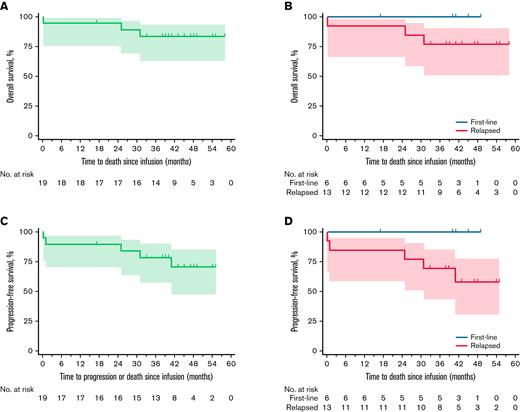
The median OS was not reached, and the estimated OS probability at 48 months was 84% (90% CI, 63-93%) (Figure 2A). The OS of patients on first-line ibrutinib compared with those enrolled in the study with relapsed or refractory disease was not significantly different (log-rank test, P = .35) (Figure 2B).
OS and PFS. (A) Shows OS among all the patients. The median OS was not reached, and the estimated OS probability at 48 months was 84% (90% CI, 63-93%). (B) Shows the OS of patients on first-line ibrutinib compared with those enrolled in the study with relapsed or refractory disease (P = .35, log-rank test). (C) Shows PFS among all the patients. Median PFS was not reached, and the estimated PFS at 48 months was 80% (90% CI, 46-85%). (D) Shows the PFS of patients receiving first-line ibrutinib compared with those enrolled in the study with relapsed or refractory disease (P = .15, log-rank test). Tick marks indicate censored data.
OS and PFS. (A) Shows OS among all the patients. The median OS was not reached, and the estimated OS probability at 48 months was 84% (90% CI, 63-93%). (B) Shows the OS of patients on first-line ibrutinib compared with those enrolled in the study with relapsed or refractory disease (P = .35, log-rank test). (C) Shows PFS among all the patients. Median PFS was not reached, and the estimated PFS at 48 months was 80% (90% CI, 46-85%). (D) Shows the PFS of patients receiving first-line ibrutinib compared with those enrolled in the study with relapsed or refractory disease (P = .15, log-rank test). Tick marks indicate censored data.
At the most recent follow-up, 1 responder had relapsed and died of progressive disease, 1 responder had relapsed and was on next-line therapy, 1 nonresponder was alive on next-line therapy, 1 patient had died of an infection in remission, and 1 had developed a second malignancy and CLL status was not evaluable. The remaining 13 patients were alive in continuous remission, of whom 4 remain on ibrutinib. Thus, PFS at 48 months was 70% (90% CI, 46-85%), and the median PFS was not reached (Figure 2C). The PFS of patients on first-line ibrutinib compared with those enrolled in the study with relapsed or refractory disease was not significantly different (P = .15) (Figure 2D).
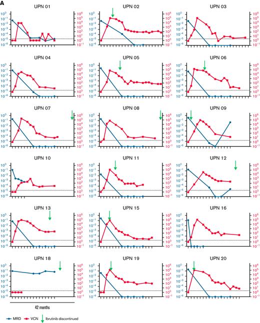
MRD over time is shown in Figure 3A. Of 15 patients with undetectable MRD at 3 or 6 months, 1 patient relapsed at 12 months and died of progressive disease, and 1 relapsed at 41 months and remains on next-line therapy 58 months after infusion. Of the 3 patients who had previously received CAR T cells, 1 patient did not respond, and 2 achieved an MRD-positive CR that lasted for ≥54 and 31 months. Of the 6 patients who received huCART-19 after first-line ibrutinib, all 6 remained in remission at the last follow-up (median, 43 months; range, 17-49 months).
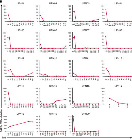
Kinetics of response and CAR T-cell expansion. (A) Shows the depth of MRD estimated from deep sequencing of the immunoglobulin loci (left y-axis) and T-cell expansion and persistence quantified by qPCR of the CAR transgene (right y-axis) for individual patients over a 42 month follow-up period. The time of ibrutinib discontinuation, where applicable, is indicated by an arrow. (B) quantifies the presence of CD19+ CD20+ B cells in the blood of individual patients over time as an indicator of CAR-T cell functional persistence. MRD, measurable residual disease; VCN, vector copy numbers per microgram of DNA.
Kinetics of response and CAR T-cell expansion. (A) Shows the depth of MRD estimated from deep sequencing of the immunoglobulin loci (left y-axis) and T-cell expansion and persistence quantified by qPCR of the CAR transgene (right y-axis) for individual patients over a 42 month follow-up period. The time of ibrutinib discontinuation, where applicable, is indicated by an arrow. (B) quantifies the presence of CD19+ CD20+ B cells in the blood of individual patients over time as an indicator of CAR-T cell functional persistence. MRD, measurable residual disease; VCN, vector copy numbers per microgram of DNA.
Expansion and persistence
Quantitation of huCART-19 cells was performed by flow cytometry and quantitative polymerase chain reaction (qPCR). Peak expansion occurred on median day 10 after infusion (range, 7-21 days), and the peak number of CAR T cells was 536 cells per ul blood (range, 0-3640 cells per ul blood). Peak expansion by qPCR occurred on median day 10 after infusion (range, 7-28 days), and the median peak vector copy number was 90 991 (range, 966-201 556). Peak CAR expansion was significantly associated with MRD response (supplemental Figure 2).
Persistence and quantitation of huCART-19 cells in individual patients are plotted against CLL molecular MRD in Figure 3A. At 12 months, 8 patients had CAR T cells detectable above the limit of quantification by qPCR (25 copies per 1 ug DNA), and 17 patients had CAR T cells above the limit of detection.
The duration of B-cell aplasia, a marker of CAR T-cell functional persistence, is shown in Figure 3B. The median B-cell percentage in the peripheral blood at month 12 after infusion was 0.05% (range, 0-64.4%). At 12 months, B cells constituted ≤1% of circulating lymphocytes in 13/18 patients.
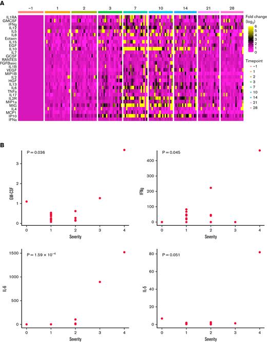
Serum cytokines were quantified over time at predetermined intervals and are represented as fold-change from the preinfusion baseline (Figure 4A). Peak cytokine production appeared to occur after the onset of clinical CRS (median day 2) and before the peak CAR-T expansion (median day 10). Of 30 measured cytokines, only granulocyte-macrophage colony-stimulating factor (GM-CSF), IFNγ, IL-6, and IL-5 correlated with CRS severity (Figure 4B).
Kinetics of cytokine production and association with CRS. (A) Expresses the production of individual cytokines (vertical axis) over time (horizontal axis) from individual patients (unique patient number not shown). Changes in cytokine levels are shown as fold-change from the day −1 baseline on a log2 scale. (B) Shows the correlation of peak cytokine production of selected cytokines out of 30 cytokines tested with CRS severity (using the Penn scale of CRS severity). Pearson correlation with a Bonferroni correction for multiple comparisons. Note that the remaining 26 cytokines tested were not found to be significantly correlated with CRS severity.
Kinetics of cytokine production and association with CRS. (A) Expresses the production of individual cytokines (vertical axis) over time (horizontal axis) from individual patients (unique patient number not shown). Changes in cytokine levels are shown as fold-change from the day −1 baseline on a log2 scale. (B) Shows the correlation of peak cytokine production of selected cytokines out of 30 cytokines tested with CRS severity (using the Penn scale of CRS severity). Pearson correlation with a Bonferroni correction for multiple comparisons. Note that the remaining 26 cytokines tested were not found to be significantly correlated with CRS severity.
Discussion
In patients not achieving CR despite ≥6 months of ibrutinib, at 3 months after huCART-19, we found an iwCLL CR rate of 43%, a marrow CR of 94%, and MRD was undetectable in 78%. This compares favorably to prior CART-19 cell studies in patients with progressive CLL without concurrent ibrutinib (iwCLL CR rates of 21-29%).11-13 CRS was frequent but mild–moderate and did not commonly require anticytokine therapy. These results suggest that the combination of huCART-19 with ibrutinib results in a high rate of sustained responses and high rates of MRD-negative marrow response in patients with CLL.
The underlying hypothesis tested in this study was that the addition of CAR T cells to ibrutinib would be safe and would deepen response rates in CLL compared with earlier studies. This hypothesis was based on our preclinical data showing that patient exposure to ≥6 months of ibrutinib can improve the function of the manufactured T-cell product, sensitize CLL to CAR T-cell killing, and impact the immune microenvironment.15,16 We also showed that ibrutinib improved survival in mice xenografted with B-cell lymphoma and leukemia cell lines that were completely resistant to ibrutinib when combined with CAR T cells, compared with CAR T-cell administration alone. Another potential mechanism by which ibrutinib can directly impact T-cell quality is through inhibition of the IL-2 inducible kinase.22
We found our approach successfully circumvented manufacturing challenges common in patients with CLL.16,23 We used a fractionated dosing strategy to mitigate toxicity, and all patients received 2 or 3 doses (2-5 × 10⁸ huCART-19 cells). This adaptive dosing approach has been of particular importance in adults with ALL and earlier trials in CLL, possibly related to toxicity in the setting of high levels of circulating disease.24 The results of this study, typically with lower levels of circulating disease, support this framework and suggest that a single dosing approach may be appropriate for most patients with CLL who have achieved some degree of disease control.
Our trial is unique for prospectively evaluating the combination of huCART-19 and an extended course of ibrutinib before CAR-T cell therapy. Several other published studies report experience with the combination in a subset of treated patients. In a trial from the Memorial Sloan-Kettering Cancer Center, 5 of 16 patients with CLL received ibrutinib at the time of T-cell collection and/or CAR T-cell administration. Consistent with our preclinical data, the ex vivo expansion of T cells was significantly greater for the 4 ibrutinib-treated patients compared with 11 ibrutinib-naïve patients.25 In a trial from the Fred Hutchinson Cancer Research Center (FHCRC) group, 19 patients with CLL with high-risk features were given ibrutinib for ≥2 weeks before apheresis until ≥3 months after infusion. Adverse events appear broadly similar to our trial, including 1 cardiac death in the context of CRS. Cardiac morbidity and mortality have been reported with ibrutinib.26 The 4-week overall response rate was 83% according to iwCLL 2018 criteria, with 61% achieving unmeasurable residual disease (uMRD) marrow responses. In all 18 evaluable patients, the 1-year OS and PFS were 64% (95% CI, 42-98%) and 38% (95% CI, 19-78%), respectively.27 In patients with uMRD, the 1-year OS and PFS rates were 86% and 59%, respectively. Their data also suggested improved outcomes compared with a group of historical control patients who received CAR T cells without ibrutinib. While the median number of prior therapies was higher in the FHCRC cohort study compared with ours, both patient populations were enriched for high-risk features. Differently from our study, the FHCRC cohort included patients who had previously progressed on ibrutinib, and 7 patients who had previously discontinued ibrutinib restarted the drug just before T-cell collection. While the FHCRC study had considerably shorter follow-up than ours, both studies support the potential role of combining ibrutinib with CAR T cells. These results may be further validated in the TRANSCEND CLL 004 study (NCT03331198), where patients with CLL receive anti-CD19 CAR T cells with or without ibrutinib in a nonrandomized fashion (different cohorts).
Notable strengths of this prospective study include the prolonged course of ibrutinib before T-cell collection, the long follow-up period, and the sensitive assessment of MRD. The median duration of follow-up among patients who received a huCART-19 infusion and survived ≥1 month was 42 months (range, 17-58 months), allowing us to state with some confidence that huCART-19 and ibrutinib-induced remissions are deep and durable. In CLL, achieving a uMRD remission has been associated with long-term remission using other modalities.28 This study was not designed to test outcomes after ibrutinib discontinuation. However, 12 patients ultimately discontinued ibrutinib for either toxicity or patient preference. Median follow-up after ibrutinib discontinuation in these patients was 26.5 months, and only 2 had CLL recurrence. Future trials are needed to formally test the ability to stop ibrutinib after CAR T–cell-induced uMRD remissions.
The results of this study are relevant to a large population of patients with CLL. The PFS of previously untreated CLL patients receiving ibrutinib with obinutuzumab or ibrutinib with rituximab is estimated to be 79% at 30 months or 89% at 3 years.29,30 However, the CR rates were approximately 19% and 17%, respectively. Patients with relapsed del(17)(p13.1) CLL treated with venetoclax showed an ORR of 77%, CR of 20%, PFS of 54% at 24 months, and uMRD at a 1-log lower sensitivity than that used in our trial was only 30%.31 When combined with ibrutinib, obinutuzumab and venetoclax led to an ORR of 88% and uMRD remission rate of 50% at 2 months after completion of treatment (the primary endpoint of the trial).32 Thus, the vast majority of patients receiving the approved novel or targeted therapies for CLL require ongoing, likely life-long treatment, which may be associated with long-term toxicity and significant cost.33
Our trial indicates that some patients with persistent CLL despite ibrutinib can receive a “one and done” treatment that leads to durable remissions with acceptable toxicity. We were encouraged to observe that all 6 patients treated with first-line ibrutinib followed by CART combination remain in remission at the last follow-up, raising the possibility of turning CLL into a rapidly curable disease.
Acknowledgments
Financial support for this article was funded by Novartis Pharmaceuticals.
Authorship
Contribution: S.G., D.L.P., J.A.F., J.L.B., M.V.M., and C.H.J. conceptualized and designed the study; S.G., V.V., N.V.F., S.M., M.O., A.L.G., W.L.G., M.S., A.L., M.R., M.M.D., and D.L.S. collected and assembled the data; S.G., E.H., W.-T.H., N.V.F., A.R.M., S.F.L., J.J.M., E.P., J.C.B., S.J.S., and C.H.J. interpreted and analyzed the data; S.G., E.H., N.V.F., V.V., and D.L.P. wrote the manuscript; B.L.L., in development, cell manufacturing, and testing: designed research, performed research, and collected data; all authors approved of the final manuscript; and S.G. and D.L.P. take accountability for all aspects of the study.
Conflict-of-interest disclosure: S.G. declares multiple patents related to CAR T cells; is a scientific founder and has equity in Carisma Therapeutics and Interius Biotherapeutics; and reports grants from Carisma Therapeutics and Interius Biotherapeutics. C.H.J. is a scientific founder and has equity in Tmunity Therapeutics and Capstan Therapeutics; reports grants from Tmunity Therapeutics; and is on the scientific advisory boards of BluesphereBio, Cabaletta, Carisma, Cellares, Celldex, ImmuneSensor, Poseida, Verismo, Viracta Therapeutics, WIRB Copernicus Group, and Ziopharm Oncology. J.L.B. is an employee of Novartis Pharmaceuticals. M.V.M. declares multiple patents related to CAR T cells. M.R. declares multiple patents related to CAR T cells. A.R.M. receives research funding from Abbvie and Pharmacyclics and consults for Pharmacyclics. J.C.B. receives honoraria from Novartis and Pharmacyclics; receives research funding from Pharmacyclics; consults for Novartis and Janssen; and has received travel and accommodation expenses from Janssen, Novartis, and Pharacyclics. S.J.S. receives research funding from Novartis, Pharmacyclics, and Janssen and consults for Novartis and Pharmacyclics.
D.L.P. is a named inventor on CART-19 related technology. B.L.L. is a consultant for Terumo and GSK; served on the Scientific Advisory Board for Akron, Avectas, Immuneel, Immusoft, In8bio, Ori Biotech, Oxford Biomedica, and Vycellix; and is a cofounder and equity holder in Tmunity Therapeutics and Capstan Therapeutics. Conflict of interest is managed in accordance with University of Pennsylvania policy and oversight. All remaining authors declare no competing financial interests.
Correspondence: Saar Gill, Cell Therapy and Transplant Program, Division of Hematology-Oncology and Abramson Cancer Center, University of Pennsylvania, 8-101 Smilow Center for Translational Research, 3400 Civic Center Blvd, Philadelphia, PA 19104; e-mail: saar.gill@pennmedicine.upenn.edu; and David Porter, Cell Therapy and Transplant Program, Division of Hematology-Oncology and Abramson Cancer Center, University of Pennsylvania 12 South Tower, 3400 Civic Center Blvd, Philadelphia, PA 19104; e-mail: David.porter@pennmedicine.upenn.edu.
References
Author notes
For original data, please contact Saar Gill at saar.gill@pennmedicine.upenn.edu.

