TO THE EDITOR:
In multiple myeloma (MM), measurable residual disease (MRD) is defined as a low-level detection of malignant plasma cells (PCs) that persist after treatment. MM MRD from bone marrow (BM) compartment characterizes the treatment efficacy, is highly predictive for outcome, and was therefore introduced as a consensus criterion for response assessment.1,2 While BM aspirate is considered the primary source of sample for MM MRD assessment, this technique is challenging in terms of repeated BM punctures, dilution by peripheral blood (PB), extramedullary disease, and heterogenous MM tumor spread throughout the BM compartment.3 To overcome these obstacles, MM MRD detection in PB samples was established.4,5 Recent studies have demonstrated that circulating malignant PCs can be detected by next-generation flow cytometry (NGF) at first diagnosis (FD) of MM.6 Circulating malignant PC detection by NGF after treatment is challenging, particular due to low frequency of malignant PCs circulating in the PB, and lacks additional studies with a sufficient number of patients.7-9
To evaluate the possibility of substituting elaborative BM aspiration with a PB sample to define the MRD status, the BM and PB MM MRD status postinduction therapy as assessed by NGF in patients with MM treated within the GMMG HD7 trial was compared (supplemental Data: Patients and Methods, supplemental Figure 1).
As a detectable number of circulating tumor cells is expected at MM FD, proof of concept (ie, general detection of circulating PCs) was performed on a small number of FD patients with MM (n = 18). For BM samples, a median of 7.35 (2.00–9.20) ×106 cells was analyzed. The analyzed median cell number in PB was 8.80 (1.00–9.60) ×106. The corresponding limit of detection (LOD, 20/total number of events acquired) and limit of quantification (50/total number of events acquired) are given in supplementary Table 1. Aberrant PCs were detected in all the analyzed BM samples (n = 18, 100%) at MM FD. PB analyses were evaluated at 2 cutoffs: 1 × 10⁻⁵ and 1 × 10⁻⁶. At the cutoff of 1 × 10⁻⁵, aberrant circulating tumor cells were found in 16 patients (89%), and at the cutoff of 1 × 10⁻⁶, they were found in 17 patients (94%, supplemental Table 2). Aberrant PCs detected in the BM of all patients resulted in a concordance of aberrant PC detection in BM and PB at MM FD of 89% and 94% for each cutoff, respectively (supplemental Figure 2). The median percentage of aberrant PC detected by NGF in MM FD BM samples was 1.100% (0.007%-13.100%), corresponding to a median tumor load (TL, number of aberrant PCs/total number of nucleated cells acquired) of 1.10 × 10⁻² (7.00 × 10⁻⁵ − 1.31 × 10⁻¹). The amount of aberrant PC detected in the MM FD PB samples was lower as compared with BM samples: median 0.017% (0%-0.540%), corresponding to a median TL of 1.21 × 10⁻⁴ (0–5.47 × 10⁻³). In both the BM and PB samples, non-aberrant PCs were detected in small percentages (supplemental Table 3). When plotting the percentage of BM and PB PCs, we found a medium-strength positive correlation between BM aberrant PCs and circulating aberrant PCs at MM FD (R2 = 0.595, supplemental Figure 3A), but not for nonaberrant PCs (R2 = 0.054, supplemental Figure 3B).
This finding, that is, an accurate detection of circulating PCs at MM FD by flow cytometry, is in line with previous reports by Sanoja-Flores et al,6 Mack et al,10 and Bertamini et al.11 Also, in accordance with Sanoja-Flores et al,6 an association between higher numbers of circulating PCs and higher levels of BM PCs was observed.
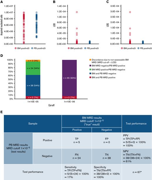
Our main focus was the concordance of NGF MRD detection in matched PB and BM samples (n = 70) obtained from patients with MM after 3 cycles of induction treatment (postInd3). In BM samples, a median of 8.05 × 10⁶ (1.70 × 10⁵–1.00 × 10⁷) cells was analyzed. The acquired median cell number in PB was 6.28 (2.10–1.00) × 106 (supplemental Table 4; Figure 1A-C). At the MRD cutoff of 1 × 10⁻⁵, 29 (41%) BM samples were MRD positive, 38 (54%) were MRD negative, and 3 cases (4%) were non-assessable. At the same MRD cutoff, 6 (9%) PB samples were MRD positive, and 64 (91%) were MRD negative (supplemental Table 5). Matching BM and PB samples at the patient level, 5 (7%) patients were concordantly BM- and PB MRD positive, while 38 (54%) were concordantly BM- and PB MRD negative. In 24 (34%) patients, BM MRD positivity was detected, but PB MRD was negative. Nonassessable BM MRD (MRD cutoff < LOD) was the reason for discordance in 3 (4%) additional patients. PB MRD positivity was not detected in any of the patients in the case of a BM MRD-negative result (Figure 1D). In the majority of samples (BM n = 37 [53%], PB n = 64 [91%]), the MRD was nonassessable and not comparable at the cutoff of 1 × 10⁻⁶ due to the low sensitivity reached in the individual samples (MRD cutoff < LOD), (Figure 1D; supplemental Table 5). Compared with BM MRD, PB MRD test performance resulted in a sensitivity of 17%, a specificity of 100%, a positive predictive value of 100%, and a negative predictive value of 61% (Figure 1E). In BM MRD-positive samples (n = 29), a median of 0.037% (<0.001%-8.5%) aberrant PCs was detected postinduction therapy. PB MRD-positive samples (n = 6) showed in median 0.003% (0.001%- 6.800%) aberrant PCs (Table 1). A linear regression analysis of BM and PB PCs was not feasible, as the postInd3 TL value was too low, and only a few (n = 5) MRD concordant positive-matched samples were detected.
BM and PB MRD NGF metrics, concordance, and test performance. Analyses were performed on matched BM/PB pairs (n = 70) postInd3. The NGF metrices: (A) acquired nucleated cell number, (B) LOD, and (C) LOQ are given for the BM and PB samples. (D) Concordance is shown of BM and PB NFG MRD results after treatment, evaluated at 2 cutoffs (1 × 10E-05 and 1 × 10E-06). (E) PB MRD test performance is calculated. Cases with discordance due to nonassessable BM MRD (n = 3) were not considered. BM, bone marrow; FN, false negative; FP, false positive; LOD, limit of detection; LOQ, limit of quantification; MRD, measurable residual disease; NGF, next-generation flow cytometry; NPV, negative predictive value; PB, peripheral blood; postInd3, after 3 cycles of induction therapy; PPV, positive predictive value; TN, true negative; TP, true positive.
BM and PB MRD NGF metrics, concordance, and test performance. Analyses were performed on matched BM/PB pairs (n = 70) postInd3. The NGF metrices: (A) acquired nucleated cell number, (B) LOD, and (C) LOQ are given for the BM and PB samples. (D) Concordance is shown of BM and PB NFG MRD results after treatment, evaluated at 2 cutoffs (1 × 10E-05 and 1 × 10E-06). (E) PB MRD test performance is calculated. Cases with discordance due to nonassessable BM MRD (n = 3) were not considered. BM, bone marrow; FN, false negative; FP, false positive; LOD, limit of detection; LOQ, limit of quantification; MRD, measurable residual disease; NGF, next-generation flow cytometry; NPV, negative predictive value; PB, peripheral blood; postInd3, after 3 cycles of induction therapy; PPV, positive predictive value; TN, true negative; TP, true positive.
Our MRD-positive rates after therapy at the 1 × 10⁻⁵ MRD cutoff are within the range of previously reported rates when assessed by flow cytometric and molecular genetic techniques like next-generation sequencing (NGS).8,10,12-18 Differences in the PB MRD-positive rates between the studies not only reflect the performance of the MRD method but also indicate the efficacy of the administered therapy and the time point of sample collection.19 These factors vary among the previously mentioned studies.
To our knowledge, only one other study has evaluated the matched BM and PB MRD results by NGF, indicating a lower sensitivity of PB MRD: Sanoja-Flores et al6 reported 26% concordantly positive results (this study, 7%), 34% concordantly negative results (this study 54%), and 40% discordant results (this study, 34%), respectively. The differences in test performance metrics, particularly in the sensitivity (40% vs 17%) and negative predictive value rates (45% versus 61%), mainly resulted from the BM MRD-positive rate being higher in the study of Sanoja-Flores et al6 (66%) than in this analysis (41%).8 In this analysis, a sufficient cell number to evaluate the MRD at the ≤ 1 × 10⁻⁵ cutoff was reached in all but one of the PB samples postInd3. However, in 50% of analyzed PB samples, postInd3 less than 1 × 10⁷ events were acquired, therefore limiting the sensitivity of the MRD analysis. This can potentially contribute to the lower rate of MRD PB samples identified in this study in comparison with Sanoja-Flores et al.6
Our results indicate that PB MRD is less sensitive than BM MRD. Furthermore, posttreatment BM- and PB-matched MM MRD analyses performed with NGS also indicate that MRD PB assessment is less sensitive than BM MRD assessment.15,16,18 On the other hand, being fast, cost-effective, and not necessarily requiring a baseline sample, NGF for MM MRD analysis in PB might be very useful when incorporated in a MM MRD flowchart. Given the high specificity and positive predictive value of PB MRD, PB MRD might serve as a minimally invasive method for MRD assessment—at least in a proportion of BM MM MRD-positive patients posttreatment (approximately 20%).
Perspectively, the MRD flowchart might start with NGF testing in PB, then proceed with NGS in the PB and NGF followed by NGS in the BM. Next steps are only taken when the result is negative. Such an algorithm could combine minimal invasiveness with cost-efficiency.
Acknowledgments: The German Speaking Myeloma Multicenter Group (GMMG) initiated and designed the HD7 trial and provided the samples for the present analysis. The investigational medicinal products isatuximab and lenalidomide were provided by Sanofi and Bristol Myers Squibb/Celgene, respectively. Sanofi funded the GMMG HD7 trial via an externally sponsored collaboration. Sanofi and Bristol Myers Squibb reviewed the manuscript.
Contribution: K.K. supervised and conceptualized the study and was responsible for validation; K.K. and M.H. were responsible for project administration; C.M., R.S., and S.H. were responsible for methodology; E.K.M., M.S.R., H.J.S., R.F., B.B., J.D., R.S., I.v.M., M.H., C.M., A.M.A., and B.H. were responsible for samples; C.M. and R.S. were responsible for software and formal analysis; K.K., C.M., and R.S. were responsible for investigation; C.M.-T., H.G., and M.H. were responsible for resources; E.P.K. curated the data; C.M. visualized the study; U.B., H.G., and M.H. were responsible for funding acquisition; K.K. and C.M. wrote and prepared the original manuscript; and all authors wrote, reviewed, and edited the manuscript.
Conflict-of-interest statement: K.K. reports research funding and honoraria from Sanofi. E.K.M. reports consulting or advisory role, honoraria, research funding, and travel accommodations and expenses from Bristol Myers Squibb/Celgene, GlaxoSmithKline, Janssen-Cilag, Sanofi, and Takeda. H.J.S. reports honoraria from AbbVie, Amgen, BMS/Celgene, Chugai, Genzyme, GSK, Janssen, Oncopeptides, Pfizer, Roche, Sanofi, Sebia, TAD Pharma, and Takeda; and travel, accommodations, expenses from Amgen, BMS/Celgene, Janssen, and Sanofi. R.S. reports speaker’s honoraria from Roche, Gilead/Kite, Janssen, Bristol Myers Squibb, and Sanofi; and consultant’s honoraria from Gilead/Kite, Janssen, BMS Bristol Myers Squibb, and Novartis. H.G. reports grants and/or provision of investigational medicinal product from Amgen, BMS, Celgene, Chugai, Dietmar-Hopp Foundation, Janssen, Johns Hopkins University, and Sanofi; research support from Amgen, BMS, Celgene, Chugai, Janssen, Incyte, Molecular Partners, Merck Sharp and Dohme, Sanofi, Mundipharma GmbH, Takeda, and Novartis; serving on advisory boards for Adaptive Biotechnology, Amgen, BMS, Celgene, Janssen, Sanofi, and Takeda; honoraria from Amgen, BMS, Celgene, Chugai, GlaxoSmithKline, Janssen, Novartis, and Sanofi. M.H. reports research funding from Sanofi, BMS, Celgene, and Novartis; and serving on the advisory board for BeiGene. The remaining authors declare no competing financial interests.
Correspondence: Katharina Kriegsmann, Department of Hematology, Oncology, and Rheumatology, Heidelberg University Hospital, Heidelberg, Germany; e-mail: katharina.kriegsmann@med.uni-heidelberg.de.
References
Author notes
The dataset might be shared upon request to Katharina Kriegsmann (katharina.kriegsmann@med.uni-heidelberg.de) after approval of an internal committee.
The full-text version of this letter contains a data supplement.


