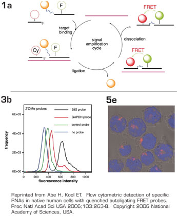
As many leukemic cells have unique genetic translocations leading to novel mRNA sequences, the ability to detect and image these molecules in live cells could find many useful applications, including rapid diagnosis and monitoring of disease, or purging cell products for transplantation. Efforts in this direction have been hampered by probes with insufficient sensitivity and signal-to-noise ratio to allow detection of moderate-to-low message levels, and need for reliable methods to deliver such probes into live cells. Recent advances in probe design by this paper's authors represent a significant step forward in this direction. Abe and Kool build on recent innovations including molecular beacon (MB) and quenched autoligating (QUAL) probes. The basic premise relies on self-directed reaction of two oligonucleotide probes which bind at adjacent positions on a target nucleic acid. One probe contains a fluorescent dye attached to one end and a quencher (non-fluorescent acceptor dye) attached to the other end. When the fluorescent dye and quencher are present in close proximity in this way, no fluorescence is detected upon excitation. The second probe contains a fluorescent acceptor dye which, upon energy transfer from a donor dye, would fluoresce BE a process called fluorescence resonance energy transfer (FRET). A unique aspect of the authors' design is the additional incorporation of a nucleophilic phosphorothioate group on one probe which could react with a 5'-electrophilic quenching dabsyl group on the other probe, causing displacement of the quencher and ligation of these two probes together (see Figure 1a). Importantly, the displacement of the quencher and the proximity of the donor (on probe one) and fluorescent acceptor dye (on probe two) would then allow fluorescence. Furthermore, since these two probes become covalently linked, the donor and acceptor dyes are locked into a fluorescent configuration, which serves as signal amplification when the probes leave the target and new probes bind. This novel combination both reduces background and increases signal to maximize signal-to-noise ratio. As proof-of-principle, these probes were delivered into live human HL-60 cells via transient permeabilization with streptolysin O (SLO). Using this novel scheme, the authors successfully detected 28S rRNA and GAPDH mRNA, which are abundant RNA species, both by flow cytometry and fluorescence microscopy (see Figures 3b and 5e). As would be expected, signal intensity in this system depends heavily on the abundance of the RNA target, as well as specific target site accessibility within each RNA molecule. As such, the authors were not able to detect S18 mRNA (estimated to be 11,000 copies per cell), but were able to detect JUN D mRNA (estimated at 5000 copies).
While more work is needed to further increase the signal-to-noise ratio to allow detection of moderate-to-low copy number RNA species, such as most translocation products, this study represents exciting proof-of-concept that RNA molecules could be imaged within live cells in a sequence-specific manner. The authors made a number of elegant innovations to reduce background and increase signal. These include the clever combination of a quencher, a nontypical FRET pair system to minimize spectral overlap, and autoligation for signal amplification. Next generation probes for a range of leukemia-related translocations, each with a different color, may be introduced into patients' blood or marrow samples and rapidly analyzed by flow cytometry for diagnosis and quantitation of disease. Leukemic cells could also be readily removed from a cell product for transplantation. For research purposes, such probes could be used to isolate pure populations of leukemic cells for detailed analyses, and to better understand the localization of target RNA molecules within leukemic cells under different conditions using laser confocal microscopy. An array of exciting clinical and research applications in leukemia will be possible as we eagerly await the further refinement of this novel technology.
Competing Interests
Dr. Lee indicated no relevant conflicts of interest.

