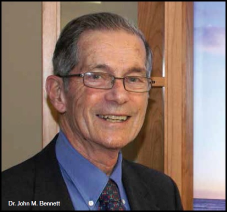
It all began with a most fortuitous interview in 1946 with William Dameshek, the founder and first editor of Blood. When I was a 13-year old student at the Roxbury Latin School in West Roxbury, Massachusetts, our class was given an assignment to interview someone famous and to present that interview to the Class of 1951. My father, a pediatrician and Tufts Medical School graduate, told me that he knew “Bill” very well and would arrange for me to interview him at his home.
I found Bill in his home office cluttered with journals and manuscripts. He told me about this new specialty that he was working on that dealt with both nonmalignant conditions and malignancies of the blood. This vivid memory remained with me for a very long time and played a significant role in my career choice.
Fast forward to 1962 when I finished my internal medicine residency at the Beth Israel Hospital (BIH) in Boston and was accepted as a hematology fellow in the Tufts/Boston City Hospital program headed by two outstanding hematologists, Willam Moloney and Jane Desforges. Several papers later (including a publication in the New England Journal of Medicine on acquired spherocytic anemia secondary to Clostridium welchii bowel infection) and after a year of learning from the masters, I was back at the BIH in charge of the diagnostic hematology laboratory. At the Yamins Research Institute at BIH, I met Al Rutenburg, a surgeon working on inflammatory bowel disorders. He taught me about the histochemistry of phosphatases, which led to the publication of a new cytochemical method for leukocyte alkaline phosphatase (low in CML!) that became widely used in the United States.
I continued to see many patients with our residents, and one, in particular, stood out. He was referred to me by none other than Bill Dameshek, for a second opinion. The patient had a refractory anemia with normal iron stores, but the erythrocytes were microcytic and hypochromic. Serum iron was elevated. My resident suggested that we assay the urine for hydroxykynurenine after tryptophan loading, which was dramatically elevated. A trial of oral pyridoxine restored his hematology profile, and another case report resulted from this treatment. I quickly appreciated that case reports were a valuable exercise for our residents and fellows, and that patients offered plentiful opportunities for new discoveries.
The 1960s was a time of monumental change for our family of three children. In 1966, I was drafted into the U.S. Army; however, with the assistance of Howard Hiatt, Chief of Medicine at the BIH, I was offered a vacancy at the Clinical Center, National Institutes of Health (NIH) in the U.S. Public Health Service (USPHS). I quickly accepted this position, and my wife and I moved to Bethesda where I headed the morphology and cytochemisty section in clinical pathology for the next two years. I had no direct patient exposure but associated with the clinical faculty of the National Cancer Institute (NCI) and all the other institutes where knowledge of bone marrow pathology was necessary. My histology, morphology and cytochemical skills were fine-tuned in Bethesda, and some 20 publications resulted.
It was there I met Henry Rappaport, the lymphoma guru. He was forming an international tutorial and NCI-supported lymphoma panel and asked me to join both. I left NIH in 1968 and moved, within a year, to the University of Rochester, where I have remained ever since, currently part-time in hematopathology.
At the first tutorial, I met Georges Flandrin, a hematopathologist at Hôpital Saint-Louis in Paris, France. He was utilizing the same cytochemical techniques that I was (peroxidase and esterases) to better define the acute leukemias. The French-American-British (FAB) leukemia working group had its genesis from initial discussions between the two of us. Each of us identified other morphologists with whom we had some contact (Drs. Daniel Catovsky, Marie-Thérese Daniel, David Galton, Harvey Gralnick, and Claude Sultan). A series of workshops was arranged where cases were reviewed and guidelines developed. The classification was based exclusively on the morphology of the bone marrow and peripheral blood cells in Romanowsky-stained air-dried smears. The initial intent was to separate acute myeloid leukemia (AML) and myelodysplastic syndromes (MDS) from acute lymphocytic leukemia (ALL), into easily identifiable and reproducible subtypes. The most widely used morphologic classification of AML, ALL, and MDS in the past four decades was published in 1976, and expanded definitions were proposed by the FAB Cooperative Group in 1982.
The FAB Working Group recognized that some patients could present with a disease that bore some resemblance to AML, but that this entity, unlike AML, did not have many leukemic blasts in the bone marrow. The disease was associated with some alteration in maturation of the three major cell lines, which resulted in pancytopenia and increased risk of infection and bleeding but did not necessarily progress to acute leukemia. Different terms were applied, including dysmyelopoietic anemia. The FAB Working Group applied the term MDS to these disorders. The progression of the disease could be highly variable: Some patients never evolved to acute leukemia, and others evolved quickly.
In 1982, a larger number of cases was reviewed with the intent of determining whether specific morphologic abnormalities, individually or in groups, would predict a different biological outcome. This larger review of cases led to an expanded definition of MDS into the well-known five subgroups with dysplastic features in common: refractory anemia (RA), RA with ring sideroblasts (RARS), RA with excess blasts (RAEB), RAEB in transformation, and chronic myelomonocytic leukemia (CMML). Investigators found that they could apply the FAB classification reasonably well. Separations in survival curves were demonstrated, ranging from five to six years for the most favorable prognostic forms of MDS, to less than one year for the least favorable forms. The FAB system served as the gold standard for more than two decades, and the Group published more than a dozen articles on various forms of acute and chronic leukemias and myeloproliferative neoplasms. The continuous need to be able to classify patients into more homogeneous subgroups led to the World Health Organization (WHO) classification.
In 1997, the WHO appointed a committee to revise and update the diagnostic categories of the lymphomas and the leukemias. I was privileged to be appointed to the subcommittee for acute leukemias and MDS. Two additional versions (published in 2008 and forthcoming later in 2016) have added molecular biology (mutations), cytogenetics, and flow cytometry as important components.
At Rochester, I became clinical director of the Cancer Center in 1974, focusing on clinical trials and new drug development, heading the hematology committee of the Eastern Cooperative Oncology Group and assuming the principal investigator role at the medical center. I was appointed editor-in-chief of Leukemia Research and served in that role for more than two decades. I also headed the Education Committee for ASH at the annual meeting.
In 1996, I decided to move into the hematopathology division to serve as an attending. The next two decades would be most rewarding ones. Continued contact with residents and fellows, connections with traveling scientists from around the world, and lasting and warm relationships with morphologists including Drs. Jean Goasguen, Barbara Bain, Dick Brunning, Ulrich Germing, Jim Vardiman, Nuket Tuzuner, Masao Tomonaga, and Yataro Yoshida have led to many pleasant trips and continued scientific efforts. It has been wonderful to witness the coming of age of trainees and fellows who have assumed important international roles, including Drs. Torsten Haferlach, Anna-Marie Storniolo, Jeff Lancet, Rami S. Komrokji, and Jane Liesveld. I have received tremendous support from the medical center, and in particular, from Drs. Richard Burack and Jonathan Friedberg, who have allowed me to continue my interests in morphology.
As I move toward my mid-80s (with more than 560 publications under my belt) I reflect on what a wonderful and full academic career I’ve had. I have been blessed with two great physician sons and one amazingly talented publishing editor daughter, as well as nine spectacular grandchildren. My wife Carol has always been at my side on most of my travel adventures (when not there, my colleagues are upset).
Teaching, clinical research, publishing results of studies, and patient care have provided me with a full academic life and no regrets.
Thoughts from a Former Protégé
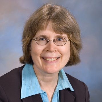
Jane Liesveld, MD
Jane Liesveld, MD
Professor of Medicine, James P. Wilmot Cancer Institute, University of Rochester Medical Center, Rochester, NY
John Bennett epitomizes the flexibility and potential for multifaceted productivity that a long academic medical career can provide. I first met John when I was a medical resident at the University of Rochester, and he was a medical oncology attending. That was before the days of extreme oncologic specialization in academic medical centers; in his clinic, one would see all solid tumor types as well as an array of hematologic malignancies. While he appeared to be up to date on all these diseases, their treatments, and available clinical trials, it was apparent even then that the microscope was one of his great loves, as he would frequently sneak in some slide reviews between clinic patients. That interest in blood and marrow morphologic interpretation led to his involvement with the FAB Working Group and countless publications and citations. While many of us always thought he was the “B” in FAB, he was really the “A” for American. Although new classifications of myelodysplasia and AML, such as the WHO, have gained ascendency, the FAB morphologic classification still influences some clinical thought processes. John’s involvement with the FAB Working Group resulted in international collaborations and friendships that he has maintained throughout his long career.
Just as John’s athletic pursuits evolved from competitive squash player (one of Rochester’s best), to tennis, and then to golf, so, too, has his academic career evolved with time. For the past two decades, he has given up direct patient care and now serves as a hematopathology attending. His “retirement” activity makes most of our prime time career efforts look anemic. He takes service in rotation; leads teaching conferences; and continues to publish, travel, and lecture. His only surrender to the concept of retirement is to spend December 25th through the month of March in Florida, and even then, his microscope travels with him and his wife, Carol, carefully packed in the back seat because it doesn’t fit in the trunk. While in the Tampa area, he is able to share his experience and expertise with Moffitt Cancer Center colleagues, and he does get in a bit of golf while Carol is on the tennis court.
John’s worldwide contacts made him an ideal editor-in-chief for Leukemia Research. He worked for many years with the Myelodysplastic Syndromes Foundation to increase awareness and support for this disease, and his interest in geriatric oncology led to involvement with the Hartford Foundation and the establishment of geriatric oncology as a subspecialty.
Outside of medicine, John’s love of music led to a long-standing involvement with the Rochester Philharmonic Orchestra, including serving as a member of its board of directors. When members of the university community gathered for a symposium to celebrate John’s 80th birthday in 2012, talks by invited hematologists and pathologists were separated by offerings from small musician groups from the Philharmonic. We could only wish that concert hall acoustics could enhance hematology lectures as well as musical performances. This event was a fitting tribute to John by not only physicians, scientists, and musicians, but also by extended family and community friends.
In his years as a pathologist, John has not lost the compassion from his internal medicine days. He recently spent time with one of my patients whom he knew only as the husband of one of Carol’s bridge partners to navigate a new diagnosis of a myeloproliferative syndrome. His energy, enthusiasm, and level of involvement in so many aspects of the hematology/oncology academic life never fail to inspire his mentees and colleagues. His career shows the diverse opportunities academic medicine can offer. That career would of course have been a poorer one without his wife Carol and their children and grandchildren. In accompanying John on his sojourns, Carol herself has become a master traveler, and if you are ever at ASH and want to skip out for a while, Carol Bennett always knows the best a city has to offer.


