Abstract
The BM microenvironment and its components regulate hematopoietic stem and progenitor cell (HSC) fate. An abnormality in the BM microenvironment and specific dysfunction of the HSC niche could play a critical role in initiation, disease progression, and response to therapy of BM failure syndromes. Therefore, the identification of changes in the HSC niche in BM failure syndromes should lead to further knowledge of the signals that disrupt the normal microenvironment. In turn, niche disruption may contribute to disease morbidity, resulting in pancytopenia and clonal evolution, and its understanding could suggest new therapeutic targets for these conditions. In this chapter, we briefly review the evidence for the importance of the BM microenvironment as a regulator of normal hematopoiesis, summarize current knowledge regarding the role of dysfunctions in the BM microenvironment in BM failure syndromes, and propose a strategy through which niche stimulation can complement current treatment for myelodysplastic syndrome.
Learning Objectives
To understand the definition and current constituents of the HSC niche in the BM
To review the role for the BM microenvironment in BM failure syndrome ontology and progression
Introduction
The BM is the exclusive site of physiologic production of all blood cells in humans. Aplastic anemia (AA) is a disorder of the BM that results in the loss of its ability to produce mature blood cells. AA treatment is focused on intense immunosuppression and/or BM transplantation. The study of therapeutic targets in AA has been limited by its rarity in the general population and the dearth of murine models of this disorder. The myelodysplastic syndromes (MDS) are also characterized by defects in the ability to form blood cells, resulting in pancytopenias. In contrast to AA, data suggest that the incidence of MDS is increasing. In fact, the Survey of Epidemiology and End Results (SEER) database underestimates the incidence of MDS by at least 3-fold.1 In MDS, the major morbidity and mortality results from the ineffective nature of the malignant clonal hematopoiesis and its suppression of residual normal hematopoiesis. All types of cytopenias are common among patients with both AA and MDS and are associated with symptomatic anemia, bleeding, and infections. A large proportion of elderly patients with MDS are either hospitalized (62%) or use the emergency department (42%) within 3 months of diagnosis.2 Although the role of the microenvironment in AA is well established, only recent studies suggest a role for the marrow microenvironment (MME) in the pathogenesis and clinical features of MDS and therapies targeting the MME in BM failure are lacking. Moreover, to date, the overwhelming majority of effort expended studying MDS has largely ignored the mechanisms by which the MDS clone alters its local microenvironment and suppresses residual normal BM function. In this chapter, we review the current understanding of the normal MME, examine evidence supporting MME disruption in BM failure syndromes, and highlight data supporting targeting the MME as a strategy for treatment. Disorders of hematopoiesis continue to have suboptimal clinical outcomes, highlighting the appeal of potential therapeutic manipulation of the MME in these situations.3
BM microenvironment in normal hematopoiesis
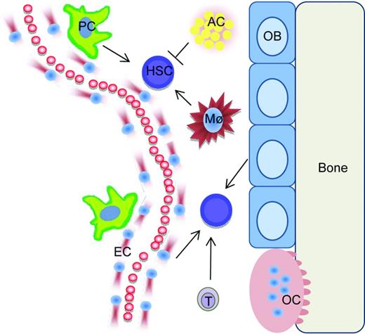
In mammals, skeletal organs are essential for normal hematopoiesis.4 Within the BM microenvironment, specific microenvironments, or niches, regulate hematopoietic stem cell (HSC) fate. Initial studies supported the central role of bone constituents in HSC regulation.5-7 As our understanding of the system has progressed, and as a result of elegant genetic studies and intravital microscopy, it became clear that the differentiation stage of mesenchymal cells is critical for their ability to support and regulate HSCs.8-10 In addition, heterogeneity of the BM endothelium has been elucidated.11-13 Currently, niche cells with mesenchymal characteristics are thought to be found in close association with arterial structures located at endosteal sites.13 These cells are likely a subset of mesenchymal stem cells (MSCs), the multipotent stromal cells that give rise to osteolineage cells, adipocytes, and chondrocytes. In the literature, this cell population is inconsistently defined, in part because of the lack of consensus on its defining features (adherence to plastic vs functional characteristics vs phenotypic markers) and the fact that the MSC abbreviation is also used to designate heterogeneous preparations of human mesenchymal stromal precursor cells.14,15 In addition to these immature cells, terminally differentiated hematopoietic cells, such as macrophages,16,17 osteoclasts,18 glia,19 and T cells20 have also been described as stimulatory components of the niche, whereas adipocytes are thought to inhibit HSCs.21 Niche composition and interactions with stem cells are coordinated by circadian rhythms,22 hormonal signals,23-25 oxygen tension,26 and likely other physiologic stimuli. Definition of the HSC niche in humans remains less understood than that in mice; however, both osteoblastic5 and mesenchymal stromal cells have been demonstrated to increase HSC support ex vivo.27 In addition to HSC niches, data support a role of the MME in both lymphopoiesis10,28 and myelopoiesis.29 The therapeutic potential of the MME in HSC regulation has already found clinical relevance, as illustrated by de Lima et al, who found that, in patients receiving cord blood transplantations for hematologic malignancies, engraftment was faster and more robust after coculturing the cord blood cells with BM mesenchymal stromal cells.27 Although our understanding of the HSC niche is likely to continue to evolve, the concept of its heterogeneity has emerged and allows for multiple targets for potential therapeutic intervention in hematopoietic regeneration (Figure 1). Moreover, the concept of the niche allows for a unique frame of reference when considering initiation of hematopoietic pathologies and interactions of malignant cells with the MME.
Schematic of HSCs within the niche. For simplicity, only some MME cell types are depicted. Pericytes (PC), macrophages (Mø), endothelial cells (EC), T cells (T), and osteoblasts (OB) are supportive of HSCs under normal conditions, whereas adipocytes (AC) have an inhibitory role.
Schematic of HSCs within the niche. For simplicity, only some MME cell types are depicted. Pericytes (PC), macrophages (Mø), endothelial cells (EC), T cells (T), and osteoblasts (OB) are supportive of HSCs under normal conditions, whereas adipocytes (AC) have an inhibitory role.
BM microenvironment and initiation of BM failure syndromes
Murine models have demonstrated that disruption of the MME can initiate myeloproliferative disease30,31 and even leukemia.32 However, until recently, the role of MME in the pathogenesis of BM failure syndromes was limited, with the exception of AA. AA was once thought to be the result of a quantitative HSC pool defect secondary to a toxin.33 In the early 1970s, however, Mathé et al observed recovery of hematopoiesis in patients with AA who failed to engraft after allogeneic stem cell transplantation with an immunosuppressive conditioning regimen, suggesting that the immune system in these patients was suppressing the growth and differentiation of HSCs.34 In addition, attempts to treat patients with AA via transplantation of HSCs from an identical twin without conditioning the recipient with total body radiation or high-dose cytotoxic agents often failed to reconstitute hematopoiesis.35 Idiopathic AA is now largely considered to be an autoimmune disease in which activated T lymphocytes induce accelerated apoptosis of HSCs.33,36 BM lymphocytes from patients with AA can inhibit hematopoiesis when cultured with normal BM.37 Oligoclonal or monoclonal expansions of CD8+ T cells have been found in patients with AA, a finding consistent with antigen-specific lymphocyte attack against hematopoietic tissue.38 Whereas the antigen(s) on HSCs that is targeted by T cells is unknown, cytokine expression by activated cytotoxic T cells may play an important role in the pathogenesis of AA. Those cytokines found to be prevalent in the BM of patients with AA include the BM-suppressive cytokine IFN-γ, as well as IL-17 and IL-27, both strong T-helper-1 cytokines.36
BM microenvironmental cells in AA have been characterized by several groups of investigators. Compared with controls, MSCs from patients with AA have aberrant morphology, decreased proliferation and clonogenic potential, and increased apoptosis. Relative to normal MSCs, those from AA patients were more difficult to induce to differentiate into osteoblasts and were more readily induced to differentiate into adipocytes. In addition to the abnormal biological features, the transcriptome of MSCs from AA patients demonstrated the down-regulation of numerous genes, including mediators of cell cycling, cell division, proliferation, chemotaxis, and hematopoietic cell interactions, and the up-regulation of genes involved in apoptosis, adipogenesis, and the immune response were up-regulated in MSCs from AA patients.39 These data suggest that MSCs represent another component of the deranged MME in AA. T lymphocytes modulate some aspects of hormonal regulation of the HSC niche.40,41 In addition, T-regulatory cells in mice are required for allogeneic HSC persistence.42 Whether altered T-cell signaling in AA contributes to abnormal MSC regulation is, to our knowledge, an unexplored and as yet unanswered question.
Frequently cited as some of the strongest evidence for MME dysfunction in MDS contributing to disease initiation and progression, Raaijmakers et al showed that deletion of Dicer1 in murine osteoprogenitors results in the development of dysplastic hematopoiesis with an MDS-like phenotype. The Dicer1 deletion leads to decreased Sbds [the gene mutated in Shwachman–Diamond syndrome (SDS)] expression in osteoprogenitors; this murine model of MDS therefore provides data indicative of a role of the MME in SDS. The Dicer1-null murine model of MDS resulted in primary MME dysfunction, leading to the secondary development of hematologic malignancy, thus supporting the concept of niche-initiated oncogenesis.43
Therefore, data support the concept that defects in the MME can be initiating steps in the pathogenesis of BM failure syndromes. AA is a paradigm for this concept because microenvironmental dysfunction, likely mediated at least in part by autoimmunity, is thought to lead to HSC pool depletion. Therefore, the MME can now be considered a targetable component of the disease process initiating BM failure syndromes.
Disruption of the BM microenvironment in inherited BM failure diseases
The rarity of the hereditary BM failure syndromes likely accounts for a dearth of studies on the MME in these diseases. Dror and Freedman studied BM stromal function in SDS in vitro using primary cells from patients. They found that SDS BM stroma had decreased ability to support and maintain both normal and SDS-associated hematopoiesis.44 More than a decade later, Andre et al studied a more select cell population, MSCs from patients with SDS. Compared with normal MSCs, SDS MSCs did not show any differences with regard to morphology, growth kinetics, expression of surface markers, differentiation, HSC support, or karyotype.45 Future studies are needed to identify the particular niche in the SDS-associated MME that contributes to dysfunctional hematopoiesis in this disease. Li et al investigated MSCs in a murine model of Fanconi anemia with deletion of the Fancg gene. They found Fancg−/− mice to have defective MSC proliferation and increased apoptosis. Introduction of human Fancg cDNA restored both normal proliferative capacity and normal cell survival to the Fancg-null MSCs. Fancg−/− MSCs had a reduced ability to support both HSC proliferation and differentiation in vitro and engraftment of transplanted HSCs in vivo. These data support the concept that Fanconi anemia proteins were required in the MME to maintain normal hematopoiesis.46 Therefore, whereas data on the role of the MME in inherited BM failure syndromes is scarce, there are some data that suggest its dysfunction in these diseases, aligning these disorders with acquired BM failure syndromes.
Disruption of the BM microenvironment in MDS
In MDS, the BM is generally hyperproliferative, but the disease is characterized by ineffective hematopoiesis and cytopenias. This paradox of myeloproliferation and cytopenias is ascribed to late precursor apoptosis in the BM. Some investigators have implicated the MME as the source of pro-apoptotic signals. Kitagawa et al found that the pro-apoptotic factor FAS ligand was produced by stromal cells, whereas its receptor was expressed in the hematopoietic cells of MDS BM.47 Stromal populations from patients with MDS are defective in their ability to support HSCs. Tennant et al demonstrated that adherent cell layers from MDS BM were defective in supporting colony formation relative to adherent cell layers from normal BM.48 Ferrer et al cocultured BM mesenchymal stromal cells from MDS patients with hCD34+ cells from healthy donors and found decreased numbers of HSCs and colony-forming units compared with cocultures using mesenchymal stromal cells from healthy donors.49 Geyh et al also reported on the diminished capacity of MDS-derived mesenchymal stromal cells to support hCD34+ HSCs in long-term culture-initiating cell assays due to altered gene expression of known mediators of interactions with HSCs, including osteopontin, Jagged-1, Kit-ligand, and angiopoietin.50
Some data support immune dysregulation in MDS. Sloand et al found expansion of cytotoxic T-cell clones in all of 34 patients with MDS cytogenetically characterized by trisomy 8. Consistent with an underlying autoimmune pathophysiology in this subset of MDS, >60% of patients with trisomy 8 as the sole cytogenetic abnormality (n = 13) who were treated with an antithymocyte globulin (ATG)-based regimen had a response, with durable reversal of cytopenias that was coincident with a stable increase in trisomy 8 BM mononuclear cells and normalization of the T-cell repertoire.51 Subsequently, Sloand et al reported on a clinical trial in which patients with MDS classified as refractory anemia, refractory anemia with ringed sideroblasts, or refractory anemia with excess blasts received immunosuppressive therapy with equine ATG, ATG plus cyclosporine, or cyclosporine alone. Of the 129 patients treated, 30% achieved a hematologic response, with a median follow-up of 3 years.52 Epling-Bernette et al found evidence of reduced natural killer (NK) cell function on the basis of in vitro assays with blood mononuclear cells from patients with MDS. Based on these results, the investigators speculated that abnormal myeloid progenitors in MDS may expand partly due to evasion of NK immunosurveillance.53 Marcondes et al similarly reported reduced NK cell function in in vitro assays using blood samples from patients with MDS.54 It has been postulated that abnormal BM stroma in MDS may play an inhibitory role in NK cell development because contact between these two cell populations is required for full NK cell differentiation and, accordingly, loss of immunosurveillance may lead to the accumulation of cells with DNA damage, contributing to MDS progression.55 Taken together, these data are supportive of immune dysregulation as a contributing factor to MDS pathogenesis, at least in some subsets of the disease.
There are multiple signals and secreted factors from MDS BM stroma that have been implicated as plausible mediators of dysfunctional interactions with hematopoietic cells. The precise roles of many of these factors, including cytokines, angiogenic factors, metalloproteinases, and adhesion molecules, are not well understood. TNFα produced by stromal cells plays a role in inducing apoptosis of maturing hematopoietic progeny.56-58 VEGF levels have been found to be increased in the BM of patients with MDS.59,60 Keith et al found increased microvascular density in the BM of patients with MDS, which correlated with increased VEGF expression.61 MDS stromal cells induce altered matrix metalloproteinase expression in clonally derived monocytes, but the significance of this finding is unclear.62 Expression of adhesion molecules, including CD166 and CD29, was altered in MDS-derived mesenchymal stromal cells; however, it is not clear whether these abnormalities influence the pathogenesis of MDS.49
Therefore, although the precise role of the MME in MDS pathogenesis is poorly characterized, the evidence is strong that it is altered in this disease and that its dysfunction contributes to disease progression.
MDS cells require their niche
Murine xenotransplantation models of MDS provide an opportunity to study this disease in an in vivo setting. However, it has been difficult to attain engraftment of clonal CD34+ cells derived from the BM of patients with MDS into NOD/SCID mice. These observations support the concept that MDS relies heavily on its MME. In fact, multiple studies demonstrate that the BM stroma is necessary for successful engraftment of clonal human MDS BM-derived CD34+ cells into mice. Kerbauy et al showed that irradiated NOD/SCID−β-2−/− mice had engraftment of clonal MDS-derived hematopoietic precursors when normal stromal cells (from cell lines derived from a healthy BM donor) were coinjected via the intramedullary route.63 Muguruma et al performed intramedullary injections of BM hCD34+ cells from MDS patients without or along with human MSCs into irradiated NOD/SCID mice with deletion of the T-cell receptor λ chain (NOG mice). Cells from 3 of 6 patients with MDS engrafted in NOG mice when coinjected with MSCs. Engraftment of MDS cells was not achieved with coinjection of stromal cells derived from sites other than BM or nonstromal cells.64
HS27a is a normal human BM stromal cell line that supports primitive hematopoietic cells, at least partially via cell-to-cell signaling through the Jagged-Notch pathway.65 This is in contrast to the HS5 human BM stromal cell line, which supports more mature colony-forming cells. Li et al found that clonal human MDS cell engraftment was achieved with intravenous coinjection of HS27a cells, but not with HS5 cells, in xenograft models.66 Highly expressed on HS27a cells, but not on HS5 cells, the cell adhesion molecule glycoprotein CD146 may be important for the engraftment of MDS clonal cells. CD146 is involved in multiple physiologic processes, including signal transduction, cell migration and development, angiogenesis, and mesenchymal cell differentiation.67 CD146+ cells are an important subset of stromal fibroblasts that express CXCL12 and contribute to the stem cell niche.68 Levels of CXCL12 and its receptor, CXCR4, are decreased in MDS cultures and this profile is associated with reduced induction of migration of CD34+ hematopoietic cells.69 Sorted CD146+ perivascular cells, but not CD146− cells, can support propagation of human HSCs with long-term reconstituting potential.70 Moreover, overexpression of CD146 in HS5 cells imparts capability to the HS5 cells to facilitate engraftment of hCD34+ clonal MDS cells in murine xenografts.66 CD146+ cells support long-term persistence of hematopoietic cells through cell-to-cell contact, with at least some reliance on Notch activation.71 Impaired Notch signaling via both anti-Notch1 antibodies and by inhibition of gamma-secretase decreased the CD146-mediated HSC support by CD146+ cells.72 These data collectively suggest that a subset of human stromal cells that are CD146+ is most likely responsible for the support of human MDS engraftment in xenograft models.
Medyouf et al recently demonstrated that engraftment of NSG mice with HSCs from patients with MDS can be significantly enhanced by cotransplantation via intramedullary injection of mesenchymal stromal cells from the same patient. They further found a unique gene expression profile in mesenchymal stromal cells from MDS patients compared with healthy control mesenchymal stromal cells, including up-regulation of LIF. Using an in vitro coculture system, they demonstrated that healthy mesenchymal stromal cells could be induced to up-regulate expression of LIF upon incubation with whole BM from MDS patients, suggesting that MDS cells are capable of inducing changes within the MME. Overall, their data support the concept that MDS cells alter mesenchymal stromal cells so that they promote clonal expansion and that mesenchymal stromal cells play an important role in disease pathogenesis.73
Therefore, much data using murine xenotransplantation models that use human stromal cells are demonstrative of a dependence of MDS HSCs on mesenchymal stromal cells.
Therapeutic targeting of the MME in BM failure syndromes
In murine models, activation of the normal HSC niche improves recovery from radiation6,20,74 and chemotherapeutic injury75 and suppresses chronic myeloid leukemia disease progression, impairing leukemic stem cell maintenance in a syngeneic model.76 In addition, a murine model of GVHD demonstrated targeting of the MME, which could be modulated to improve hematopoiesis.77 This body of work provides a rationale to consider MME modulation as a strategy to treat BM failure syndromes.
The notion that the underlying pathophysiology in AA results from an MME that is suppressive to HSCs largely due to activated T lymphocytes was confirmed through the success of trials of immunosuppressive therapy (with ATG alone or in combination with glucocorticoids, cyclosporine, or cyclophosphamide).33 AA is the only BM failure syndrome in which successful therapeutic targeting of the MME (BM T lymphocytes) has been realized.
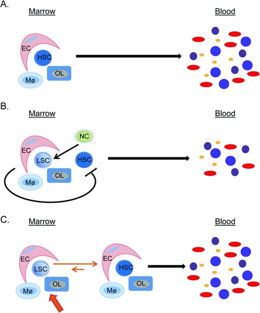
Because MDS can be cured by allogeneic HSC transplantation and stromal cells remain of patient origin after this procedure, it has been postulated that alterations in MDS stroma must be secondary to interactions with clonal MDS cells and reversible upon their eradication.69 Therefore, identification and targeting of MDS-initiated signals that modulate the MME represent an appealing therapeutic target for the treatment of MDS. The relatively recent availability of transgenic murine models of MDS78,79 will allow for the definition of the response of the normal MME to the development and progression of MDS in an immunocompetent system. These models could also be helpful in elucidating the degree of support for hematopoiesis by the MDS MME and determining whether it has a role in propagation of the disease. If so, then these model systems could be suitable as preclinical models of MME manipulation in MDS (Figure 2). Because strategies have already been advanced identifying components of the MME that can be stimulated in the normal BM and in certain myeloablative conditions (as well as chronic myeloid leukemia), defects in the MME of MDS become appealing therapeutic targets for the improvement of hematopoiesis.
Conceptual models of MME–HSC interactions are illustrated. (A) Normally, the HSC is supported by various niche cells, including endothelial cells (EC), macrophages (Mø), and osteolineage cells (OL). (B) In hematologic malignancies, clonal neoplastic cells (NC) alter the MME so that it becomes supportive of leukemic stem cells (LSC) and becomes less supportive of normal HSCs, ultimately leading to decreased normal hematopoiesis. (C) There is a strong rationale for therapeutic targeting of the MME in hematologic malignancies to push the MME into becoming less supportive of LSCs and more supportive of HSCs in an effort to restore normal hematopoiesis.
Conceptual models of MME–HSC interactions are illustrated. (A) Normally, the HSC is supported by various niche cells, including endothelial cells (EC), macrophages (Mø), and osteolineage cells (OL). (B) In hematologic malignancies, clonal neoplastic cells (NC) alter the MME so that it becomes supportive of leukemic stem cells (LSC) and becomes less supportive of normal HSCs, ultimately leading to decreased normal hematopoiesis. (C) There is a strong rationale for therapeutic targeting of the MME in hematologic malignancies to push the MME into becoming less supportive of LSCs and more supportive of HSCs in an effort to restore normal hematopoiesis.
Conclusion
The regulatory role of the MME in determining normal HSC fate and supporting hematopoiesis, although still being described, provides a rationale for studying the role of the MME in hematologic disorders. Disruption of the MME in inherited and acquired BM failure syndromes has long been reported. Given that T cells are considered a component of the MME, AA is the first BM failure syndrome in which therapeutic targeting of the MME in the form of immunosuppressive therapy has met with success. More and larger studies are needed to further delineate whether an altered MME is involved in the pathogenesis and progression of inherited BM failure syndromes. Recent progress has been made in our understanding of the role of the MME in disease pathogenesis and in the support of MDS. Further definition of the role of the MME could facilitate its therapeutic targeting in BM failure syndromes to exploit the biology of the niche by interfering with its disruption and by stimulating its supportive components as an additional tool for improvement of hematopoiesis and mitigation of its associated morbidities.
Acknowledgments
This work was supported by the National Institutes of Health (National Institute of Diabetes and Digestive and Kidney Diseases Grant DK081843, National Institute of Allergy and Infectious Diseases Grants AI091036 and AI107276, National Cancer Institute Grant CA166280, and National Institute on Aging Grant AG046293 to L.M.C.) and the Department of Defense (Grant BM110106 to L.M.C.). S.R.B. is supported by a Wilmot Cancer Research Fellowship. The authors thank Drs. Michael Becker and Marshall Lichtman for helpful discussion.
Disclosures
Conflict-of-interest disclosures: L.M.C. holds patents with or receives royalties from Fate Therapeutics. S.R.B. declares no competing financial interests. Off-label drug use: None disclosed.
Correspondence
Laura M. Calvi, MD, Professor of Medicine, Pharmacology and Physiology, Department of Neurologic Surgery, Wilmot Cancer Center, University of Rochester School of Medicine, 601 Elmwood Ave, Box 693, Rochester, NY 14642; Phone: (585)275-5011; Fax: (585)276-2576; e-mail: laura_calvi@urmc.rochester.edu.


