Abstract
VWD is the most common inherited bleeding disorder and is due to a deficiency and/or abnormality of VWF. VWD is inherited in an autosomal-dominant or autosomal-recessive pattern, but women are apparently more symptomatic. Three main criteria are required for correct diagnoses of VWD: (1) positive bleeding history since childhood, (2) reduced VWF activity in plasma, and (3) history of bleeding in the family. The bleeding score, together with baseline VWF levels and family history, have been proposed as more evidence-based criteria for VWD. Measurements of a reduced VWF activity in plasma are essential for the diagnosis of VWD; assays for the evaluation of the interactions between VWF and platelet glycoprotein Ib receptor with or without ristocetin, as well as VWF collagen binding, are currently in use. However, other tests such as VWF antigen, factor VIII, ristocetin-induced platelet agglutination, multimeric analysis, VWF propeptide, VWF/FVIII binding assay, and assessment of biological response to desmopressin are necessary to characterize VWD types. Levels of VWF activities <30 U/dL have been associated with a bleeding phenotype and the presence of mutations in the VWF gene.
Learning Objectives
To identify patients at risk for VWD according to their history of bleeding using a specific questionnaire to calculate BS
To use appropriate laboratory tests to classify VWD patients
To predict the bleeding phenotype of VWD and the need for replacement therapy using BS and baseline VWF levels
Introduction
VWD is considered the most common inherited bleeding disorder, even though its prevalence varies considerably according to the setting of diagnosis.1-5 In population-based studies, prevalence was estimated to be as high as 0.6%–1.3%,6,7 ∼2 orders of magnitude higher than in specialized centers (0.005%–0.01%), where symptomatic patients with VWD are usually referred.8-12 VWD is due to quantitative and/or qualitative defects of VWF, a multimeric glycoprotein synthesized by endothelial cells and megakaryocytes that mediates platelet adhesion/aggregation and stabilizes factor VIII (FVIII) in the circulation.1-5 In VWD, bleeding events are caused not only by impaired platelet glycoprotein Ib receptor–VWF interactions that are usually assessed in plasma in the presence or absence of ristocetin (VWF:RCo or VWF:GPIb), but also by reduced FVIII levels that often accompany the VWF defect.1-12
The current classification of VWD has proposed 6 different types: VWD1, VWD3, VWD2A, VWD2B, VWD2M, and VWD2N.2 A partial quantitative defect marks VWD1, whereas VWD3 is characterized by the nearly total absence of VWF in plasma and platelets. VWD2A and VWD2B are marked by the absence of high-molecular-weight VWF multimers in plasma, but in VWD2B, there is also an increased affinity of VWF for its platelet receptor, the glycoprotein Ib alpha (GpIbα). The identification of qualitatively abnormal variants with decreased platelet-dependent function and a normal multimeric structure marks VWD2M. VWD2N shows a full array of multimers, the defect being in the N-terminal region of the VWF where the binding domain for FVIII is located. This type is distinguishable from mild hemophilia A only by the abnormal binding of FVIII to VWF (see “Second-level laboratory tests”). Correct classification of different types by clinical and laboratory parameters is important for the management of patients with VWD.1-12
Clinical and laboratory diagnosis
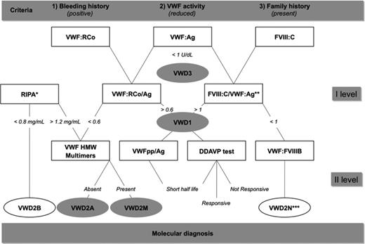
Three main criteria are required for correct diagnoses of VWD: (1) positive bleeding history since childhood, (2) reduced VWF activity in plasma, and (3) history of bleeding in the family with autosomal-dominant or autosomal-recessive inheritance. The clinical, laboratory, and molecular parameters useful for VWD diagnosis and classification are listed in Table 1; the use of these parameters is shown in a flowchart (Figure 1). The clinical parameters include both personal and family history of bleeding; the presence of other affected members within the family is important to determine whether the inheritance is autosomal-dominant or autosomal-recessive.
Clinical and laboratory tests with molecular parameters used for VWD diagnosis

For the use of these tests, see the diagnostic flowchart in Figure 1.
Flowchart proposed for the correct diagnosis and classification of different VWD types. After bleeding history of suspected patients with VWD is collected and family history of bleeding investigated (Table 1), a reduced level of VWF activity should be measured using VWF:RCo. First level of diagnosis: VWD3 can be diagnosed in case of undetectable VWF:Ag. FVIII:C is always reduced in VWD3 and in VWD2N; it can be reduced or normal in all the other VWD types. VWD2B can be identified in cases of heightened RIPA (<0.8 mg/mL), whereas VWD1, VWD2A, and VWD2M cause low ristocetin-induced platelet agglutination (>1.2 mg/mL). A proportionate reduction of both VWF:Ag and VWF:RCo with a VWF:RCo/Ag ratio >0.6 suggests VWD1. If the VWF:RCo/Ag ratio is <0.6, VWD2A, VWD2B, or VWD2M should be suspected. VWD2N can be suspected if FVIII:C/VWF:Ag ratio <1, whereas a FVIII:C/VWF:Ag ratio >1 can be associated with VWD1. Second level of diagnosis: Multimeric analysis in plasma is necessary to distinguish between VWD2A (lack of the largest and intermediate-sized multimers) and VWD2M (all multimers present). Patients with VWD2B can sometimes show all multimers. VWFpp/VWF:Ag is increased in VWD1 with a short half-life of VWF. A DDAVP challenge test can identify patients with no biological response, short biological response, or adequate response to this drug. VWF:FVIIIB should be performed to confirm VWD2N. After phenotypic diagnosis is performed, mutations should be sought to confirm VWF defects within the family of VWD patients. More detailed information is provided in Federici and Canciani.3 *Ristocetin-induced platelet agglutination is the specific assay used to identify VWD2B because aggregation always occurs with low concentrations (<0.8) of ristocetin. Similar findings can be found in PT-VWD. Additional testing, for example, with a binding assay of patient VWF to normal platelets in the presence of various doses of ristocetin, is needed to distinguish the 2 disorders. **FVIII:C/VWF:Ag ratio <1 is suggestive of VWD2N. ***To distinguish VWD2N from mild hemophilia A, the specific VWF:FVIII:B assay should be always performed.
Flowchart proposed for the correct diagnosis and classification of different VWD types. After bleeding history of suspected patients with VWD is collected and family history of bleeding investigated (Table 1), a reduced level of VWF activity should be measured using VWF:RCo. First level of diagnosis: VWD3 can be diagnosed in case of undetectable VWF:Ag. FVIII:C is always reduced in VWD3 and in VWD2N; it can be reduced or normal in all the other VWD types. VWD2B can be identified in cases of heightened RIPA (<0.8 mg/mL), whereas VWD1, VWD2A, and VWD2M cause low ristocetin-induced platelet agglutination (>1.2 mg/mL). A proportionate reduction of both VWF:Ag and VWF:RCo with a VWF:RCo/Ag ratio >0.6 suggests VWD1. If the VWF:RCo/Ag ratio is <0.6, VWD2A, VWD2B, or VWD2M should be suspected. VWD2N can be suspected if FVIII:C/VWF:Ag ratio <1, whereas a FVIII:C/VWF:Ag ratio >1 can be associated with VWD1. Second level of diagnosis: Multimeric analysis in plasma is necessary to distinguish between VWD2A (lack of the largest and intermediate-sized multimers) and VWD2M (all multimers present). Patients with VWD2B can sometimes show all multimers. VWFpp/VWF:Ag is increased in VWD1 with a short half-life of VWF. A DDAVP challenge test can identify patients with no biological response, short biological response, or adequate response to this drug. VWF:FVIIIB should be performed to confirm VWD2N. After phenotypic diagnosis is performed, mutations should be sought to confirm VWF defects within the family of VWD patients. More detailed information is provided in Federici and Canciani.3 *Ristocetin-induced platelet agglutination is the specific assay used to identify VWD2B because aggregation always occurs with low concentrations (<0.8) of ristocetin. Similar findings can be found in PT-VWD. Additional testing, for example, with a binding assay of patient VWF to normal platelets in the presence of various doses of ristocetin, is needed to distinguish the 2 disorders. **FVIII:C/VWF:Ag ratio <1 is suggestive of VWD2N. ***To distinguish VWD2N from mild hemophilia A, the specific VWF:FVIII:B assay should be always performed.
Clinical parameters
Clinical manifestations are excessive mucocutaneous bleeding and prolonged oozing after surgical procedures. In women, menorrhagia may be the only clinical manifestation. Soft tissue and joint bleeding are rare, except in patients with VWD3, characterized by severe deficiencies of both VWF and FVIII. The clinical expression of the disease is usually mild in most patients with VWD1 and VWD2N, whereas severity increases in VWD2M, VWD2B, VWD2A, and particularly in VWD3. Although in classical hemophilia, there is an excellent relationship between plasma levels of FVIII and the frequency and severity of clinical bleeding, such a relationship is less clear and straightforward in VWD. A plasma VWF level of 30 IU/dL has been suggested as a threshold to distinguish patients with a bleeding tendency from healthy subjects with low-borderline plasma levels of VWF.13-15 Usually, the bleeding history is an essential criterion for the diagnosis of inherited bleeding disorders, including VWD.16-18 A bleeding score (BS) based upon bleeding symptoms and calculated using the questionnaire proposed by Tosetto et al19 was used to confirm the diagnosis in a large cohort of European families with type 1 VWD20 ; it was subsequently applied with some modifications to other clinical studies on VWD.21,22 More recently, in a limited number of patients with some VWD types (1, 2A, 2B, 2M), attempts were made to use the BS not just for diagnostic purposes, but also to evaluate the patients' tendency to bleed.23-25 It has been suggested that BS >3 and BS >5 in males and females, respectively, constitute useful cutoffs to identify individuals for whom measuring VWF activities is worthwhile. Pediatric cases should be evaluated using less stringent criteria.26,27 However, because a young child may have had no hemostatic challenges at all, in most cases, the correct diagnosis of VWD requires repeated assessment of VWF activities and an accurate family history. More recently, a more comprehensive bleeding assessment tool (ISTH-BAT) has been recommended by the International Society on Thrombosis and Haemostasis.28 The role of the BS in evaluating bleeders and bleeding rate has been described recently.29
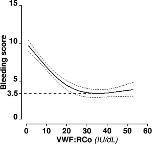
In VWD patients, the severity of bleeding correlates with the degree of reduction of VWF activities: the most common test for VWF activity measures in plasma the VWF:RCo or VWF:GPIb. The correlation between VWD severity and VWF levels has been assessed in 1234 VWD patients included in the retrospective Italian Registry (RENAWI-1)30 and confirmed in a large prospective cohort study (RENAWI-2). Among the 796 cases included in RENAWI-2, only 75 (9.4%) had at least 1 spontaneous bleeding event requiring treatment during the 1-year follow-up period.31 Bleeders had median values of BS and baseline VWF:RCo/FVIII:C levels higher and lower than nonbleeders, respectively. Considering the baseline levels of VWF:RCo, all the patients could be classified as mild, moderate, and severe according to the levels of VWF activity.31 The proportion of mild (31-56 IU/dL), moderate (10-30 IU/dL), and severe (<10 IU/dL) cases included in RENAWI-2 was 34%, 28%, and 38%, respectively, with different distributions within the VWD1, VWD2A, VWD2B, and VWD2M types. Indeed, the BS measured at the time of inclusion was inversely related to baseline levels of VWF:RCo, reaching a plateau at a mean value of 3.5, corresponding to VWF:RCo levels >30 U/dL (Figure 2).
Restricted cubic spline curve showing the age- and sex-adjusted relationship between VWF:RCo plasma levels and BS in all RENAWI-2 patients with VWD. Dotted lines represent 95% confidence intervals. A plateau was found at a mean BS value of 3.5 (dashed horizontal line) that was reached for VWF:RCo levels >30 IU/dL. More detailed information is provided in Federici et al.31
Restricted cubic spline curve showing the age- and sex-adjusted relationship between VWF:RCo plasma levels and BS in all RENAWI-2 patients with VWD. Dotted lines represent 95% confidence intervals. A plateau was found at a mean BS value of 3.5 (dashed horizontal line) that was reached for VWF:RCo levels >30 IU/dL. More detailed information is provided in Federici et al.31
Among other general diagnostic tools used in the past, the bleeding time (BT), the original hallmark of the disease, is not always prolonged and may be normal in patients with mild forms, such as those with VWD1 and VWD2N.1-5 Therefore, it is not particularly useful for diagnosis. Evaluation of closure time (CT) with a platelet function analyzer (PFA-100) gives a rapid and simple measure of VWF-dependent platelet activity at high shear stress; it can be performed on whole blood and therefore can be used instead of the BT in children or when the BT is not feasible. This system is sensitive and reproducible for VWD screening, but the CT is normal in VWD2N and cannot be modified in VWD3 after the administration of VWF/FVIII concentrates.32 Based on these observations, BT and CT are not in the flowchart in the differential diagnosis of VWD types (Figure 1).
First-level laboratory tests
In contrast to hemophilia A, which requires only 2 parameters for diagnosis: a prolonged partial thromboplastin time and low levels of FVIII, several laboratory tests are always necessary to diagnose VWD types (Table 1). Among the tests that are performed sequentially at the first and second levels (Figure 1), the ristocetin cofactor activity (VWF:RCo) explores the interaction of VWF with platelet GPIbα and is still the standard method for measuring VWF activity. VWF:RCo is based on the property of the antibiotic ristocetin to agglutinate formalin-fixed normal platelets in the presence of VWF. This method is specific for VWF abnormalities, but it is not very sensitive (values <15 U/dL not reliable) and not always reproducible (inter-assay and intra-assay CV of 8%–15%). Among several other methods developed more recently, the novel ELISA VWF:RCo assays using recombinant or plasma-derived GPIb offer increased (as low as <1 U/dL) sensitivity and a lower CV (5%–8%).33
VWF antigen (VWF:Ag) is a very sensitive assay as measured by ELISA. VWF:Ag is undetectable in VWD3, is reduced in VWD1, and can be normal in most VWD2A, VWD2B, VWD2M, and VWD2N cases. In patients with normal VWF structure (VWD1 and VWD2N), VWF:RCo values are similar to VWF:Ag (VWF:RCo/Ag ratio >0.6); VWF:RCo/Ag ratios <0.6 are characteristic of VWD2A, VWD2M, and most cases with VWD2B.3 In the past, VWD1 was reported to be the most frequent form of VWD, accounting for ∼70% of cases. A reappraisal of VWD diagnoses after 10 years (1998-2008) in 1234 Italian patients showed only 671/1234 (55%) patients with VWD1 because many cases previously diagnosed as VWD1 were rediagnosed as VWD2A or VWD2M due to discrepant VWF:RCo/Ag ratios.30 The presence of qualitative defects of VWF in previously diagnosed VWD1 has been also reported in 154 families evaluated prospectively by a European study20 ; in this cohort of patients, a VWF:RCo/Ag ratio <0.6 was predictive of structural abnormalities and mutations within specific regions of VWF gene.20 Therefore, we have introduced the cutoff level of 0.6 for the VWF:RCo/Ag ratio in the flowchart (Figure 1).
The procoagulant activity of factor VIII (FVIII:C) is usually very low (1-5 U/dL) in patients with VWD3, who are characterized by undetectable levels of VWF:Ag. In patients with VWD2A, VWD2B, and VWD2M, FVIII:C is normal in most cases. VWF is the carrier of FVIII; in healthy subjects, the proteins are found in the circulation as the FVIII/VWF complex with a FVIII:C/VWF:Ag ratio of 1. The FVIII:C/VWF:Ag ratio can be a useful laboratory marker because a ratio >1 suggests VWD1 and <1 suggests VWD2N.1-5 However, additional tests should be always performed to confirm the diagnosis of patients with VWD1 and VWD2N.
Ristocetin-induced platelet agglutination is measured by mixing different concentrations of ristocetin and patient platelet-rich plasma in the aggregometer. Results are expressed as the concentrations of ristocetin (in milligrams per milliliter) able to induce 30% agglutination. Most VWD types show a low response to ristocetin (>1.2 mg/mL of ristocetin concentration), but an important exception is VWD2B, in which there is hyperresponsiveness to ristocetin (<0.8 mg/mL) due to a higher than normal affinity of VWF for platelet GPIbα.23 A similar enhanced ristocetin-induced platelet agglutination can be found in platelet-type VWD.2 Both VWD2B and platelet-type VWD can be associated with thrombocytopenia.
Second-level laboratory tests
Normal VWF is composed of a complex series of multimers with molecular weights ranging from 800 to 20 000 kDa, which can be analyzed by agarose gel electrophoresis. Low-resolution agarose gels distinguish VWF multimers, which are conventionally indicated as high, intermediate, and low molecular weight. In VWD1, VWD2M, and VWD2N, all multimers are present, whereas in VWD2A, the high- and intermediate-molecular-weight multimers are missing. Most VWD2B show the loss of high-molecular-weight multimers, although there are patients with relatively normal multimers.23 VWF multimeric analysis with high-resolution agarose gels can be useful to further characterize patients with VWD2A (VWD2A subtypes IIC, IID, IIE, IIF, IIG, and IIH), as described previously.1-5
The VWF propeptide (VWFpp) and VWF proteins remain noncovalently associated and stored in alpha-granules of megakaryocytes/platelets or Weibel-Palade bodies in endothelial cells for regulated release. In plasma, VWFpp and mature multimers dissociate and circulate independently. VWFpp circulates in plasma as a homodimer with a half-life of 2-3 hours, whereas mature VWF circulates with a half-life of 8-12 hours.2 For these reasons, the ratio between VWFpp and VWF:Ag has been proposed to identify VWD1 patients with reduced VWF survival.34,35
Desmopressin (1-deamino-8-D-arginine vasopressin, DDAVP) is a synthetic analog of vasopressin that is relatively inexpensive and carries no risk of transmitting blood-borne infectious agents. DDAVP, infused intravenously at a dose of 0.3 μg/kg diluted in 50 mL of saline over 30 minutes, usually increases plasma VWF and FVIII 3-5 times above baseline levels within 30-60 minutes; in general, high VWF and FVIII levels last for 6-8 hours.36,37 A test dose of DDAVP is recommended in VWD patients at the time of diagnosis to establish the individual patterns of biological response and to predict clinical efficacy during bleeding because the responses in a given patient are consistent on different occasions.36,37 A DDAVP challenge test is an important tool for VWD management because VWD patients can be divided according to their biological response into 3 different groups: short half-life, responsive, not responsive.37 An increased ratio of VWFpp/VWF:Ag usually correlates with a short half-life of VWF activity after DDAVP.34,35 Knowledge of the biological response after such an infusion at the time of diagnosis is important because VWD patients can be identified as being unresponsive or having a short-lived responsive to DDAVP. Such patients should be shifted to the use of VWF/FVIII concentrates.1-5
The VWF binding assay to FVIII (VWF:FVIIIB) measures the affinity of VWF for FVIII. In this assay, anti-VWF antibody is coated on wells of a microtiter plate and test plasma is added to the wells. The FVIII/VWF complex in plasma is bound by the antibody, after which FVIII is removed from the complex by a high-ionic-strength buffer. Excess recombinant FVIII (rFVIII) is then added and, after removal of unbound rFVIII, the VWF and the bound rFVIII are assayed.1-5 This assay allows VWD2N to be distinguished from mild to moderate hemophilia A.
Additional, automatic, and novel assays for phenotypic diagnosis
The VWF collagen binding assay (VWF:CB) is particularly sensitive to VWD variants characterized by the absence of the larger VWF multimers38-40 Therefore, VWF:CB is often used as an alternative to multimeric analysis and VWF:CB/Ag ratios are useful in distinguishing VWD2A from VWD2M. However, in rare patients with mutations in the A3 domain (W1745C and S1783A) with normal multimeric structure, the VWF:CB/Ag is abnormal in the presence of normal VWF:RCo.40
Several modifications to the original VWF:RCo assay have been published. Several diagnostics companies have produced more reliable reagents and assays that can be automated on common photo-optical coagulation analyzers. This allows turbidimetric measurements and faster availability. The first commercially available automated VWF:RCo was produced by Siemens (BC VWF reagent) using Siemens BCS analyzers. Instrumentation Laboratory has developed an improved version of the VWF:RCo assay; recombinant wild-type GPIb has been coupled to uniform beads, making the assay completely platelet free. The assay is available in 2 versions based on turbidimetric or chemiluminescence detection; both systems are precise and suitable for VWD diagnosis.41-44 More recently, automatic VWF:GPIb-binding assays independent of ristocetin have been developed and introduced into general use with promising results.45,46
Role of genotype in VWD diagnosis
Molecular diagnosis can be useful to confirm specific VWF defects in VWD families, especially those with VWD2A, VWD2B, VWD2M and VWD2N because mutations are clustered in specific exons of VWF gene.1-5 In VWD3 patients, no specific mutations can be used as molecular markers for the disease because gene defects are spread throughout the entire VWF gene. However, large deletions should be sought because they can be associated with the appearance of allo-antibodies against VWF. In VWD1, the probability of finding mutations within the entire VWF gene is high only when VWF levels are below 30 U/dL. It is still not clear whether most mild VWD1 patients really have a mutation in the VWF locus and the possibility of external modifiers of VWF levels should be considered.1-5
Clinical definition of severe versus mild VWD: bleeding phenotype and score
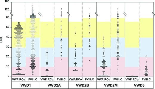
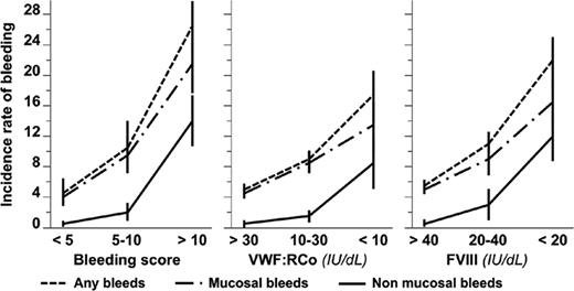
VWD3 is always classified as severe by definition because VWF levels are undetectable in both plasma and platelets with relatively low amounts of FVIII:C (<20 U/dL) in plasma.1-5 Conversely VWD1, VWD2A, VWD2B, VWD2M, and VWD2N can be very heterogeneous and their clinical presentation is strictly correlated with the circulating levels of functional VWF activity. Using such a definition of “clinical severity” based on the levels of defective VWF:RCo and FVIII:C, 3 different groups of VWD could be identified in the 796 Italian patients according to the inclusion criteria of RENAWI-2,31 as shown in Figure 3. Considering the extreme heterogeneity of VWD, the “severe forms of VWD” might represent the “tip of the iceberg” overlying a large number of patients with moderate to mild VWF defects (Figure 4). Although no diagnostic problems occur in moderate to mild VWD with levels of VWF:RCo <30 U/dL, a definite diagnosis of VWD is often difficult to make in patients with very mild VWD forms and VWF:RCo levels >30 U/dL. In fact, it is well known that the physiological changes of VWF levels and the variability of the VWF:RCo assays can obscure mild defects of VWF. In the very mild VWD, the limit between “disease” and “reduced levels of VWF in a normal individual” can be difficult in the absence of bleeding history in other members of the family, as described previously.13-15 In contrast, many mild VWF defects remain undiagnosed in the absence of well-documented personal and family bleeding histories. The use of the analytic approach suggesting that all the 3 major criteria (bleeding, reduced VWF activity, and affected family members) should be satisfied might help to distinguish mild VWD from healthy individuals with low VWF.46 BS, together with threshold levels of VWF:RCo and/or FVIII, has not only been useful to confirm the diagnosis, but should also be considered a predictor of clinical outcomes, as observed recently in a large cohort of Italian patients with different VWD types.31 Indeed, in RENAWI-2, the bleeding rates (any, mucosal, and/or nonmucosal) were different according to different BS, VWF:RCo, and FVIII:C levels (Figure 5). BS >10 was associated with the highest incidence of both mucosal (20.63 per 100 patient-years; 95% confidence interval, 12.20-29.06) and nonmucosal bleeding (12.54 per 100 patient-years; 95% confidence interval, 6.90-19.95).
Distribution of levels of VWF:RCo and FVIII:C activities in the entire cohort of 796 patients included in RENAWI-2.31 Patients with VWD1, VWD2A, VWD2B, VWD2M, or VWD3 (from left to right) are distributed within 3 different groups from the bottom to the top according to their baseline levels of VWF:RCo and FVIII:C. Severe cases (pink area) are characterized by the lowest levels of activities (VWF:RCo <10 and FVIII:C <20 IU/dL); moderate cases (light blue) by the moderately reduced levels of activities (VWF:RCo = 10-30 and FVIII:C = 20-40 IU/dL); mild cases (yellow) by the less reduced levels of activities (VWF:RCo = 31-55 and FVIII:C >40 IU/dL). Note the extreme heterogeneity within VWD1, VWD2A, VWD2B, and VWD2M. More detailed information is provided in Federici et al.31
Distribution of levels of VWF:RCo and FVIII:C activities in the entire cohort of 796 patients included in RENAWI-2.31 Patients with VWD1, VWD2A, VWD2B, VWD2M, or VWD3 (from left to right) are distributed within 3 different groups from the bottom to the top according to their baseline levels of VWF:RCo and FVIII:C. Severe cases (pink area) are characterized by the lowest levels of activities (VWF:RCo <10 and FVIII:C <20 IU/dL); moderate cases (light blue) by the moderately reduced levels of activities (VWF:RCo = 10-30 and FVIII:C = 20-40 IU/dL); mild cases (yellow) by the less reduced levels of activities (VWF:RCo = 31-55 and FVIII:C >40 IU/dL). Note the extreme heterogeneity within VWD1, VWD2A, VWD2B, and VWD2M. More detailed information is provided in Federici et al.31
Pictorial representation of the 3 different degrees of VWD severity according to levels of VWF and FVIII activities: severe, moderate, and mild VWD. In the upper part of the pyramid, “severe VWD” cases (VWD3, VWD2A, and VWD1) are included with levels of VWF:RCo <10 U/dL and/or FVIII:C <20 U/dL; the “moderate VWD” cases (VWD2B, VWD2M, VWD2N, and VWD1) with levels of VWF:RCo 10-30 U/dL and/or FVIII:C 20-40 U/dL; and the “mild VWF” (VWF:RCo 30-50 U/dL and/or FVIII:C 40-60 U/dL) are described in the middle portion and in the base of the pyramid.
Pictorial representation of the 3 different degrees of VWD severity according to levels of VWF and FVIII activities: severe, moderate, and mild VWD. In the upper part of the pyramid, “severe VWD” cases (VWD3, VWD2A, and VWD1) are included with levels of VWF:RCo <10 U/dL and/or FVIII:C <20 U/dL; the “moderate VWD” cases (VWD2B, VWD2M, VWD2N, and VWD1) with levels of VWF:RCo 10-30 U/dL and/or FVIII:C 20-40 U/dL; and the “mild VWF” (VWF:RCo 30-50 U/dL and/or FVIII:C 40-60 U/dL) are described in the middle portion and in the base of the pyramid.
Incidence rate of bleeding. Incidence rate is shown per 100 patient-years as follows: any type (dashed line), mucosal (dash-dotted line), and nonmucosal (solid line) according to BS (left), VWF:RCo (middle), and FVIII (right). Vertical bars represent 95% confidence intervals. More detailed information is provided in Federici et al.31
Incidence rate of bleeding. Incidence rate is shown per 100 patient-years as follows: any type (dashed line), mucosal (dash-dotted line), and nonmucosal (solid line) according to BS (left), VWF:RCo (middle), and FVIII (right). Vertical bars represent 95% confidence intervals. More detailed information is provided in Federici et al.31
Conclusions and future perspectives
VWD is the most common inherited bleeding disorder due to the heterogeneity of VWF defects. The clinical diagnosis and classification of VWD can be difficult because of the widely variable phenotype. The likelihood of diagnosing VWD correctly improves when the clinical assessment of bleeding is correlated with appropriate laboratory studies. Molecular diagnosis can be useful to confirm specific VWF defects in VWD families. It is still not clear whether most mild VWD1 patients really have a mutation in the VWF locus. Despite its complex and heterogeneous nature, VWD can now be efficiently diagnosed and classified in most Western countries. A correct diagnosis and classification of VWD provides the best therapeutic approach to VWD patients.
Acknowledgments
Data on the diagnosis and management of VWD are reported from the Italian retrospective (RENAWI1) and prospective (RENAWI-2) registries of VWD sponsored by a grant from the Italian Ministry of Health. The author thanks all of the members of the Italian Association of Hemophilia Centers who participated in RENAWI-1 and RENAWI-2 and acknowledges the work of Luigi Flaminio Ghilardini, who prepared the figures reported in this manuscript.
Disclosures
Conflict-of-interest disclosure: The author is on the board of directors or an advisory committee for Octapharma, LFB, Instrumentation Laboratories, Grifols, Baxter, and CSL-Behring; has consulted for Octapharma, LFB, Instrumentation Laboratories, Grifols, Baxter, and CSL-Behring; has received honoraria from Octapharma, LFB, Instrumentation Laboratories, Grifols, Baxter, and CSL-Behring; and has been affiliated with the speakers' bureau for Octapharma, LFB, Instrumentation Laboratories, Grifols, Baxter, and CSL-Behring. Off-label drug use: None disclosed.
Correspondence
Augusto B. Federici, MD, Hematology and Transfusion Medicine, L. Sacco University Hospital, Department of Clinical Sciences & Community Health, University of Milan, Via Francesco Sforza 35, 20122 Milan, Italy; Phone: 39-02-5031-9895; Fax: 39-02-5031-9897; e-mail: augusto.federici@unimi.it.


